Coach
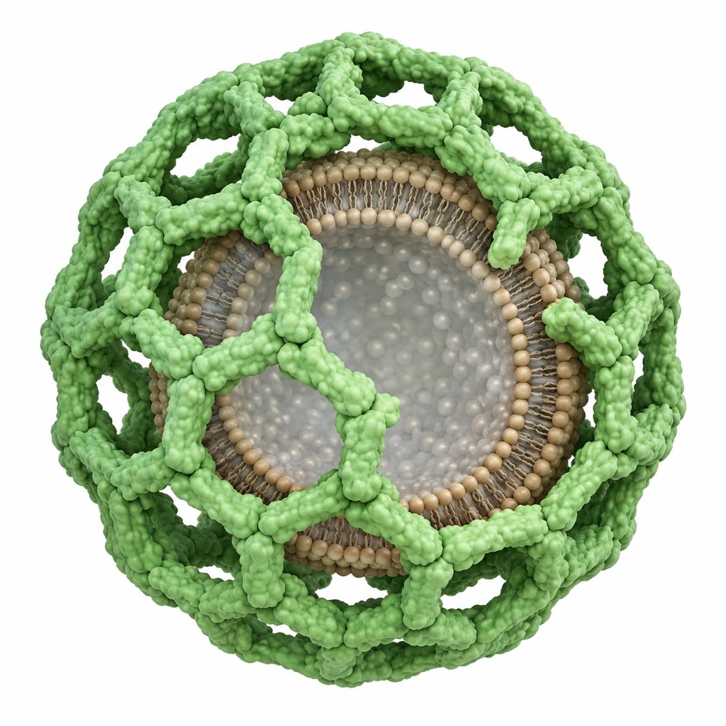
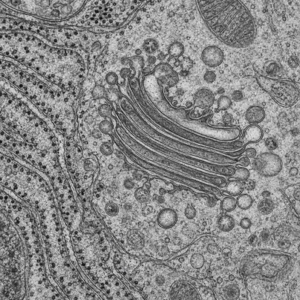
CoachA transport vesicle is shaped by a coat of proteins assembled on the cytosolic face of a membrane, which curves the membrane into a bud of , collects cargo through receptors that span the membrane, and is shed after the vesicle pinches off. Three coats serve the main routes: COPII for vesicles leaving the reticulum for the Golgi, COPI for the return traffic from the Golgi to the reticulum and between Golgi cisternae, and clathrin — a three-legged protein that polymerises into a cage of hexagons and pentagons — for vesicles leaving the trans-Golgi for endosomes and for endocytosis at the plasma membrane, where adaptor proteins link the cage to the receptors being internalised. Each coat is recruited by a small GTPase (Sar1 for COPII, Arf1 for COPI and clathrin) that inserts into the membrane in its GTP form and, on hydrolysing GTP after budding, lets the coat fall off. Retrieval signals keep the compartments distinct: escaped reticulum proteins carrying KDEL are bound in the Golgi by a receptor and carried back in COPI vesicles.