University Biology — Year 3 · Bachelor Year 3
8Membrane Traffic and Protein Sorting
A cell of the pancreas makes some ten million molecules of digestive enzyme a minute, packs them into granules, and releases them into a duct on a signal from the gut — without ever letting a molecule of trypsin loose in its own cytoplasm. A liver cell takes cholesterol out of the blood by swallowing the particles that carry it, whole, at a rate of thousands a minute, and returns each receptor to the surface for the next. A newly made protein destined for the mitochondrion, the nucleus, the lysosome, the membrane or the outside world reaches its address among a dozen compartments with an error rate that would shame a postal service. This chapter is about the sorting: the signals written into proteins, the machines that read them, the vesicles that carry membrane and cargo between compartments, the coats that shape the vesicles and the proteins that make them fuse with the right target, and the two-way traffic — secretion out, endocytosis in — that a eukaryotic cell keeps up every second of its life.
8.1 Signals and addresses
Definition 8.1 (Sorting signals)
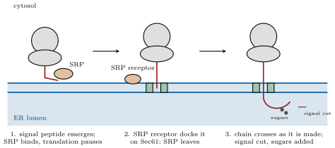
A sorting signal is a segment of a protein, or a modification of it, read by a receptor that delivers the protein to one compartment. The signal peptide, a stretch of residues at the amino terminus with a hydrophobic core, sends a protein into the endoplasmic reticulum as it is being made; from there the default route leads through the Golgi to the plasma membrane or the outside, and further signals divert to the lysosome (a phosphorylated sugar) or back to the reticulum (the carboxy-terminal sequence KDEL). A nuclear localisation signal, a short basic patch such as PKKKRKV, is bound by importins that carry the folded protein through the nuclear pore; a mitochondrial presequence, an amphipathic helix of residues, is recognised by receptors on the outer membrane and threaded, unfolded, through translocases of both membranes; peroxisomal enzymes end in SKL. Proteins with none of these stay in the cytosol. Every signal was found the same way: deleting it leaves the protein in the cytosol, grafting it onto a cytosolic protein sends that protein to the compartment.
Evidence. Blobel and Dobberstein (1975) translated the messenger RNA of an antibody light chain in a cell-free system. Without membranes the product was longer by about twenty residues than the secreted protein; with fragments of endoplasmic reticulum added during translation, the product had the secreted size and was protected from added protease — it was inside the vesicles — whereas membranes added after translation did nothing. The extra amino-terminal segment, the signal peptide, is needed to enter the reticulum, is removed inside it, and works only while the chain is being made: the signal hypothesis, confirmed in every detail when the receptor (the signal-recognition particle, Walter and Blobel 1981) and the channel (Sec61) were purified. ∎
Proposition 8.2 (Membrane proteins and topology)
A hydrophobic stretch of about twenty residues in the middle of a chain being translocated acts as a stop-transfer sequence: the channel opens sideways and releases it into the bilayer as a transmembrane helix, with the part already translocated in the lumen and the rest made into the cytosol. A second such stretch restarts translocation, and a protein with alternating signals ends with membrane-spanning helices. The orientation set in the reticulum is never changed: what faces the lumen of the reticulum faces the lumen of the Golgi and of every vesicle after it, and the outside of the cell after fusion with the plasma membrane. Hence sugars, added only in the lumen, are always on the extracellular face of a surface protein, and the topology of a receptor can be read from where its glycosylation sites lie.
Method 8.3 (Pulse–chase)
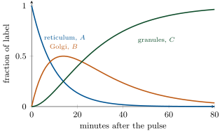
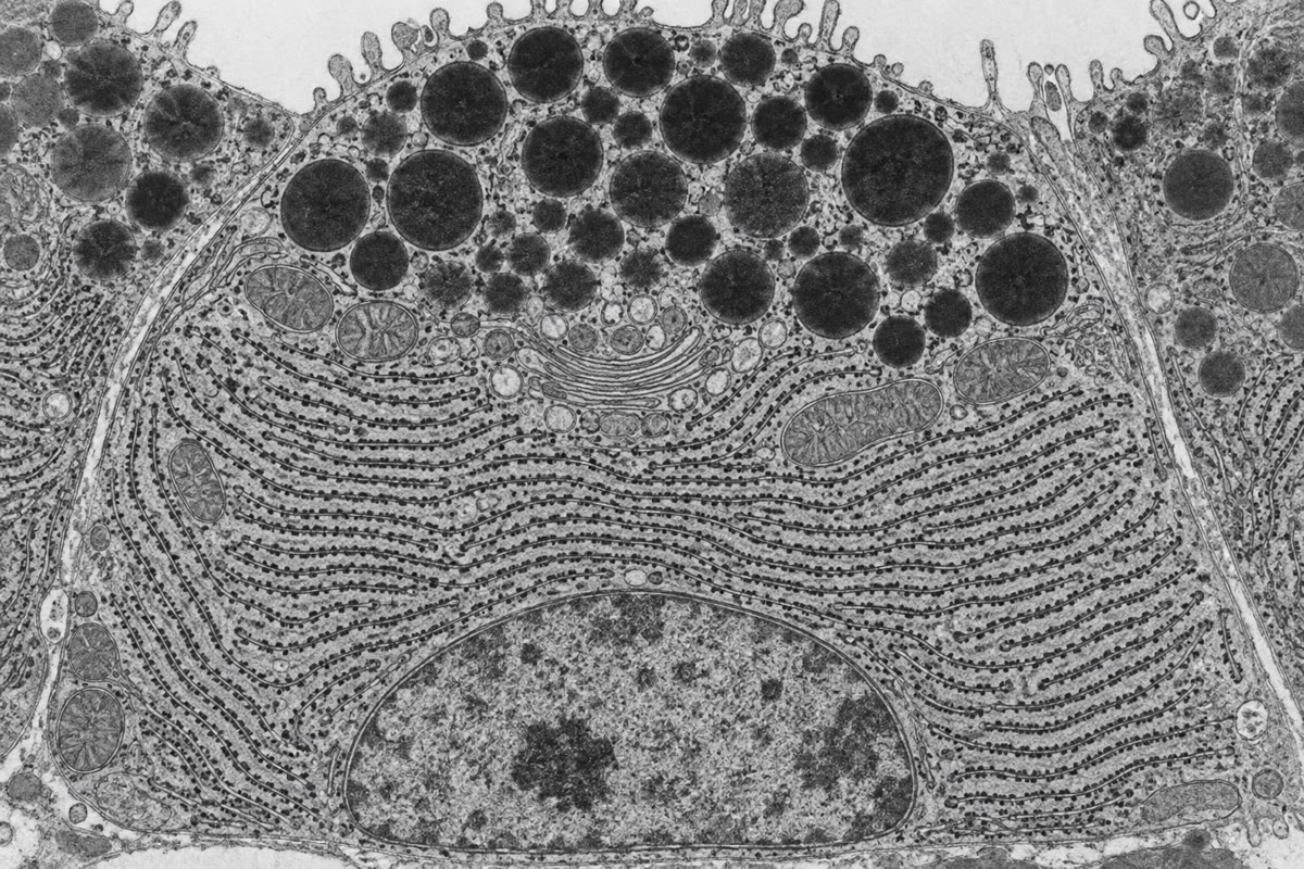
To follow a population of newly made molecules through the cell: (1) expose the cells briefly (the pulse, a few minutes) to a radioactive or otherwise tagged precursor — tritiated leucine for proteins; (2) replace it with an excess of unlabelled precursor (the chase), so that no new label is incorporated; (3) fix samples of cells at intervals and locate the label by autoradiography in electron micrographs, or by isolating the compartments and counting; (4) plot the fraction of label in each compartment against time. The order in which compartments fill and empty is the route; the timing gives the residence time in each. Palade’s laboratory (1960s) applied it to the pancreatic acinar cell and found the label in the rough reticulum at , in the Golgi at , in condensing vacuoles and granules at , and in the duct after an hour and on a signal: the secretory pathway, laid out in time.
Theorem 8.4 (Sequential compartments and the pulse–chase curves)
Let a pulse of labelled protein leave compartment for with first-order rate and leave for with rate , retaining it. With all the label in at ,
and the label in peaks at . The mean residence time in is and in is , whatever the order of the two rates.
Proof. gives the exponential. For , ; try : the derivative is and , so the equation requires , , and as required. Setting : , whence . The mean time a molecule spends in a compartment it leaves at rate is . ∎
Example 8.5 (Palade’s cell in numbers)
Take (ten minutes in the reticulum) and (twenty in the Golgi). The Golgi peaks at , holding then : half the label. At , , , : three quarters of the protein has reached the granules. The observed curves of Palade’s laboratory have this shape, and fitting them was how the residence times were first measured.
8.2 The endoplasmic reticulum: folding, sugars and quality
Definition 8.6 (N-glycosylation and quality control)
As the chain enters the lumen a preassembled oligosaccharide of fourteen sugars (two N-acetylglucosamines, nine mannoses, three glucoses) is transferred from a lipid carrier onto asparagines in the sequence Asn-X-Ser/Thr — N-glycosylation. The three glucoses are then trimmed one by one, and the trimming is a folding clock: a protein still carrying one glucose is held by the chaperone calnexin; a protein whose last glucose has been removed but which is not yet folded is re-glucosylated by an enzyme that recognises exposed hydrophobic patches, and returns to calnexin — the calnexin cycle. Proteins that fail repeatedly are pulled back into the cytosol, ubiquitinated and destroyed by the proteasome: ER-associated degradation (ERAD). When unfolded proteins accumulate faster than they can be folded or destroyed, sensors in the reticulum membrane launch the unfolded protein response: translation is slowed, chaperone genes induced, the reticulum expanded, and, if the stress persists, the cell killed. The commonest mutation of cystic fibrosis (F508 in CFTR) makes a channel that would work but folds too slowly, is caught by ERAD and never reaches the surface; drugs that help it fold are now a treatment.
8.3 Vesicles: budding, targeting, fusion
Definition 8.7 (Coats and vesicle budding)
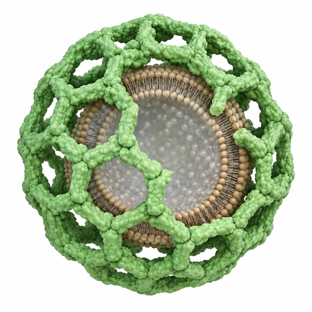
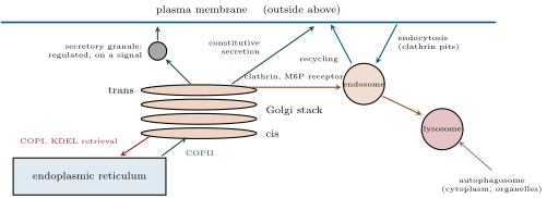
A transport vesicle is shaped by a coat of proteins assembled on the cytosolic face of a membrane, which curves the membrane into a bud of , collects cargo through receptors that span the membrane, and is shed after the vesicle pinches off. Three coats serve the main routes: COPII for vesicles leaving the reticulum for the Golgi, COPI for the return traffic from the Golgi to the reticulum and between Golgi cisternae, and clathrin — a three-legged protein that polymerises into a cage of hexagons and pentagons — for vesicles leaving the trans-Golgi for endosomes and for endocytosis at the plasma membrane, where adaptor proteins link the cage to the receptors being internalised. Each coat is recruited by a small GTPase (Sar1 for COPII, Arf1 for COPI and clathrin) that inserts into the membrane in its GTP form and, on hydrolysing GTP after budding, lets the coat fall off. Retrieval signals keep the compartments distinct: escaped reticulum proteins carrying KDEL are bound in the Golgi by a receptor and carried back in COPI vesicles.
Definition 8.8 (Targeting and fusion)
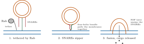
A vesicle finds its target by two layers of recognition. Rab GTPases — some sixty in a human cell, each on the membranes of one compartment — recruit long tethering proteins that catch an incoming vesicle carrying the matching Rab. Then the SNAREs act: a v-SNARE on the vesicle and t-SNAREs on the target, helical proteins anchored in their membranes, wind around each other from their distal ends toward the membranes, and the energy of the four-helix bundle they form pulls the two bilayers together until they fuse. Each SNARE pairing is specific, a second check on the address. After fusion the bundle is pried apart by the ATPase NSF for reuse. Fusion can be made to wait for a signal: in the nerve terminal the SNAREs are held half-zipped by complexin until synaptotagmin, binding calcium that enters when an action potential arrives, completes the zippering in under a millisecond — the release of neurotransmitter treated in the Year 2 volume. The neurotoxins of tetanus and botulism are proteases that cleave SNAREs.
Evidence. Novick and Schekman (1980) isolated temperature-sensitive yeast mutants that stopped secreting at and accumulated protein in whichever compartment their defect blocked — reticulum, Golgi or vesicles piled under the surface; the sec genes defined the steps and, cloned, turned out to encode the coats, the GTPases and the SNAREs. Rothman reconstituted transport between Golgi cisternae in a test tube with purified membranes, cytosol and ATP, and from it purified NSF and the SNAREs; the two approaches converged on the same proteins, and the yeast and mammalian machines proved to be the same. ∎
Proposition 8.9 (The Golgi)
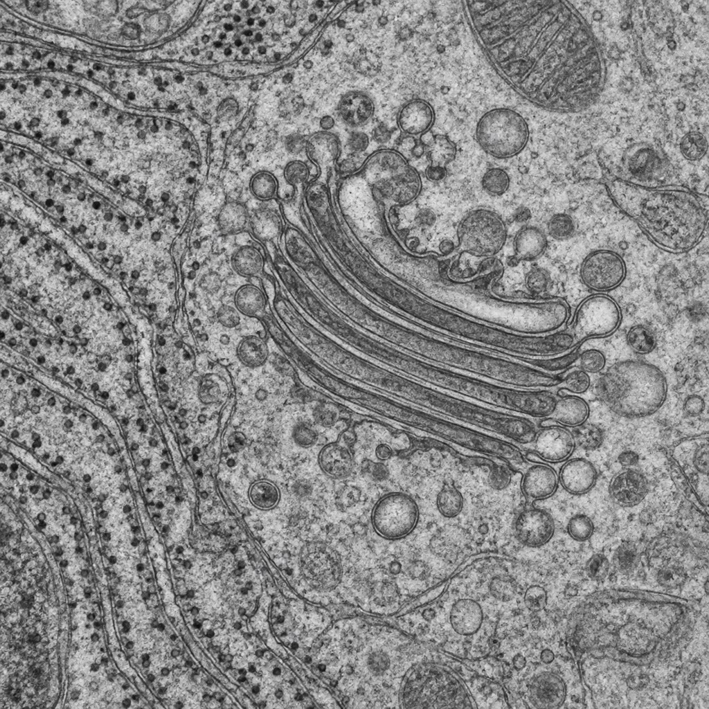
The Golgi is a stack of four to eight flattened cisternae, cis face toward the reticulum, trans face toward the plasma membrane, each cisterna housing a different set of enzymes that trim and rebuild the sugars of passing glycoproteins and add O-linked sugars, sulfate and phosphate. Cargo advances chiefly by cisternal maturation: a new cisterna forms at the cis face from arriving vesicles, moves through the stack as the ones before it do, and matures as COPI vesicles carry each cisterna’s enzymes backward to the younger one behind it; at the trans face it breaks up into vesicles bound for their destinations. There the sorting is decided: lysosomal hydrolases, tagged in the cis Golgi with mannose-6-phosphate, are bound by their receptor and packed into clathrin vesicles for the endosome; regulated secretory proteins aggregate at the mildly acid pH into condensing granules; everything else leaves in the constitutive stream for the surface. In I-cell disease the phosphorylating enzyme is missing, the hydrolases are secreted instead of delivered, and the lysosomes fill with undigested material.
8.4 Endocytosis and the lysosome
Definition 8.10 (Endocytosis)
Receptor-mediated endocytosis concentrates a ligand bound to surface receptors into clathrin-coated pits, which pinch off (the GTPase dynamin constricts the neck) as vesicles of about — a thousand per minute in a fibroblast, turning over the whole surface membrane in an hour. The vesicles fuse into early endosomes, whose lumen is acidified to pH 6 by a proton pump; most ligands release their receptors there, the receptors return to the surface in recycling vesicles, and the ligand travels on as the endosome matures into a late endosome (pH 5.5) and fuses with a lysosome: a compartment at pH 4.5–5 holding some sixty hydrolases — proteases, nucleases, lipases, glycosidases — active only at that pH, so that a leak into the neutral cytosol does little harm. The lysosome also digests the cell’s own material delivered by autophagy: a double membrane grows around a portion of cytoplasm or a worn-out organelle, closes into an autophagosome, and fuses with the lysosome; the products are returned to the cytosol. Autophagy is induced by starvation (it recycles a cell’s own substance for energy) and clears aggregates and damaged mitochondria; its failure is implicated in neurodegeneration.
Evidence. Brown and Goldstein (1970s) followed low-density lipoprotein (LDL), the particle that carries cholesterol in blood, into cultured fibroblasts. LDL bound saturably to some surface sites per cell, was internalised within minutes into coated pits, and its cholesterol was released in lysosomes and switched off the cell’s own cholesterol synthesis. Fibroblasts from patients with familial hypercholesterolaemia bound no LDL (receptor absent) or bound it but did not internalise it (a mutation in the receptor’s cytosolic tail that prevents its capture by clathrin adaptors): the disease was a defect of endocytosis, the receptor’s tail contained the internalisation signal, and the recycling receptor — each making hundreds of round trips — was established as the general mechanism of uptake. ∎
Proposition 8.11 (Uptake through recycling receptors)
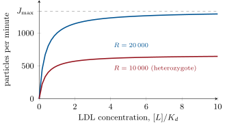
Let a cell have receptors in all, of which are at the surface and inside on their way back. A surface receptor is occupied with probability , an occupied receptor is internalised at rate , and an internalised receptor returns at rate . At steady state the flux of ligand into the cell is
which saturates, as the ligand concentration rises, at : the uptake of a cell is limited not by its receptors alone but by how fast it can bring them back.
Proof. Surface receptors are lost at rate and regained at rate ; at steady state these balance, so . Each internalisation carries one ligand, so , which gives the formula. As the flux tends to — the receptors cycle as fast as the two rates allow, with a fraction of them at the surface at any moment. ∎
Example 8.12 (Cholesterol by the particle)
A fibroblast with LDL receptors, an internalisation time of () and a return time of () takes up, at saturating LDL, particles per minute, with a third of its receptors at the surface at any moment; each particle brings some molecules of cholesterol, two million a minute, enough to build about a tenth of a square micrometre of membrane. A heterozygote for familial hypercholesterolaemia, with half the receptors, takes up half as much; blood cholesterol doubles, and heart disease comes twenty years early. The statins raise by starving the liver cell of its own cholesterol.
Remark 8.13 (One membrane, many compartments)
Every membrane of the secretory and endocytic system is, topologically, one membrane: the lumen of the reticulum is continuous, through vesicles, with the outside of the cell, and a lipid or protein made in the reticulum can reach the surface without ever crossing a bilayer. What keeps the compartments distinct is not walls but traffic — selective packaging out, selective retrieval back, and a specific pair of recognition molecules at every fusion. The compartments are steady states of flows, like the pools of a river, and a cell that stops trafficking loses its geography within hours.
8.5 Exercises
Exercise 8.1 ★
Give the signal and the destination for each: a stretch of twenty hydrophobic residues at the amino terminus; PKKKRKV; an amphipathic amino-terminal helix; KDEL at the carboxy terminus; a mannose-6-phosphate on the sugars.
Solution
Solution of Exercise 8.1.
Hydrophobic amino-terminal stretch: signal peptide, into the endoplasmic reticulum (and by default on to the surface). PKKKRKV: nuclear localisation signal, into the nucleus through the pore. Amphipathic amino-terminal helix: mitochondrial presequence, into the mitochondrial matrix. KDEL: retrieval from the Golgi back to the reticulum. Mannose-6-phosphate: from the trans-Golgi to the endosome and lysosome.
Exercise 8.2 ★
Describe the three key observations of the Blobel–Dobberstein experiment and what each showed.
Solution
Solution of Exercise 8.2.
(1) Without membranes the translated chain was longer than the secreted protein by about twenty residues: a segment is removed during secretion. (2) With membranes present during translation the product had the mature size and was protected from protease: it had entered the vesicles and lost the segment inside them. (3) Membranes added after translation did neither: entry is coupled to synthesis — cotranslational — and the segment, the signal peptide, works only on a nascent chain.
Exercise 8.3 ★
Name the coat and the direction of transport for: reticulum to Golgi; Golgi to reticulum; trans-Golgi to endosome; plasma membrane to endosome.
Exercise 8.4 ★
Why are the sugars of a plasma-membrane protein always on the outside of the cell?
Solution
Solution of Exercise 8.4.
Sugars are added only inside the lumen of the reticulum and Golgi. The lumenal face of every vesicle stays lumenal through budding and fusion, and when a vesicle fuses with the plasma membrane its lumenal face becomes the outside of the cell; the orientation is never inverted, so what was glycosylated inside ends up outside.
Exercise 8.5 ★★
In the model of Theorem 8.4 with and , find when the Golgi label peaks and how much is there at the peak; then the fraction in granules at .
Solution
Solution of Exercise 8.5.
; . At , , so .
Exercise 8.6 ★★
A membrane protein has hydrophobic stretches of residues at positions 30, 90, 150 and 210 and an amino-terminal signal peptide. Draw or describe its topology: how many helices, and which side each segment between them faces. Where can it be glycosylated?
Solution
Solution of Exercise 8.6.
The signal peptide puts the amino terminus into the lumen; each hydrophobic stretch is a stop-transfer or start-transfer alternately, so there are four transmembrane helices (about residues 30–52, 90–112, 150–172, 210–232). Segments: amino terminus lumenal; 52–90 cytosolic; 112–150 lumenal; 172–210 cytosolic; carboxy terminus lumenal. Glycosylation only on the lumenal segments — the amino terminus, 112–150 and the carboxy terminus — which become extracellular at the surface.
Exercise 8.7 ★★
Using Proposition 8.11 with , , , compute and the fraction of receptors at the surface at saturation. What single change would double the uptake?
Solution
Solution of Exercise 8.7.
per minute; surface fraction . Doubling doubles exactly; the return rate is the bottleneck (most receptors are inside), and doubling would give , a factor .
Exercise 8.8 ★★
Predict the fate of lysosomal hydrolases, and the phenotype, in (a) a cell lacking the mannose-6-phosphate receptor, (b) a cell lacking the phosphotransferase that adds the tag, (c) a cell treated with a drug that neutralises endosomal pH.
Solution
Solution of Exercise 8.8.
(a) No receptor: the tagged hydrolases follow the default route and are secreted; lysosomes lack enzymes and fill with undigested material — a storage disease. (b) No phosphotransferase: the same phenotype by a different lesion (I-cell disease), the enzymes untagged and secreted. (c) Neutralised endosomes: the receptor cannot release its cargo, so it is not recycled and the hydrolases are misdelivered or secreted; LDL and other receptors also stop cycling.
Exercise 8.9 ★★
The JD mutation in the LDL receptor changes a tyrosine in the cytosolic tail. Cells binding LDL normally fail to take it up. Explain the mechanism, and predict where the receptors are found on the cell surface relative to coated pits.
Solution
Solution of Exercise 8.9.
The tyrosine belongs to the tail motif that the clathrin adaptor recognises; without it the receptor is not gathered into coated pits. The receptors bind LDL normally but are spread uniformly over the surface, excluded from the pits, and the ligand stays outside — a disease of sorting, not of binding.
Exercise 8.10 ★★★
Solve the pulse–chase model for the case (the formula of the theorem is singular there): show that and find the peak. Then explain why measuring only the granule fraction cannot determine both rates separately.
Solution
Solution of Exercise 8.10.
With , try : , as required, with . Peak where : , . In general , which is symmetric under exchange of and : the granule curve alone cannot say which of the two compartments is the fast one.
Exercise 8.11 ★★★
A fibroblast internalises clathrin vesicles of diameter per minute and has a surface of . How long to internalise the equivalent of the whole surface? Why does the cell not shrink, and what does the balance imply about the rate of exocytosis and the volume of fluid taken in per hour?
Solution
Solution of Exercise 8.11.
Each vesicle has area ; a thousand a minute is , so the whole surface in . The cell does not shrink because membrane returns by exocytosis (recycling vesicles) at the same rate — a balance of flows. Each vesicle holds L; a thousand a minute for an hour is L, about of a cell’s volume per hour.
Exercise 8.12 ★★★
Botulinum toxin cleaves a SNARE in motor nerve terminals; tetanus toxin cleaves the same SNARE in inhibitory interneurons of the spinal cord. Explain why the first causes flaccid and the second spastic paralysis, and why a toxin that blocks fusion is useful in medicine at tiny doses.
Solution
Solution of Exercise 8.12.
Botulinum toxin acts in the motor terminal: no acetylcholine is released, the muscle receives no command and lies limp — flaccid paralysis. Tetanus toxin is carried up the motor axon and across to the inhibitory interneurons that normally restrain motor neurons; with their SNARE cleaved no glycine or GABA is released, the motor neurons fire unopposed and every muscle contracts — spastic paralysis. A few nanograms of botulinum toxin injected into an overactive muscle silence it locally for months (until new SNARE is made and the terminal recovers): a treatment for dystonias, spasticity, squint and wrinkles.
8.6 Problem: A Pancreatic Cell’s Day
Problem 8.1
Weekend problem — a secretory cell’s throughput counted in molecules, vesicles and square micrometres, its pulse–chase curves solved, a liver cell’s cholesterol uptake computed through recycling receptors, and a lysosome acidified proton by proton, ending on the time of the Golgi peak, the vesicles budding per minute and the LDL particles taken up per hour
Data: an acinar cell makes enzyme molecules per minute, each , across. Transport vesicles are spheres of diameter; a granule is a sphere of . Membrane area per lipid molecule , two leaflets. The cell’s reticulum has an area of . Pulse–chase: , . A liver cell: LDL receptors, , , , plasma LDL particle concentration ; cholesterol molecules per particle. A lysosome: sphere of , pH from to , buffering capacity such that protons must be pumped for each one that stays free; a proton pump moves protons per second.
Part I — Throughput.
- What mass of enzyme does the cell make per minute, and per day? Compare with the cell’s own protein content, about .
- How many enzyme molecules fit in a vesicle if they fill of its volume (sphere of )? How many vesicles per minute leave the reticulum?
- How much membrane area do those vesicles carry per minute, and in how long would they consume the whole reticulum if none came back?
- How many lipid molecules is that per minute? What must the cell do to keep its reticulum?
- How many enzyme molecules fit in a granule at the same packing, and how many granules does the cell fill per hour?
- A meal empties granules in ten minutes by fusion with the apical membrane, adding their membrane to a surface of . By what factor does the apical surface grow, and what must follow?
Part II — Timing.
- With the given rates, when does the Golgi label peak, and what fraction is there?
- What fraction of a pulse is in the reticulum, Golgi and granules at ?
- Mean residence times in the two compartments? What is the mean total time from synthesis to granule?
- A drug slows exit from the Golgi to . New peak time and peak fraction; what does the Golgi look like in the microscope?
- Explain why the Golgi peak fraction is raised to a power (give the power) and hence always below .
- If the reticulum step were much faster than the Golgi step, what would approach? Interpret.
Part III — Cholesterol.
- Compute at the plasma LDL concentration, and the uptake in particles per minute and per hour.
- How many cholesterol molecules per hour? The cell’s membranes hold cholesterol molecules; how long to replace them all from LDL alone?
- Compute the fraction of receptors at the surface, and the number of round trips each receptor makes in its lifetime.
- A heterozygote has : recompute . Plasma LDL rises until uptake matches production: by what factor, if is well below saturation?
- A statin doubles in the heterozygote’s liver cells. What happens to plasma LDL, on the same reasoning?
- Explain why the receptor-null homozygote, with plasma LDL five times normal, is still worse off than the arithmetic of question 16 alone suggests.
Part IV — Acid.
- Compute the lysosome’s volume in litres and the number of free protons at pH and at pH .
- How many protons must be pumped in all, with the buffering? How long does that take with pumps?
- The pump moves protons in against the gradient. What electrical problem arises, and how is it solved (think of a counter-ion)?
- Why are the hydrolases made active only at pH 5, and what does this protect?
- Why does the LDL receptor let go of LDL at pH 6 while the mannose-6-phosphate receptor lets go of its cargo at the same pH — what must both proteins have in common?
- Chloroquine is a weak base that crosses membranes uncharged and is trapped once protonated. By what factor does it concentrate in a lysosome at pH relative to the cytosol at pH (the ratio of proton concentrations), and what does that do to the lysosome?
- Summarise: the Golgi peak time (question 7), the vesicles leaving the reticulum per minute (question 2), and the LDL particles taken up per hour (question 13).
Solution
Solution of Problem 8.1.
1. g per minute, a day — twice its own protein content. 2. Vesicle volume nm, of it ; one enzyme molecule nm: about molecules per vesicle; vesicles per minute. 3. Area each, per minute: the reticulum’s in about an hour if nothing came back. 4. nm, two leaflets at : lipids per minute. The cell returns membrane by COPI vesicles and recycling, and synthesises lipid to replace what leaves for good in granules. 5. Granule volume nm, of it : molecules per granule; molecules an hour fill about granules. 6. Each granule adds ; add to : twentyfold. The membrane must be retrieved by compensatory endocytosis within minutes. 7. , . 8. ; ; . 9. and ; mean total . 10. ; : the Golgi swells, engorged with three quarters of the cargo. 11. At the peak ; substituting into gives : the power is . For the exponent is negative and the base above ; for the base is below and the exponent positive — in both cases the value is below (check: here). 12. For , : the label is in the Golgi at once and leaves at — the reticulum step is invisible and the Golgi curve is a simple decay. 13. ; particles per minute, an hour. 14. cholesterol molecules an hour; hours. 15. Surface fraction ; one round trip takes : about trips in twenty hours. 16. per minute. Below saturation , so with half the receptors the plasma LDL must double for the same clearance. 17. back to : clearance restored, plasma LDL falls back toward normal — the statin effect. 18. With no receptors the LDL is cleared only by slow non-specific routes, so it circulates for days instead of hours, is oxidised, and is taken up by the scavenger receptors of macrophages, which become the foam cells of atherosclerotic plaques; and statins, which work by inducing receptors, have nothing to induce. 19. L. At pH : mol — about free protons. At pH : mol, about . 20. protons; pumps at per second move a second: about . 21. Pumping positive charge in makes the lumen positive, and a few thousand charges would stop the pump; chloride enters through a channel (or cations leave) to neutralise the charge. 22. Enzymes inactive at neutral pH do no harm if a lysosome leaks or if they are missorted to the cytosol or the surface: the acid requirement confines digestion to the compartment built for it. 23. Both must carry a pH sensor — histidines, whose protonation near pH 6 changes the binding surface: the LDL receptor folds a domain over its own binding site when protonated, the mannose-6-phosphate receptor loses affinity likewise. 24. -fold concentration; the trapped base consumes protons, raises the lysosomal pH, and inactivates the hydrolases — which is how chloroquine kills malaria parasites in their food vacuole and how it blocks autophagy in experiments. 25. Golgi peak at ; some vesicles leave the reticulum per minute; LDL particles taken up per hour.