 Coach
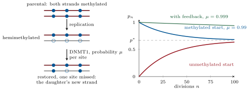
CoachIn vertebrates a methyl group is added to carbon 5 of cytosine almost only in the dinucleotide CpG (a C followed by a G on the same strand), a sequence that is its own complement, so that a methylated CpG is normally methylated on both strands. Some of the CpGs of a human cell are methylated, and methylated promoters are silent: the methyl groups are read by methyl-CpG-binding proteins (MeCP2, MBD1–4) that recruit HDACs and H3K9 methylases, and they block several transcription factors directly. The exceptions are the CpG islands, stretches of about a kilobase, rich in G and C and in CpG, that sit at the promoters of most housekeeping genes and stay unmethylated in normal cells. The methyl group is written by DNA methyltransferases: DNMT3A and DNMT3B perform de novo methylation of unmethylated sites, and DNMT1, which prefers a hemimethylated CpG — one strand methylated, the other not — performs maintenance methylation behind the replication fork. Removal is passive, by replication without maintenance, or active, by the TET enzymes, which oxidise 5-methylcytosine to 5-hydroxymethylcytosine and beyond until base-excision repair replaces it with a plain cytosine.
Examples
Example 1.9 (Why the genome is short of CpG)
The human genome is GC, so if bases were independent a CpG would occur with frequency , about times in the diploid genome. The observed number is about , one fifth of that. The reason is the mark itself: a methylated cytosine that loses its amino group becomes thymine, a normal base that repair cannot recognise as wrong, whereas an unmethylated cytosine deaminates to uracil, which is excised. Over evolutionary time methylated CpGs have mutated into TpG and CpA, and only the unmethylated islands have kept their CpGs. The mark has left its signature in the sequence.
Example 1.11 (Numbers)
Measured values for mammalian cells are and per division at a typical CpG. Then , close to the genome-wide methylated fraction, and : a fully methylated site left to the machinery alone would be half-way back to the average after divisions. A silenced promoter that stays silent for the hundreds of divisions of a lifetime therefore needs more than DNMT1: the methyl-CpG readers recruit H3K9 methylation and the H3K9 readers recruit DNMT3, so that a dense island of marks raises its own toward and lowers the neighbouring toward . Conversely, a cell that loses DNMT1 loses the mark passively: with the fraction of methylated strands halves at each division.