Coach
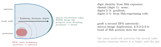
CoachVertebrate embryos are cellular from the start, so their gradients are made by secreted proteins rather than a syncytium’s diffusion, and the same few families do everything: Wnt, Hedgehog, BMP and other TGF- relatives, FGF, Notch and retinoic acid. Spemann and Mangold (1924) grafted a piece of the dorsal lip of a newt gastrula onto the belly of another and obtained a second embryo, head to tail, made mostly of host cells: the graft was an organiser, inducing its neighbours to form a nervous system and axis, and its molecules were found seventy years later to be secreted antagonists (Chordin, Noggin) that block BMP, whose absence lets ectoderm become neural — the default. The neural tube is then patterned dorsoventrally by sonic hedgehog from the notochord and floor plate (high: motor neurons; low: interneurons) against BMP from the roof; and the limb bud by two centres, the zone of polarising activity at its posterior margin secreting Shh (its graft to the anterior margin gives a mirror-image duplication of the digits, six fingers meeting thumb to thumb) and the apical ectodermal ridge at its tip secreting FGFs that keep the underlying cells dividing and specify the proximal-to-distal sequence of humerus, forearm, hand. Grafting, ablation and beads soaked in protein remain the tools; the logic — a local source, a gradient, thresholds, a code of transcription factors — is the fly’s.