University Biology — Year 3 · Bachelor Year 3
23Developmental Genetics and Evolution of Form
A fruit fly’s egg is half a millimetre long, and in the three hours after fertilisation, before a single cell membrane has formed between its six thousand nuclei, it decides which end will be the head, which the tail, where each of fourteen segments will lie, and which of them will carry legs, wings or antennae. It does so with a few dozen genes, turned on in a hierarchy of ever finer stripes, each stripe read from the concentrations of the products of the stripes before it. In 1980 two young scientists in Heidelberg screened thousands of mutant flies for larvae with missing or duplicated parts, and found the whole hierarchy: genes whose loss deletes a block of segments, genes whose loss deletes every other segment, genes whose loss turns every segment back to front. The same year’s work on a gene that turns a fly’s third segment into a copy of its second — giving it four wings, as its ancestors had — opened a set of master genes that, it turned out, lay in the same order along the chromosomes of a mouse and a human, and did the same job. This chapter is about how an embryo computes its own geometry, how a few hundred regulatory genes build every animal body, and how changing when and where they act has produced the diversity of animal form.
23.1 The fly’s coordinates
Definition 23.1 (The segmentation hierarchy)
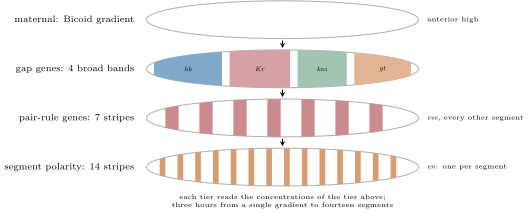
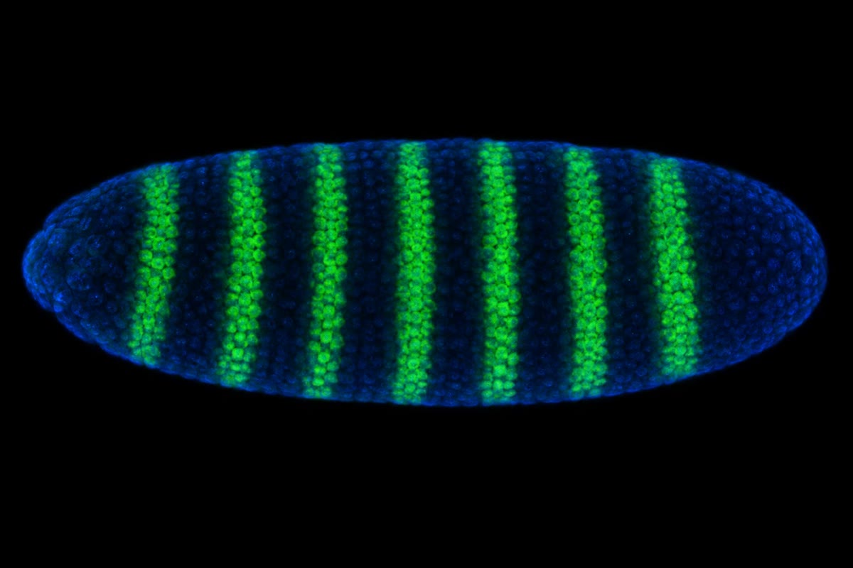
The Drosophila embryo develops for its first thirteen nuclear divisions as a syncytial blastoderm, a single cell whose nuclei share one cytoplasm, so that proteins diffuse freely from where they are made. Four gradients laid down by the mother’s cells set the axes: bicoid mRNA is tethered at the anterior pole and its protein diffuses back to form an exponential gradient from head to tail; nanos mRNA at the posterior pole makes the mirror gradient; hunchback and caudal mRNAs are uniform, but Bicoid protein represses caudal translation and Nanos represses hunchback, so their proteins form gradients too. These maternal-effect products, whose mutant phenotype depends on the mother’s genotype and not the embryo’s, switch on the gap genes (hunchback, Krüppel, knirps, giant) in broad bands, each responding to a range of Bicoid concentration and repressing its neighbours; the gap proteins, in combination, switch on the pair-rule genes (even-skipped, fushi tarazu, hairy) in seven stripes each, alternating; and the pair-rule proteins switch on the segment polarity genes (engrailed, wingless, hedgehog) in fourteen stripes, one per segment, which fix each segment’s front and back and are maintained, after the cells form, by signalling between them. Three hours, four tiers, from a single gradient to fourteen segments — and the mutant names record the screen: hunchback lacks the thorax, Krüppel (“cripple”) the middle, fushi tarazu (“not enough segments”) every other segment, hedgehog has a lawn of bristles.
Theorem 23.2 (A morphogen gradient from synthesis, diffusion and decay)
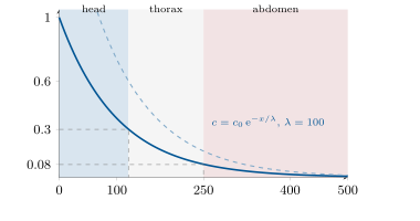
A protein made at one end of a tissue at rate (per unit area), diffusing with coefficient and degraded with rate constant , reaches a steady state in which its concentration along the axis obeys
The decay length sets the gradient’s reach; a cell reads its position by comparing with thresholds — the French flag of Wolpert (1969): above blue, between and white, below red — and the boundary for threshold lies at . Two consequences: doubling the source shifts every boundary back by the same , and a fractional error in reading the concentration is a positional error , so a gradient can place a boundary to within one cell only if the cells read it to within a few per cent. Bicoid has in an egg of : the gradient spans a factor , and the hunchback boundary sits at mid-egg where Bicoid has fallen to about a twelfth of its peak.
Proof. Conservation in a slab between and : the net diffusive inflow balances the degradation at steady state, giving the equation; the solution bounded as is the decaying exponential with . The source: the diffusive flux at , , equals , so . Threshold: gives ; doubling adds to every regardless of . Error: differentiating , . The steady state is reached on the time scale , which for Bicoid () is comparable to the two hours available, so the gradient is read while still settling. ∎
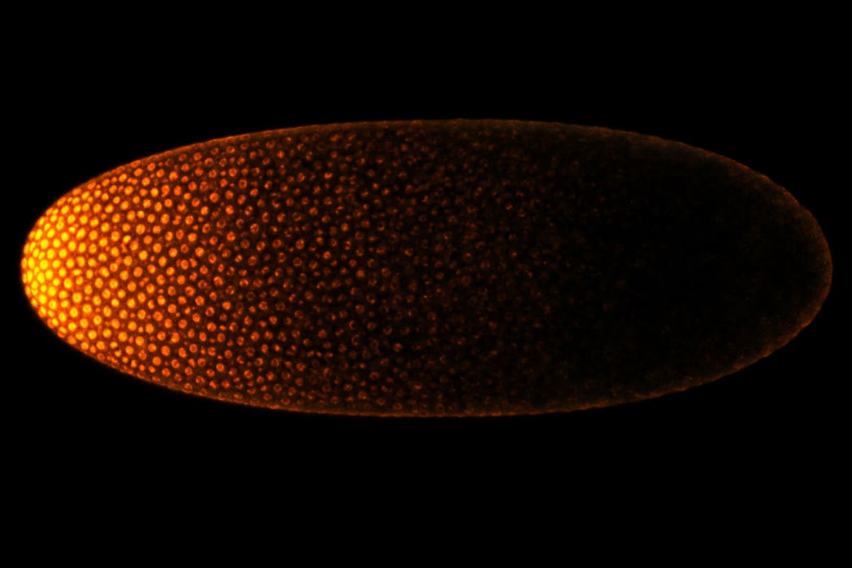
Evidence. Nüsslein-Volhard and Wieschaus (1980) fed flies a mutagen, bred the descendants to homozygosity, and looked at the cuticles of some dead larvae under the microscope for missing or repeated pattern: about a hundred and twenty genes, sorted by their phenotypes into the gap, pair-rule and segment-polarity classes — a hierarchy visible before any molecule was known. Driever and Nüsslein-Volhard (1988) stained embryos for Bicoid protein and saw the exponential gradient from the anterior pole; embryos from mothers with one, two, four or six copies of the gene had gradients of proportionate height, and the head fold and the hunchback boundary moved posteriorly with the dose, as the threshold model predicts and by roughly the per doubling it requires. Injecting bicoid mRNA into the middle of an egg produced a head there. ∎
23.2 Identity: the Hox genes
Definition 23.3 (Homeotic genes and colinearity)
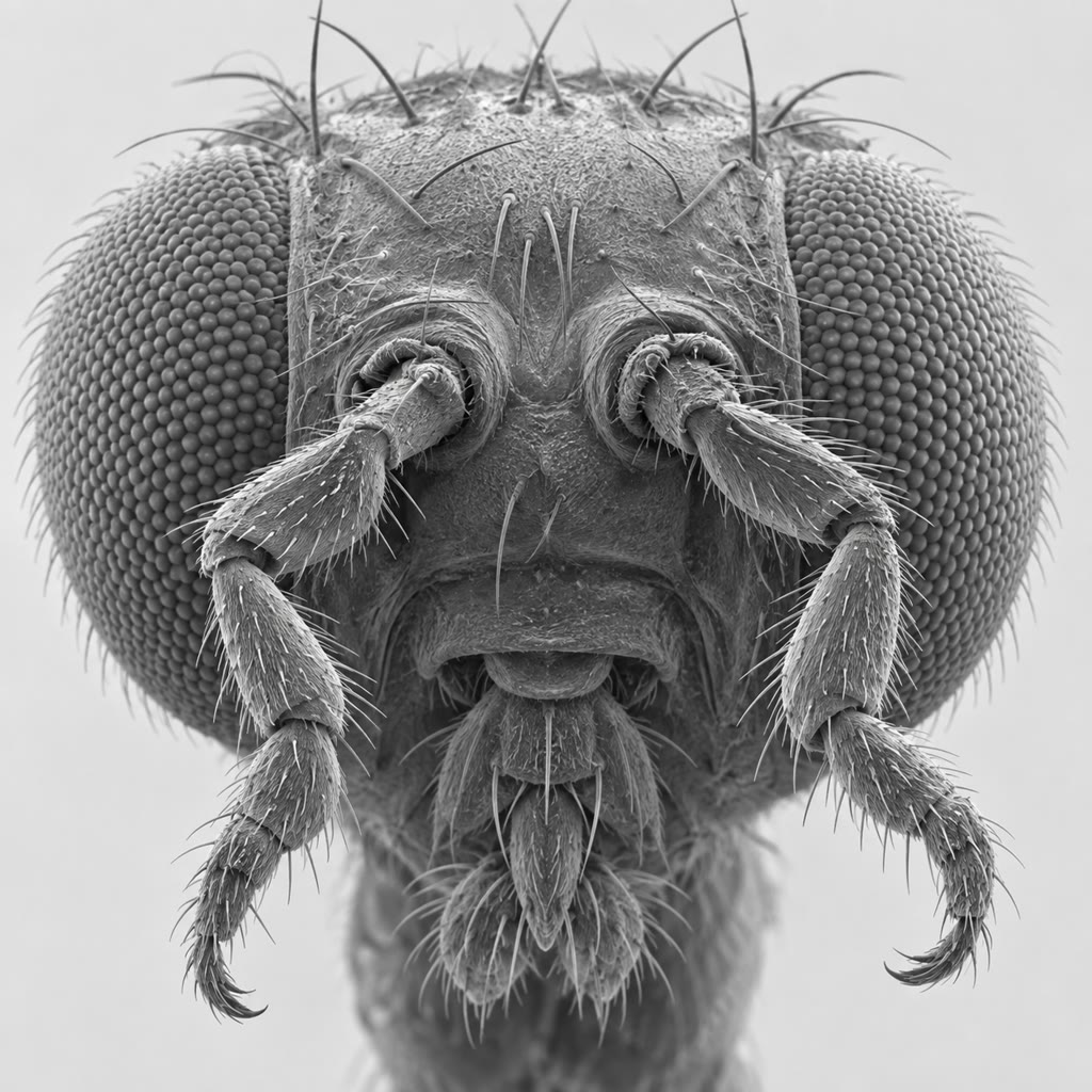
A homeotic mutation turns one body part into another: Bateson’s word (1894) for the abnormalities that “substitute one member of a series for another.” In the fly, Antennapedia dominant mutants grow legs where antennae should be; bithorax mutants (Lewis, 1978) turn the third thoracic segment into a copy of the second, so that the balancers (halteres) become a second pair of wings. The eight genes responsible, the Hox genes, lie in two clusters on one chromosome, and Lewis noticed that their order along the DNA matches the order along the body of the segments they control — colinearity, still not fully explained. Each encodes a transcription factor with the same 60-amino-acid DNA-binding homeobox domain (McGinnis, Gehring, 1984), which was then found in every animal: mice and humans carry four clusters of thirteen genes each, colinear in the same way, expressed in nested domains along the body axis, the later genes in the cluster more posterior. A cell’s identity along the axis is given by the combination of Hox genes it expresses, the Hox code; posterior genes dominate anterior ones (posterior prevalence), so losing a posterior gene lets the segment adopt the identity of the one in front. Hox genes do not build a segment; they tell an already-built segment which one it is, by switching on the batteries of downstream genes appropriate to a leg, a wing, a rib or a lumbar vertebra.
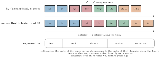
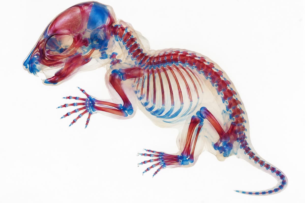
Evidence. Lewis (1978) mapped the bithorax complex as a series of mutations each transforming one segment into the one in front, arranged on the chromosome in body order, and proposed that the genes were sequentially activated along the axis. McGinnis and colleagues (1984) found the homeobox by cross-hybridisation between Antennapedia and Ultrabithorax and then in frogs and mice. The functional equivalence was shown directly: a mouse Hoxb6 gene expressed in a fly produces the Antennapedia phenotype, legs from antennae (1990s); a mouse lacking Hoxc8 has an extra rib on its first lumbar vertebra, the segment having taken the identity of the one in front; and altering Hox expression boundaries in chick and mouse shifts the position where forelimbs form, the neck’s length in a swan being matched by a posterior shift of the same boundary. ∎
23.3 Building a vertebrate
Definition 23.4 (Organisers and signalling centres)
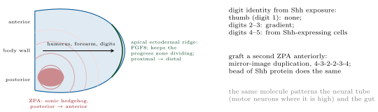
Vertebrate embryos are cellular from the start, so their gradients are made by secreted proteins rather than a syncytium’s diffusion, and the same few families do everything: Wnt, Hedgehog, BMP and other TGF- relatives, FGF, Notch and retinoic acid. Spemann and Mangold (1924) grafted a piece of the dorsal lip of a newt gastrula onto the belly of another and obtained a second embryo, head to tail, made mostly of host cells: the graft was an organiser, inducing its neighbours to form a nervous system and axis, and its molecules were found seventy years later to be secreted antagonists (Chordin, Noggin) that block BMP, whose absence lets ectoderm become neural — the default. The neural tube is then patterned dorsoventrally by sonic hedgehog from the notochord and floor plate (high: motor neurons; low: interneurons) against BMP from the roof; and the limb bud by two centres, the zone of polarising activity at its posterior margin secreting Shh (its graft to the anterior margin gives a mirror-image duplication of the digits, six fingers meeting thumb to thumb) and the apical ectodermal ridge at its tip secreting FGFs that keep the underlying cells dividing and specify the proximal-to-distal sequence of humerus, forearm, hand. Grafting, ablation and beads soaked in protein remain the tools; the logic — a local source, a gradient, thresholds, a code of transcription factors — is the fly’s.
Proposition 23.5 (Spontaneous pattern: Turing’s mechanism)
Not every pattern is read from a pre-existing gradient. Turing (1952) showed that two diffusing substances that react — an activator that stimulates its own production and that of an inhibitor, and an inhibitor that suppresses the activator and diffuses much faster — can turn a uniform field into a regular array of peaks spontaneously: a small local excess of activator grows by self-enhancement while the inhibitor it produces spreads out and prevents new peaks forming nearby, so peaks appear at a spacing set by the inhibitor’s range, , and not by any external coordinate. The mechanism, admitted here in its mathematics, is the best account of the spots and stripes of animal coats, the spacing of hair follicles and feather buds, the ridges of the palate, the digits of the hand (whose number rises when the pattern’s wavelength is shortened by lowering Hox activity), and the stripes of zebrafish, where the interacting cells — pigment cells that repel neighbours of one kind and attract those of another at a distance — have been identified and their stripes made to re-form after laser ablation exactly as the theory predicts.
Proof. Admitted at this level. ∎
23.4 Evolution of form
Definition 23.6 (Evo-devo)
The genes that build animals are an ancient and shared toolkit: a few hundred transcription factors and signalling pathways present in the common ancestor of all animals, and animal diversity comes mostly from changes in when, where and how much they are expressed rather than in what they encode. Deep homology: the gene Pax6 directs eye formation in flies, squid and mice, whose eyes are not homologous as organs — a mouse Pax6 expressed on a fly’s leg makes an ectopic fly eye there (Halder, Gehring, 1995). Cis-regulatory evolution: the loss of pelvic spines in freshwater sticklebacks is the deletion of one enhancer of the Pitx1 gene, the protein untouched and still serving elsewhere; the loss of wing spots in some fruit flies, a change in an enhancer of yellow; the loss of legs in snakes, a broken enhancer of sonic hedgehog in the limb. Hox shifts: the boundary of Hoxc6 expression marks the first thoracic vertebra in mouse, chick, goose and snake alike, at vertebra 7, 14, 23 and 3; insects’ loss of abdominal legs was the Hox protein Ubx acquiring a repressive domain that switches off the leg gene Distal-less, where in crustaceans it does not. Heterochrony, a change in the timing of a developmental programme, made the axolotl a permanent larva and the human face a juvenile ape’s. The old question of how new forms arise becomes a question about regulatory DNA: most of the genome that matters for form is not gene but switch.
Example 23.7 (Four wings)
The ancestors of flies had four wings, as dragonflies and bees still do; flies have two, the hind pair reduced to the balancers. In flies, Ultrabithorax is expressed in the third thoracic segment and represses the wing programme there — some hundred target genes, a haltere being a wing with those genes switched off. Lewis’s triple-mutant fly, lacking Ubx function in the third segment, grows a second pair of wings: not a new structure but the release of an old one. In butterflies Ubx is expressed in the hindwing as well, and there it does not suppress the wing but changes its pattern: the same protein, a different set of targets. Four hundred million years of dipteran evolution turned on one gene’s decision in one segment, and a single mutation undoes it in a generation — which is why the history of form is legible in the genes that build it.
Remark 23.8 (Geometry from chemistry)
An embryo has no blueprint and no surveyor. It has a few gradients made by diffusion and decay, cells that read them against thresholds and switch on transcription factors, and a code of those factors that tells each region what to become; it has local organisers that induce their neighbours, and reactions that pattern themselves. The mathematics is that of first-order kinetics and diffusion — an exponential, a square root, a threshold — and it explains not only how a fly’s head is placed but why doubling a gene shifts a boundary by a fixed distance, why six fingers appear when a signal is duplicated, and why a snake has three hundred ribs. Evolution edits the switches, not the machine: the toolkit of a sponge and a human is nearly the same, and what differs is the wiring.
23.5 Exercises
Exercise 23.1 ★
Name the four tiers of the fly’s segmentation hierarchy, one gene of each, and the mutant phenotype that placed it in its class.
Solution
Solution of Exercise 23.1.
Maternal-effect: bicoid, embryos from mutant mothers lack head and thorax. Gap: Krüppel, a contiguous block of segments missing. Pair-rule: fushi tarazu (or even-skipped), every other segment missing. Segment polarity: hedgehog (or engrailed), part of each segment replaced by a mirror image of the rest.
Exercise 23.2 ★
What is a maternal-effect gene? A female homozygous for a bicoid null mutation is mated to a wild-type male: describe her embryos and explain why their own genotype does not save them.
Solution
Solution of Exercise 23.2.
A gene whose product is deposited in the egg by the mother, so that the embryo’s phenotype follows her genotype. Her embryos lack head and thorax and carry posterior structures at both ends; they die. The paternal wild-type allele is in the zygote, but bicoid mRNA is made during oogenesis by the mother’s nurse cells and anchored at the pole before fertilisation; the zygote’s own copy would be transcribed hours later, after the gradient’s time has passed.
Exercise 23.3 ★
State colinearity and posterior prevalence. Predict the phenotype of a mouse lacking Hoxc8 and of a fly expressing Antennapedia in its head.
Solution
Solution of Exercise 23.3.
Colinearity: the order of Hox genes along the chromosome matches the order of their expression domains along the body. Posterior prevalence: where several are expressed, the most posterior one sets the identity. A mouse lacking Hoxc8 shows an anterior transformation — its first lumbar vertebra takes the identity of a thoracic one and carries a rib. Antennapedia in the head converts antennae to legs, the identity of the second thoracic segment.
Exercise 23.4 ★
What did the Spemann–Mangold graft show, and what were the organiser’s molecules found to be? Why is neural fate called the “default”?
Solution
Solution of Exercise 23.4.
A grafted dorsal lip induced a second complete axis made mostly of host cells: the graft instructed its neighbours — an organiser. Its molecules are secreted BMP antagonists (Chordin, Noggin, Follistatin). Ectoderm becomes neural when BMP signalling is absent and epidermis when BMP is present, so neural is what ectoderm does when told nothing: the organiser works by silencing an instruction.
Exercise 23.5 ★★
Bicoid: , half-life . Compute , , and the ratio of concentrations at the anterior pole and at mid-egg ().
Solution
Solution of Exercise 23.5.
; ; ratio .
Exercise 23.6 ★★
With , a boundary sits at . Where does it sit if the mother carries four copies of bicoid instead of two? One copy? Express the shifts as a fraction of a egg.
Solution
Solution of Exercise 23.6.
Four copies double : the boundary moves back by to ; one copy halves it: forward by to . Each shift is of egg length — larger than observed, which is itself evidence for mechanisms that damp the dose dependence.
Exercise 23.7 ★★
Nuclei at the blastoderm are apart. To place a boundary to within one nucleus with , how precisely must a nucleus read the Bicoid concentration? If the nucleus counts molecules over a time and the count is Poisson, how many must it count?
Solution
Solution of Exercise 23.7.
: for , . A Poisson count has relative error , so molecules must be counted — a few per cent of the molecules present in a nucleus, or a single count integrated over time.
Exercise 23.8 ★★
The gradient approaches steady state on the time scale . With a half-life of , what fraction of the steady-state concentration is reached at , and ? What does this imply for an embryo that must read the gradient by ?
Solution
Solution of Exercise 23.8.
: gives at , at , at , at . The gradient is still rising by several per cent while it is read, which would shift a boundary by , about one nucleus, over the reading window: the embryo must either read at a fixed time or use a read-out insensitive to the overall level.
Exercise 23.9 ★★
A ZPA is grafted to the anterior margin of a chick limb bud. Predict the digit pattern, and the pattern if instead a bead releasing a low dose of Shh is implanted there.
Solution
Solution of Exercise 23.9.
A second ZPA gives a mirror-image duplication, 4-3-2-2-3-4: the anterior cells now see high Shh and become posterior digits. A bead releasing little Shh gives a partial duplication — an extra anterior digit 2, say 2-2-3-4 — because the anterior cells see only the low concentration that specifies digit 2.
Exercise 23.10 ★★★
A morphogen is made throughout a tissue of length at rate per unit volume, degraded at rate , and destroyed at the boundary (an absorbing sink), with no flux at . Solve for and sketch it. How does this gradient’s shape differ from the source-at-one-end case, and what happens when ?
Solution
Solution of Exercise 23.10.
Particular solution ; homogeneous solutions , ; no flux at kills the ; fixes the amplitude: . The profile is a plateau near that falls to zero in a boundary layer of width at the sink — not an exponential from a point, but a level field with an edge; the highest concentration is farthest from the sink. For , and : a parabola independent of , since molecules reach the sink before they can decay.
Exercise 23.11 ★★★
Two thresholds at and divide a gradient into three regions. Show that the widths of the first two regions do not depend on , and explain why an embryo whose length varies between individuals ( to ) needs something more than a single exponential to scale its pattern. Propose one mechanism.
Solution
Solution of Exercise 23.11.
and : the first region is wide, the second , both independent of , which shifts them together. The third region, , absorbs all the variation in egg length: in a egg with the abdomen is , in a egg — the pattern is not scaled. Mechanisms: a second gradient from the posterior pole (Nanos, Caudal, or the terminal system) read together with Bicoid, so that positions are fixed by the ratio; or an “expander” molecule made where the morphogen is low that lengthens until the gradient fills the egg.
Exercise 23.12 ★★★
Sticklebacks lost pelvic spines by deleting a Pitx1 enhancer, snakes lost limbs by breaking a sonic hedgehog enhancer. Explain why regulatory changes rather than coding changes are the usual route to a lost or altered structure, in terms of pleiotropy and of what the same protein does elsewhere.
Solution
Solution of Exercise 23.12.
A coding change alters the protein wherever it acts: Pitx1 also builds the pituitary and jaw, sonic hedgehog also patterns the neural tube, gut and teeth, so a broken protein is lethal or pleiotropically crippling. An enhancer drives expression in one tissue at one time; deleting it removes the structure that tissue would have built and leaves every other use of the protein intact. Enhancers are modular and often partly redundant, so intermediate steps are viable, and the same route — a switch lost — has been taken independently in many stickleback lakes.
23.6 Problem: The French Flag
Problem 23.1
Weekend problem — an egg’s coordinate system computed from first-order kinetics: the Bicoid gradient’s length, source and time to form, the boundaries it draws and their precision, what gene dosage and egg size do to them, and the Hox code read downstream, ending on the decay length, the shift per doubling and the positional precision
Data: egg length ; Bicoid , half-life ; source flux such that with two maternal copies; nuclear spacing at cycle 14; thresholds (head/thorax) and (hunchback boundary) for the two-copy case. Read-out noise for a single nucleus. Avogadro’s number ; a nucleus is a sphere of radius .
Part I — The gradient.
- Compute from the half-life, in , and the lifetime in minutes.
- Compute in micrometres. How many decay lengths is the egg?
- The concentration falls by what factor from pole to pole? By what factor over one nuclear spacing at mid-egg?
- Compute the source flux in , and the number of molecules entering a patch of the anterior pole per second.
- Number of Bicoid molecules in a nucleus at the pole () and at mid-egg.
- Fraction of the steady state reached at and (). Is the gradient steady when the boundaries are drawn, at about ?
Part II — Boundaries.
- Positions of the two boundaries, in and as fractions of egg length.
- The mother has four copies: new , and the two boundary positions. By how much did each move? Six copies?
- One copy: positions. Which regions grew, which shrank?
- Positional error from a read-out noise, in micrometres and in nuclear spacings.
- If the error must be one nucleus, what read-out precision is needed? If the nucleus averages over independent readings and the noise falls as , how many readings?
- A nucleus at the hunchback boundary holds the number computed in question 5. What is the Poisson noise of a single instantaneous count? Compare with and comment.
Part III — Scaling and robustness.
- Eggs vary from to . If and are fixed, where does the hunchback boundary fall as a fraction of egg length in each? Is the pattern scaled?
- Suppose instead that scales as (the same lifetime, but a source whose spread grows with the egg). Show that is then constant and the pattern scales. Is this plausible?
- The hunchback gene responds to Bicoid with a Hill coefficient of : its expression is . Over how many micrometres does expression go from to around the boundary? Compare with one nuclear spacing.
- Explain why a steep response sharpens a boundary but does not by itself reduce the positional error due to concentration noise.
- Mutual repression between gap genes sharpens and stabilises their boundaries. Argue in a sentence why two genes that repress each other cannot both be on in the same nucleus at steady state.
- Temperature raises by and by per degree. Change in per degree, and the boundary shift for a warmer room. Why might embryos need a compensating mechanism?
Part IV — Identity.
- The Hox code: with eight fly Hox genes each on or off, how many codes are possible? How many are used (fourteen segments)? Why does posterior prevalence reduce the effective number?
- A fly lacks Ubx in its third thoracic segment. What does the segment become, and why is the result four wings and not a new structure?
- A mouse’s Hoxc6 boundary lies at vertebra 7, a goose’s at 23. What does this predict about their necks, and what changed in evolution: the gene, or its expression?
- A ZPA graft gives digits 4-3-2-2-3-4. Explain the pattern from two Shh gradients meeting in the middle.
- Halder and Gehring expressed mouse Pax6 in a fly’s leg and obtained a fly eye there. What does this show about the gene, and what does it not show about the eyes of flies and mice?
- The sixth finger: shortening Turing’s wavelength adds a digit. With five digits across a hand plate, what wavelength change gives six? Which parameter of the reaction–diffusion system would produce it?
- Summarise: (question 2), the boundary shift per doubling of the source (question 8), and the positional error from noise (question 10).
Solution
Solution of Problem 23.1.
1. ; . 2. ; the egg is . 3. from pole to pole; , an change per nucleus. 4. . One nM is molecules per , so about molecules per per second. 5. Nuclear volume . At , molecules per : molecules. At mid-egg : about molecules. 6. at : ; at : . The gradient is within of steady when read — still rising by an amount that would move a boundary by , a nucleus. 7. (); (). 8. Four copies: ; both boundaries move back by , to and . Six copies: , to and . 9. One copy: forward by , to and . The head region shrank from to ; the thorax kept its width () and moved forward; the abdomen grew from to . 10. , nuclear spacings. 11. One nucleus: . From , : two independent readings (or the average of two neighbouring nuclei) suffice. 12. : a single instantaneous count is already more precise than the assumed, so the observed noise is not counting noise alone but comes from binding kinetics and the read-out machinery — and averaging over time improves on it. 13. and of egg length: an discrepancy, six nuclei. The pattern does not scale with a fixed . 14. If with fixed, and is the same in every egg: perfect scaling. But is a property of the molecule and the cytoplasm, not of the egg’s size, so this is implausible as stated; scaling in real embryos is partial and comes from other read-outs. 15. at and at : a factor in concentration, — eleven nuclei, far wider than the observed boundary of two or three; the gradient’s read-out alone is not sharp enough. 16. Steepness maps a given concentration to on or off more decisively, so the transition region is narrower; but a error in the concentration still moves the point where the threshold is crossed by : the response can be a perfect step and the step still lands in the wrong place. 17. If both were on, each would be repressing the other, so at least one would be driven off; with cooperative repression the only stable states are one-on-one-off, and a boundary between them is a switch, not a slope. 18. per degree: falls per degree, for , to ; the hunchback boundary moves from to , three nuclei forward. Flies develop between and with normal proportions, so something compensates — a source or read-out that shifts with temperature the other way. 19. codes; fourteen used. With posterior prevalence only the most posterior gene on matters, so at most nine distinct identities (eight genes, or none): the code is a ranking, not a binary word. 20. The third thoracic segment becomes a second: Ubx repressed the wing programme that a thoracic segment runs by default, and removing the repressor releases the ancestral state — four wings, as in the fly’s four-winged ancestors. 21. The goose’s cervical region runs to vertebra 22: a long neck of many vertebrae, the mouse’s seven. The gene is the same; the anterior limit of its expression moved posteriorly — a regulatory change in where a Hox gene is switched on. 22. Each margin is now a source; Shh is highest at both edges (digit 4), falls toward the centre (3, then 2), and never reaches the low level that specifies digit 1, so the middle holds two digits 2 back to back: 4-3-2-2-3-4. 23. Pax6 is a conserved master regulator that can trigger whatever eye programme the host carries: the fly built a fly eye, so the mouse gene carries no mouse-eye information, only the command “build an eye here.” It shows deep homology of the gene network, not homology of the organs: the compound eye and the camera eye evolved separately from an ancestor that had a Pax6 photoreceptor patch. 24. Five digits across is a wavelength of ; six needs , a shortening of a sixth. Since , raise the inhibitor’s decay by or lower its diffusion by ; in the hand plate, reducing the dose of the posterior Hox genes does it, and the mutant mice have six, seven or more thin digits. 25. ; a doubling of the source shifts every boundary by ; noise in the concentration is , about one nucleus, of positional error.