 Coach
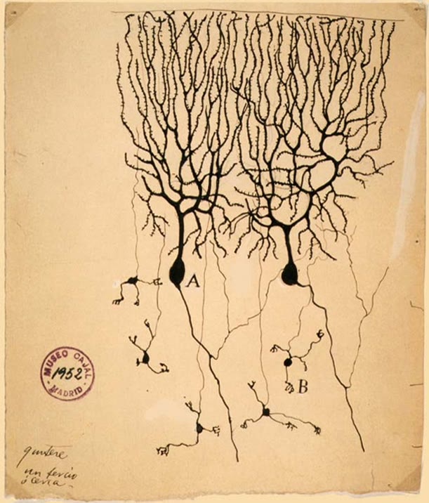
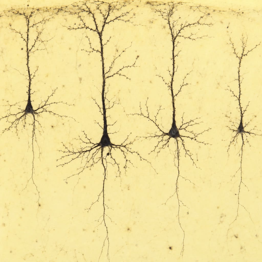
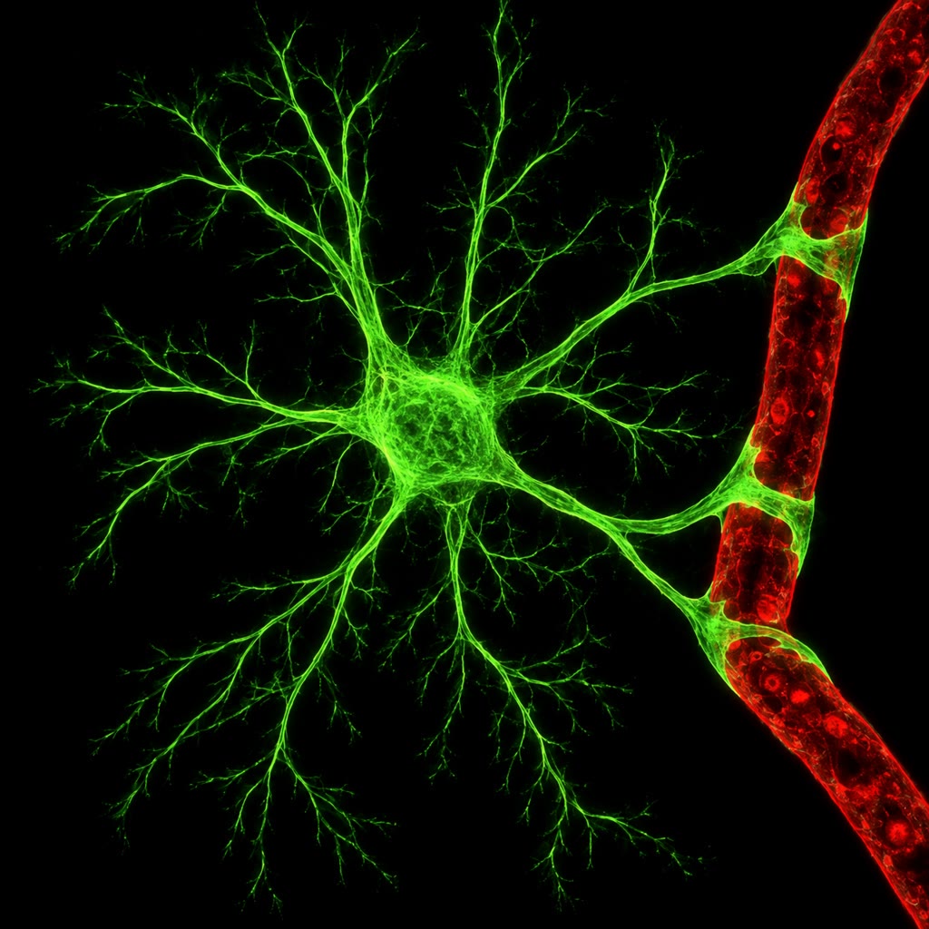
CoachNeurons come in a few architectures that recur across the brain: pyramidal cells, the excitatory projection neurons of the cortex, with a long apical dendrite and an axon that may travel a metre; Purkinje cells of the cerebellum, whose flat dendritic tree receives two hundred thousand synapses; sensory neurons with one process to the periphery and one to the cord; and interneurons, short-axon cells that stay within a region, most of them inhibitory. About four in five cortical neurons release glutamate and excite; one in five release GABA and inhibit. The glia, as numerous as the neurons, do the rest. Astrocytes wrap synapses and blood vessels: they take up the potassium that firing neurons release and the glutamate that synapses spill, supply lactate, regulate blood flow locally, and induce and maintain the blood–brain barrier. Oligodendrocytes in the central nervous system and Schwann cells in the periphery wrap axons in myelin, dozens of layers of membrane that insulate the axon between the nodes where the channels sit. Microglia are the brain’s resident macrophages, pruning synapses in development and clearing debris. Ependymal cells line the ventricles and make the cerebrospinal fluid.
Examples
Example 17.3 (Numbers for a dendrite and two axons)
A dendrite of with and : — a synaptic potential at the tip of a millimetre-long dendrite reaches the cell body at a third of its size, which is why the geometry of a dendritic tree is part of its computation. , the window within which inputs must arrive to sum. A squid giant axon of , unmyelinated, conducts at about ; to do the same a vertebrate uses a myelinated axon of , fifteen thousand times smaller in cross-section — the reason a vertebrate nerve can carry ten thousand fast fibres in the space of one squid axon. A motor axon conducts at ; when multiple sclerosis strips its myelin the velocity falls tenfold and conduction may fail altogether at the bare stretches.