Biology · Glossary

What is Structural methods?

Definition 7.6 University Biology — Year 3 · Chapter 7 — Structural Biology of Proteins

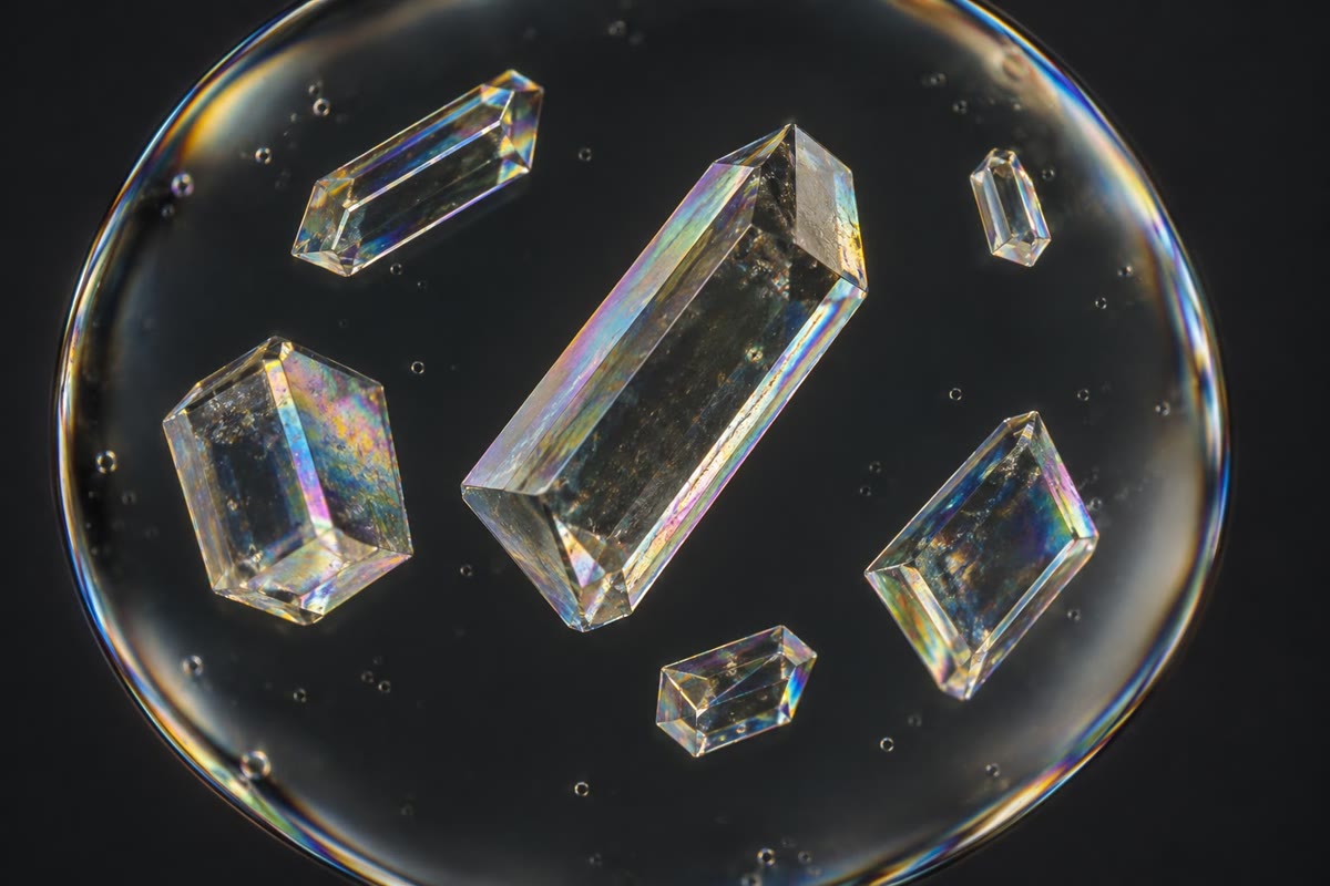
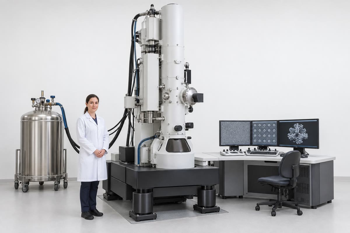
X-ray crystallography grows a crystal of the protein, exposes it to a beam of X-rays and records the diffraction pattern; the positions of the spots give the lattice and their intensities, once the phases of the waves are recovered, give the electron density of the unit cell, into which the chain is built. Nuclear magnetic resonance (NMR) measures, in solution, the couplings between the nuclei of atoms close in space, from which distances and hence a structure are computed; it works for proteins under about 40kDa40\,\mathrm{kDa} and reports their motions. Cryo-electron microscopy images thousands to millions of single molecules frozen in a thin film of vitreous ice, each in a random orientation, and reconstructs the three-dimensional density by combining them; since direct electron detectors (2013) it reaches atomic resolution and needs no crystal, so that large complexes, membrane proteins and flexible assemblies that never crystallised are now solved routinely. The resolution of a structure is the smallest spacing at which two features are separated: at 0.35nm0.35\,\mathrm{nm} the backbone and large side chains are placed, at 0.2nm0.2\,\mathrm{nm} every atom, at 0.1nm0.1\,\mathrm{nm} hydrogens.

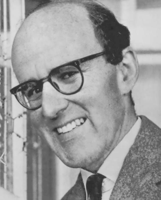
Max Perutz in 1962, the year of his Nobel prize for the structure of haemoglobin (Associated Press, public domain). Centre: protein crystals in a hanging drop, the starting point of crystallography. Right: a cryo-electron microscope, the instrument that made crystals unnecessary. Max Perutz in 1962, the year of his Nobel prize for the structure of haemoglobin (Associated Press, public domain). Centre: protein crystals in a hanging drop, the starting point of crystallography. Right: a cryo-electron microscope, the instrument that made crystals unnecessary. Max Perutz in 1962, the year of his Nobel prize for the structure of haemoglobin (Associated Press, public domain). Centre: protein crystals in a hanging drop, the starting point of crystallography. Right: a cryo-electron microscope, the instrument that made crystals unnecessary.
Max Perutz in 1962, the year of his Nobel prize for the structure of haemoglobin (Associated Press, public domain). Centre: protein crystals in a hanging drop, the starting point of crystallography. Right: a cryo-electron microscope, the instrument that made crystals unnecessary.
Read in context →