Coach
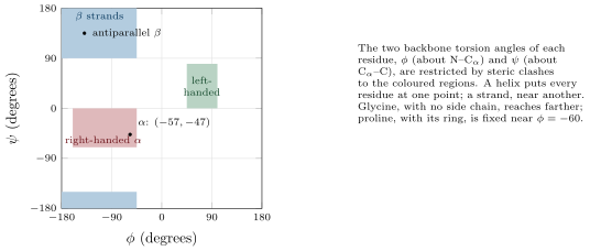
CoachThe primary structure is the sequence of residues. The secondary structure is the local regular conformation of the backbone, stabilised by hydrogen bonds between backbone carbonyls and amides: the -helix, a right-handed spiral of residues per turn, rising per residue ( per turn), each carbonyl bonded to the amide four residues on; and the -sheet, in which extended strands lie side by side, parallel or antiparallel, bonded to their neighbours, the side chains alternating above and below the sheet. The tertiary structure is the fold of a whole chain in three dimensions; a compact, independently folding unit of residues within it is a domain, and the arrangement of a domain’s secondary elements is its fold. The quaternary structure is the assembly of several chains: haemoglobin’s . About folds account for all known domains, and a few dozen of them — the Rossmann fold, the TIM barrel, the immunoglobulin fold — for a large fraction of all proteins: nature reuses folds and varies their sequences.