University Biology — Year 3 · Bachelor Year 3
7Structural Biology of Proteins
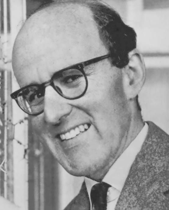
Max Perutz began work on the structure of haemoglobin in 1937 and published it in 1960: twenty-two years to see, at a resolution of half a nanometre, how four chains of a hundred and fifty amino acids fold around four haems. A haemoglobin molecule folds itself in a few milliseconds. Between these two facts lies the subject of this chapter: what a protein’s shape is, why a chain of amino acids has one shape rather than another, how it finds that shape so fast among an astronomical number of alternatives, what goes wrong when it does not, how the shapes are seen — by X-rays, by magnetic resonance, by electrons, and now by computation — and how a protein moves between shapes to do its work, with haemoglobin’s switch between a tense and a relaxed form as the model for every allosteric protein since.
7.1 What a protein’s structure is
Definition 7.1 (Levels of structure)
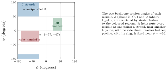
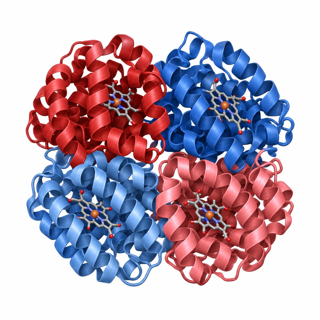
The primary structure is the sequence of residues. The secondary structure is the local regular conformation of the backbone, stabilised by hydrogen bonds between backbone carbonyls and amides: the -helix, a right-handed spiral of residues per turn, rising per residue ( per turn), each carbonyl bonded to the amide four residues on; and the -sheet, in which extended strands lie side by side, parallel or antiparallel, bonded to their neighbours, the side chains alternating above and below the sheet. The tertiary structure is the fold of a whole chain in three dimensions; a compact, independently folding unit of residues within it is a domain, and the arrangement of a domain’s secondary elements is its fold. The quaternary structure is the assembly of several chains: haemoglobin’s . About folds account for all known domains, and a few dozen of them — the Rossmann fold, the TIM barrel, the immunoglobulin fold — for a large fraction of all proteins: nature reuses folds and varies their sequences.
Proposition 7.2 (What holds a fold together)
A folded protein is stabilised by the hydrophobic effect — burying non-polar side chains in a core away from water, which frees the water molecules that would otherwise be ordered around them — by the hydrogen bonds of the secondary structure and of buried polar groups, by van der Waals packing of a core as dense as a crystal, by occasional salt bridges, and, in secreted proteins, by disulfide bonds. The hydrophobic effect supplies most of the drive; the hydrogen bonds mostly pay for themselves (a backbone bonded to water in the unfolded state is bonded to itself in the folded one) and instead dictate which fold. The net free energy of folding is small, to — the strength of a handful of hydrogen bonds for a molecule with hundreds — because the loss of conformational entropy on folding nearly cancels the gain in enthalpy. Proteins are marginally stable: a few degrees, a change of pH, a single mutation in the core can unfold them, and this marginality is what lets them move.
Evidence. Anfinsen (1961) unfolded ribonuclease A with urea and a reducing agent, breaking its four disulfides and destroying its activity; on removing both, the enzyme refolded, re-formed the correct four of the possible disulfide pairings, and recovered full activity. No template, no enzyme, no information beyond the sequence was present: the native structure is the thermodynamically most stable state accessible to the chain, and the sequence encodes it. Reoxidising the disulfides while the chain was still in urea gave a scrambled mixture with activity, which then unscrambled to the native form when allowed to fold: the chain finds the minimum by itself. ∎
7.2 How a chain finds its fold
Theorem 7.3 (Levinthal’s paradox)
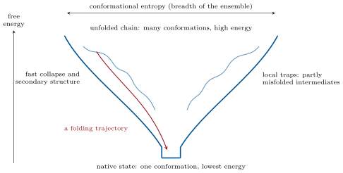
If each of the residues of a protein could adopt backbone conformations independently, the chain would have conformations. With and that is ; sampling them at one per picosecond ( per second) would take seconds, about years. Proteins of this size fold in milliseconds to seconds. Therefore folding is not a search through conformations at random: the energy surface is a funnel, in which almost every partial conformation is biased toward the native state, and the chain descends by local moves that are each downhill on average.
Proof. The count is the multiplication principle; the time is the count divided by the sampling rate, s, and a year is s. The conclusion is the contrapositive: a random search of that size is impossible in the observed time, so the search is not random. What makes it non-random is that an interaction formed in one part of the chain persists while others form (hydrophobic collapse and helix formation happen in microseconds and reduce the space to be searched), and that partially correct structures are on average lower in energy than wrong ones, so that the descent is guided — a funnel rather than a golf course with a single hole. ∎
Definition 7.4 (Chaperones)
A molecular chaperone is a protein that assists the folding of others without becoming part of them or supplying information about the fold: it prevents the aggregation that competes with folding in the crowded cytoplasm. Hsp70 binds exposed hydrophobic segments of nascent or unfolded chains, releasing them on ATP hydrolysis for another attempt; the chaperonin GroEL (Hsp60 in mitochondria, TRiC in the eukaryotic cytosol) is a double barrel of fourteen subunits whose central cavity, capped by GroES, encloses a single chain of up to in isolation for about ten seconds — an Anfinsen cage — and ejects it whether folded or not, to try again. About a tenth of newly made bacterial proteins pass through GroEL, and a cell whose chaperones are overwhelmed — by heat, which is why most are heat-shock proteins induced by a rise in temperature — fills with aggregates.
Definition 7.5 (Amyloid)
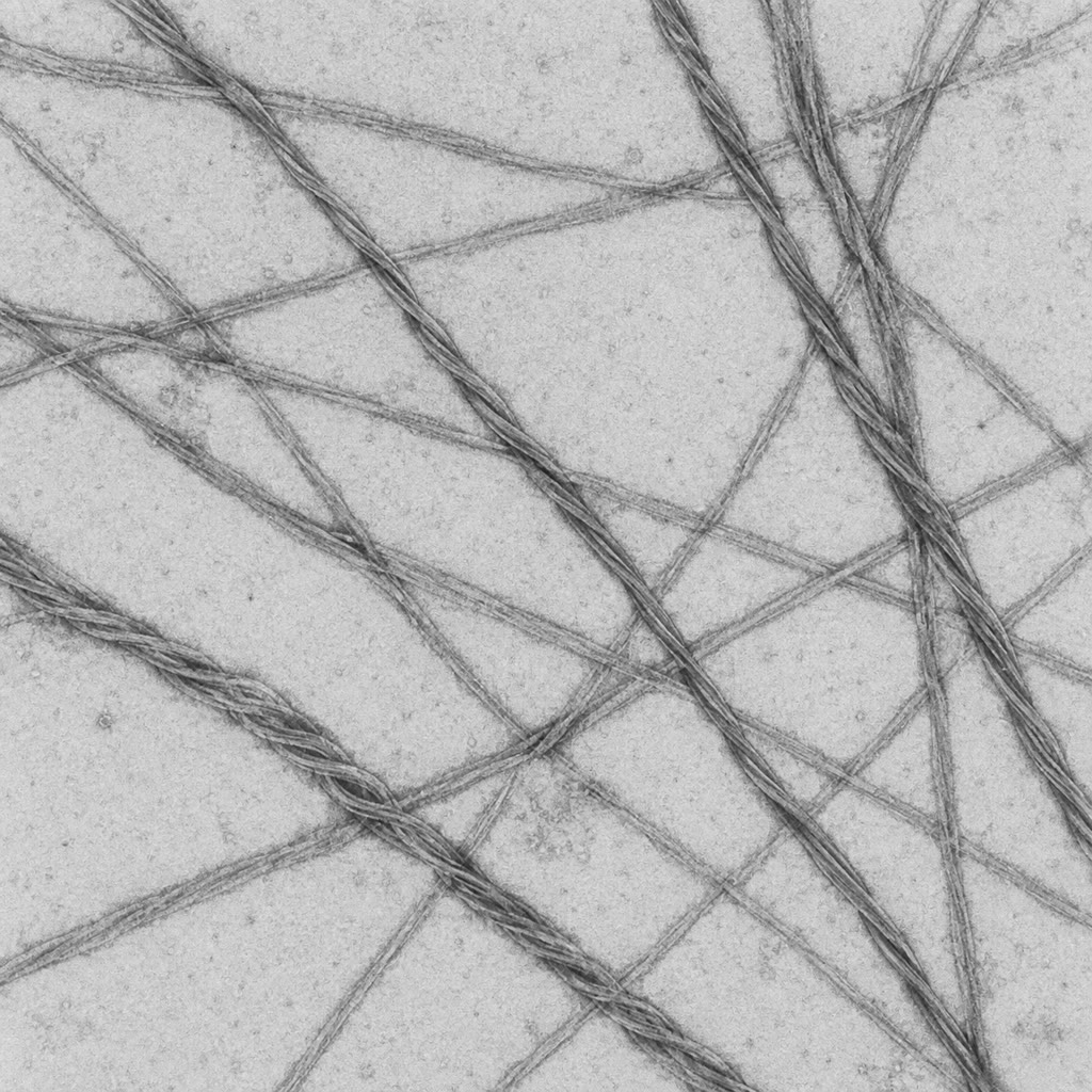
Amyloid is an ordered aggregate of protein in which the chains stack into unbranched fibrils wide, the strands of each chain running perpendicular to the fibril axis and hydrogen-bonded to the strands of the next — the cross- structure, the same whatever the protein. Almost any protein can form it under destabilising conditions; in the misfolding diseases particular proteins do so in the body: amyloid- and tau in Alzheimer’s disease, -synuclein in Parkinson’s, huntingtin, transthyretin, islet amyloid in type 2 diabetes. A prion is an amyloid that propagates: the misfolded form of the prion protein PrP converts the normal form on contact into more of itself, so that the “infection” carries no nucleic acid, only a shape — the mechanism of scrapie, of bovine spongiform encephalopathy and of Creutzfeldt–Jakob disease, established by Prusiner from 1982 against long resistance.
7.3 Seeing a protein
Definition 7.6 (Structural methods)
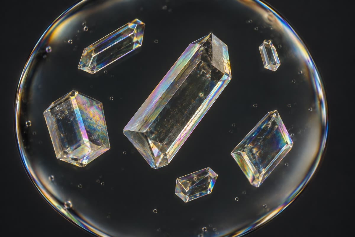
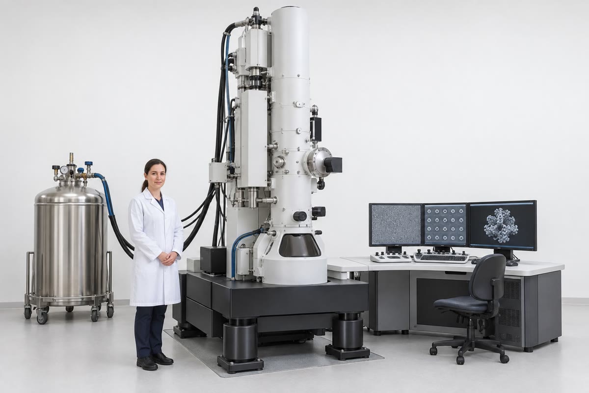
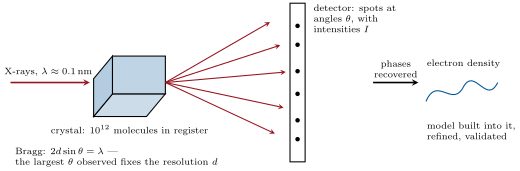
X-ray crystallography grows a crystal of the protein, exposes it to a beam of X-rays and records the diffraction pattern; the positions of the spots give the lattice and their intensities, once the phases of the waves are recovered, give the electron density of the unit cell, into which the chain is built. Nuclear magnetic resonance (NMR) measures, in solution, the couplings between the nuclei of atoms close in space, from which distances and hence a structure are computed; it works for proteins under about and reports their motions. Cryo-electron microscopy images thousands to millions of single molecules frozen in a thin film of vitreous ice, each in a random orientation, and reconstructs the three-dimensional density by combining them; since direct electron detectors (2013) it reaches atomic resolution and needs no crystal, so that large complexes, membrane proteins and flexible assemblies that never crystallised are now solved routinely. The resolution of a structure is the smallest spacing at which two features are separated: at the backbone and large side chains are placed, at every atom, at hydrogens.
Evidence. Kendrew (1958) obtained the first structure of a protein, sperm-whale myoglobin, at — a sausage of unexpected irregularity, “more complicated and less symmetrical than anyone had predicted” — and at in 1960, when the -helices Pauling had proposed from model-building in 1951 were seen for the first time. Perutz (1960) solved haemoglobin at using the heavy-atom method he had devised to obtain the phases: mercury atoms attached to the protein change the intensities in a way that reveals the missing information. Comparing the oxygenated and deoxygenated forms (1970) he saw the subunits rotate against one another by on binding oxygen, the first allosteric transition observed at atomic scale. ∎
Method 7.7 (Solving a structure by crystallography)
(1) Purify milligrams of the protein and screen hundreds of conditions (precipitant, pH, salt) for crystals; (2) flash-freeze a crystal and collect diffraction images at a synchrotron as it rotates in the beam; (3) index the spots to obtain the unit cell and symmetry; the highest angle at which spots are seen sets the resolution by Bragg’s law, ; (4) solve the phase problem — the detector records intensities but not phases — by heavy atoms, by the anomalous scattering of selenium in selenomethionine, or, for a protein similar to a known one, by molecular replacement, placing the known model in the cell; (5) compute the electron density, build the chain into it and refine the model against the data until the disagreement (-factor) is small; (6) validate: geometry, Ramachandran outliers, and the fit to data the model was not refined against.
Remark 7.8 (Prediction has joined the methods)
Since 2020 a protein’s structure is predicted from its sequence with an accuracy that often rivals a experimental structure (Chapter 5). The prediction is a hypothesis about a static native state; the experimental methods remain the arbiter, and the only source of information about ligands, conformational changes, complexes and the dynamics discussed below. Their roles have changed — a predicted model solves the phase problem by molecular replacement, and directs which experiments are worth doing — rather than one replacing the other.
7.4 Allostery: haemoglobin as the model
Definition 7.9 (Allostery and cooperativity)
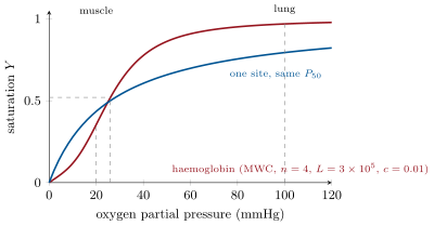
A protein is allosteric when the binding of a ligand at one site changes the affinity of another site, through a change of conformation transmitted across the molecule. When the two sites bind the same ligand and the effect is positive, binding is cooperative: the fractional saturation rises with ligand concentration as a sigmoid rather than a hyperbola, described empirically by the Hill equation with a Hill coefficient (haemoglobin: for its four sites; myoglobin, one site: ). Haemoglobin exists in two quaternary conformations, the T state (tense, low affinity, favoured when no oxygen is bound) and the R state (relaxed, high affinity), and switches between them as a whole; protons, carbon dioxide and 2,3-bisphosphoglycerate stabilise T and so lower the affinity — the Bohr effect, by which working tissue, acid and warm, takes more oxygen from the blood.
Theorem 7.10 (The Monod–Wyman–Changeux model)
Let a protein of identical sites exist in two conformations, and , in equilibrium in the absence of ligand, every site of binding the ligand with dissociation constant and every site of with , . All sites of a molecule switch together (the transition is concerted). Then with the fractional saturation is
and the fraction of molecules in the state is . For and the curve is sigmoid: the first ligands bind to the scarce, high-affinity molecules and tip the equilibrium, so that binding recruits more and the affinity of the remaining sites rises — cooperativity without any direct interaction between sites.
Proof. The species with ligands bound in the form has concentration (each of the choices of occupied sites contributes by the law of mass action), and in the form , since . Summing over by the binomial theorem, the total protein is and the total ligand bound is , using . Dividing bound ligand by times total protein gives ; the fraction is the terms over the total. When is small the terms dominate the denominator (because ) and , the low affinity; when is large enough that the terms take over and the affinity is : the switch between the two regimes is the sigmoid. ∎
Example 7.11 (Haemoglobin in numbers)
Take , , (so ) and . At the partial pressure of oxygen in arterial blood, , and ; at , the venous value at rest, ; at , — the model reproduces the half-saturation pressure of ; at , in an exercising muscle, . A non-cooperative carrier of the same half-saturation pressure would give in the lungs and in the muscle, unloading of its capacity where haemoglobin unloads . Cooperativity is what makes a carrier that loads fully at one pressure and unloads mostly at a pressure only five times lower.
Proposition 7.12 (Mechanism of the switch)
In deoxyhaemoglobin the iron sits slightly out of the haem plane, pulled toward the proximal histidine, and the haem is domed. Oxygen binding flattens the haem and draws the iron, and with it the histidine and the helix it belongs to, by about toward the haem; that shift is amplified at the interface between the and dimers, which rotate by and break the salt bridges that hold the T state. The effectors act on those same bridges: protons bind histidines whose bridges exist only in T (the Bohr effect), carbon dioxide forms carbamates with the amino termini that stabilise T, and 2,3-bisphosphoglycerate, a strongly negative small molecule, sits in the central cavity between the chains, which is wide enough only in T. Fetal haemoglobin, with chains that bind it less well, has a higher affinity than the mother’s and draws oxygen across the placenta.
7.5 Proteins in motion
Definition 7.13 (Disorder and dynamics)
An intrinsically disordered region has no stable fold on its own and exists as an ensemble of rapidly interconverting conformations, often folding only when it meets its partner; about a third of human proteins carry a disordered region of more than thirty residues, and they are enriched in signalling and regulation, where a flexible segment can be phosphorylated at many sites, bind several partners in turn, and act as a linker. Folded proteins move too: side chains rotate in picoseconds, loops open in nanoseconds to microseconds, domains hinge in microseconds to milliseconds, and an enzyme’s catalytic cycle is a choreography of such motions rather than a static lock-and-key. A crystal structure is one snapshot of a conformational ensemble; NMR and molecular simulation see the rest. Multivalent disordered proteins and RNAs can also demix from the cytoplasm into liquid droplets — biomolecular condensates such as nucleoli, stress granules and P bodies — that concentrate reactions without a membrane, and whose ageing into solid aggregates is one route to the amyloid diseases.
Remark 7.14 (Structure and function)
The lesson of sixty years of structures is double. A fold is a solution to a physical problem — bind this, catalyse that, transmit a signal over four nanometres — and knowing the structure often makes the mechanism plain, as it did for the haem’s iron and for the oxygen-driven rotation of haemoglobin’s dimers. And a fold is a historical object: the same few hundred architectures, tinkered with, duplicated and fused, serve for the whole diversity of protein function, so that the structure of a protein from a bacterium will tell one, often, how the human cousin works.
7.6 Exercises
Exercise 7.1 ★
Define the four levels of protein structure and give the example of each in haemoglobin.
Solution
Solution of Exercise 7.1.
Primary: the sequence of the residues of an chain. Secondary: its eight -helices. Tertiary: the globin fold of one chain wrapped around its haem. Quaternary: the tetramer and the way its dimers rotate against each other.
Exercise 7.2 ★
An -helix contains residues. How long is it, how many turns does it make, and how many backbone hydrogen bonds hold it (each residue’s carbonyl bonds to the amide four residues on)?
Solution
Solution of Exercise 7.2.
; turns; hydrogen bonds.
Exercise 7.3 ★
What did Anfinsen’s refolding of ribonuclease show, and why was it important to do the experiment without any cellular component present?
Solution
Solution of Exercise 7.3.
That the native structure, including the correct pairing of four disulfides out of possibilities, is recovered spontaneously from the unfolded chain: the sequence contains all the information needed, and the native state is the thermodynamic minimum. Doing it in a test tube with pure protein excluded any template, enzyme or cellular machinery as the source of the information.
Exercise 7.4 ★
Rank crystallography, NMR and cryo-electron microscopy by the size of protein each handles best, and say which gives information about motion.
Solution
Solution of Exercise 7.4.
NMR: small proteins, below about , in solution. Crystallography: any size that crystallises, from peptides to ribosomes. Cryo-electron microscopy: large complexes, above about , without crystals. NMR reports motion directly (and cryo-EM can sort molecules into conformational classes); a crystal structure is a static average.
Exercise 7.5 ★★
Redo Levinthal’s count for a protein of residues with , and for a peptide of residues with . For which of the two is a random search conceivable within a second at trials per second?
Solution
Solution of Exercise 7.5.
conformations, s years. , ms: the peptide could search exhaustively within a second; the protein could not in the age of the universe.
Exercise 7.6 ★★
Using the MWC equation with , , , compute at , and , and compare with a single site of dissociation constant (). Where does the cooperativity show?
Solution
Solution of Exercise 7.6.
. : (single site ). : (). : (). The MWC curve lags far behind the single site at low ligand, then rises steeply — from to over two decades where the single site goes from to : that steepness is the cooperativity.
Exercise 7.7 ★★
Explain, with the MWC model, why 2,3-bisphosphoglycerate lowers the oxygen affinity of haemoglobin, which parameter of the model it changes, and why stored blood, which loses the compound, delivers oxygen poorly when transfused.
Solution
Solution of Exercise 7.7.
Bisphosphoglycerate binds only the T state (in the central cavity), so it stabilises T: in the model it raises , the ratio of the unliganded protein, and so shifts the curve to the right — lower affinity, more oxygen released at tissue pressures. Stored red cells lose the compound, falls, the curve shifts left, and transfused blood holds its oxygen instead of giving it up in the tissues, until the cells resynthesise it over a day.
Exercise 7.8 ★★
A crystal diffracts to a maximum angle with X-rays of wavelength . What is the resolution? What features of the protein will be visible? Which angle would be needed for ?
Solution
Solution of Exercise 7.8.
: the backbone and every side chain are placed, individual atoms resolved. For : , .
Exercise 7.9 ★★
A mutation replaces a buried leucine by a lysine. Predict its effect on stability, using Proposition 7.2, and explain why the same substitution on the surface would be harmless. Then explain why a protein with is not “very stable” although the number looks large.
Solution
Solution of Exercise 7.9.
A charged lysine buried among non-polar side chains costs tens of kilojoules per mole (its charge unsolvated, the leucine’s hydrophobic burial lost), more than the whole of folding: the protein unfolds or the core rearranges. On the surface the lysine is hydrated as it would be in the unfolded state and nothing is lost. means : one molecule in is unfolded at any moment, and the whole margin equals two or three hydrogen bonds out of hundreds — one mutation’s worth.
Exercise 7.10 ★★★
Derive the fraction of molecules in the state, , from the species counted in the proof of Theorem 7.10, and show that for the ligand concentration at which half the molecules are in satisfies . Compute this for haemoglobin’s parameters and compare with .
Solution
Solution of Exercise 7.10.
The species total and the species , so . For the term is , and when , i.e. . For haemoglobin , so : , close to the of — the conformational switch and half-saturation nearly coincide.
Exercise 7.11 ★★★
Prions transmit a shape. Explain why the “protein-only” hypothesis was resisted, what experiment would refute it, and what evidence (resistance of infectivity to nucleases and ultraviolet light, sensitivity to protein denaturants, synthetic prions) supports it. Why does a prion need a seed?
Solution
Solution of Exercise 7.11.
It was resisted because every known infectious agent replicated through nucleic acid, and a protein cannot copy its sequence. A nucleic acid found to be required, or purified protein shown unable to transmit, would refute it. Support: infectivity survives nucleases, ultraviolet and ionising radiation at doses that destroy any genome, but is destroyed by protein denaturants and proteases; recombinant PrP folded in vitro into fibrils transmits disease to animals, with the same strain properties on passage. A seed is needed because the conversion of a normal monomer is prohibitively slow alone — the nucleus of the cross- structure must first form, which is the rate-limiting event — whereas a pre-formed fibril end converts monomers rapidly: the shape is copied by templated growth.
Exercise 7.12 ★★★
A protein family has kept its fold while its sequences have diverged to identity. Explain why structure is more conserved than sequence, what kind of residues are the ones conserved, and how this is exploited both by profile methods (Chapter 5) and by the design of new proteins.
Solution
Solution of Exercise 7.12.
Many sequences fold to the same structure: what a fold needs is a pattern of hydrophobic and polar positions, a few glycines and prolines at the turns, and the residues that do the job; everything else on the surface can change without penalty, so mutations accumulate there while selection preserves the fold and function. The conserved residues are therefore the core (as a pattern more than as identities), the catalytic and binding residues, and structural glycines, prolines and cysteines. Profile methods weight exactly those columns and so recognise family members at identity; protein design runs the logic backwards, choosing a hydrophobic pattern for a target fold and letting the surface be anything.
7.7 Problem: The Fold of a Globin
Problem 7.1
Weekend problem — a globin’s helices measured, its folding timed against Levinthal, its oxygen binding computed from the two-state model, and its structure solved from a crystal, ending on the saturation difference between lung and muscle, the Levinthal time and the resolution of the map
Data: a globin chain of residues, mean residue mass , of residues in eight -helices; helix rise per residue, residues per turn. Partial specific volume of protein . MWC parameters for haemoglobin: , , , . X-ray wavelength ; unit cell a cube of edge ; crystal a cube of edge .
Part I — Anatomy of a chain.
- How many residues are in helices? What total length of helix is that, and how many turns?
- How many backbone hydrogen bonds do the helices contain, counting one per residue beyond the first four of each helix?
- The chain’s mass and its volume from the partial specific volume; the radius of a sphere of that volume.
- The extended chain would be per residue long. Compare its length with the sphere’s diameter: by what factor does folding compact the chain?
- The eight helices average residues. If the residues were laid end to end, what fraction of the extended length is helical, and how does the helix shorten it?
- A globin buries about non-polar side chains in its core. Taking of hydrophobic stabilisation per buried side chain and a conformational entropy cost of per residue on folding (all ), estimate the net of folding and compare with the range in Proposition 7.2.
Part II — Finding the fold.
- Compute the Levinthal count for residues with and the time to sample it at per second.
- The globin folds in . How many conformations, at most, can it have visited? What fraction of the count is that?
- Suppose instead each helix forms independently in , and the eight formed helices then find their packing among orderings sampled at per second. Estimate the folding time. Is this a funnel?
- A chaperonin encloses a chain for per cycle and releases it folded with probability per cycle. What is the expected number of cycles, and the expected time, to fold a difficult substrate? What fraction is still unfolded after a minute?
- A destabilising mutation raises the unfolded fraction at from to . With and , by how much did change?
- Explain why a hundredfold more unfolded protein can be the difference between health and amyloid disease, using the need for a seed.
Part III — Binding oxygen.
- Compute from the MWC equation at (lung) and (working muscle).
- Compute the fraction of molecules in the state at those two pressures.
- How much of the loaded oxygen is unloaded between lung and muscle? Compare with a single-site carrier of half-saturation pressure .
- Blood carries of haemoglobin per litre, each gram binding of oxygen. How many millilitres of oxygen per litre does haemoglobin deliver to the muscle, from question 15? A resting body consumes : what blood flow does that require at rest (unloading from to )?
- Bisphosphoglycerate raises to . Recompute at and say what this does to delivery at rest.
- Fetal haemoglobin has, in effect, . Compute its at the placental pressure of , compare with the mother’s, and explain the transfer.
Part IV — Seeing it.
- How many unit cells does the crystal contain? If each cell holds four globin chains, how many molecules diffract together?
- Spots are seen to . What is the resolution, and will the side chains be placed?
- What angle would give ? Why does a poorly ordered crystal diffract only to low angles?
- In cryo-electron microscopy the signal of the averaged image grows as the square root of the number of particles. If particles give , and resolution improves as the inverse of the signal, how many particles give ?
- Cryo-electron microscopy needs no crystal. Name two kinds of protein or complex for which this decides whether a structure can be obtained at all, and explain why.
- A predicted model of the globin differs from the crystal structure by on average over the backbone. Name two things the prediction cannot supply that the crystal did.
- Summarise: the unloading between lung and muscle (question 15), the Levinthal time for residues (question 7), and the resolution of the map (question 20).
Solution
Solution of Problem 7.1.
1. helical residues; ; turns. 2. hydrogen bonds. 3. Mass g; volume cm ; radius . 4. Extended against a diameter of : a factor of . 5. The helical residues are of the extended length, ; the helix compresses them to , a factor . 6. gained, lost: net , in the marginal range. 7. ; s years. 8. conformations, a fraction of the count. 9. Helices in (in parallel); packings at per second take : total about , the observed order of magnitude. Yes: solving the parts independently and then the whole is a funnel — the search space is factorised, not exhausted. 10. Expected cycles , about ; after six cycles remains unfolded. 11. with : from to , a loss of of stability (, ). 12. Aggregation begins with a nucleus, whose formation rate rises as a high power of the concentration of the aggregation-prone unfolded species; a hundredfold more of that species raises the chance of a seed forming within a lifetime by orders of magnitude, and once a seed exists growth is fast and self-templating. 13. : . : numerator , denominator : . 14. in the lung; in the muscle. 15. of capacity unloaded (two thirds of the load). Single site: . 16. Capacity ; to the muscle . At rest ; of blood flow — the order of a resting cardiac output. 17. , : numerator , denominator : instead of — far more oxygen unloaded at rest, the adaptation of blood to altitude and of muscle to work. 18. Fetal, , : numerator , denominator : . Maternal at the same pressure: numerator , denominator : . At equal pressure the fetal blood holds more oxygen, so oxygen flows from mother to fetus across the placenta. 19. cells; molecules. 20. : yes, every side chain and atom placed. 21. , . In a disordered crystal the molecules are not in exact register, the waves scattered at high angle — which encode fine detail — no longer add in phase, and the spots fade at high angle; only the low-resolution spots survive. 22. Halving needs twice the signal, hence four times the particles: about (more in practice, as other errors enter). 23. Membrane proteins, which rarely crystallise from detergent solutions and can be imaged in a lipid nanodisc; large flexible complexes — ribosomes, spliceosomes, ion channels with their regulators — whose heterogeneity prevents crystallisation but whose particles can be sorted into conformational classes. 24. The bound haem and oxygen and their exact geometry, the ordered water and ions, the second conformation (T against R) and the proof that the fold is right — a prediction is a single unliganded model with no experimental check. 25. Unloading of capacity (single site ); the Levinthal time about years; a map at .