Coach
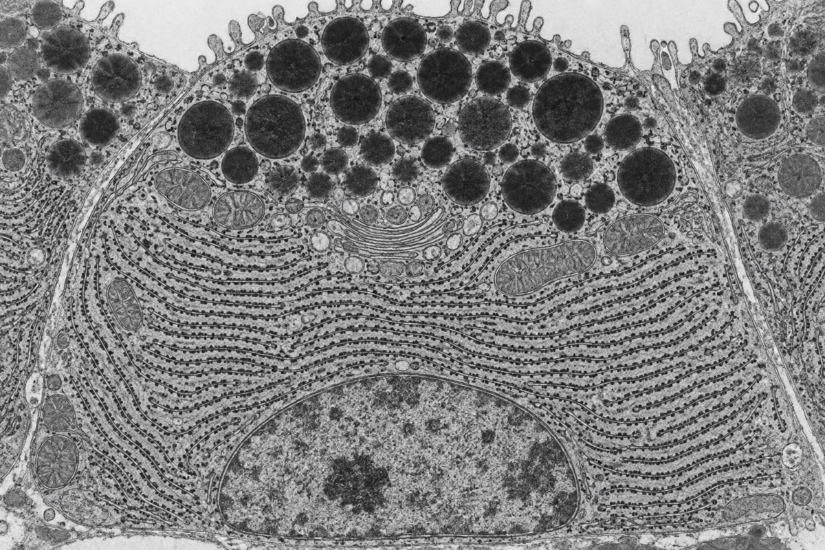
CoachAs the chain enters the lumen a preassembled oligosaccharide of fourteen sugars (two N-acetylglucosamines, nine mannoses, three glucoses) is transferred from a lipid carrier onto asparagines in the sequence Asn-X-Ser/Thr — N-glycosylation. The three glucoses are then trimmed one by one, and the trimming is a folding clock: a protein still carrying one glucose is held by the chaperone calnexin; a protein whose last glucose has been removed but which is not yet folded is re-glucosylated by an enzyme that recognises exposed hydrophobic patches, and returns to calnexin — the calnexin cycle. Proteins that fail repeatedly are pulled back into the cytosol, ubiquitinated and destroyed by the proteasome: ER-associated degradation (ERAD). When unfolded proteins accumulate faster than they can be folded or destroyed, sensors in the reticulum membrane launch the unfolded protein response: translation is slowed, chaperone genes induced, the reticulum expanded, and, if the stress persists, the cell killed. The commonest mutation of cystic fibrosis (F508 in CFTR) makes a channel that would work but folds too slowly, is caught by ERAD and never reaches the surface; drugs that help it fold are now a treatment.