 Coach
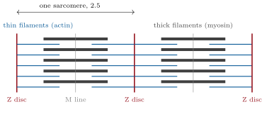
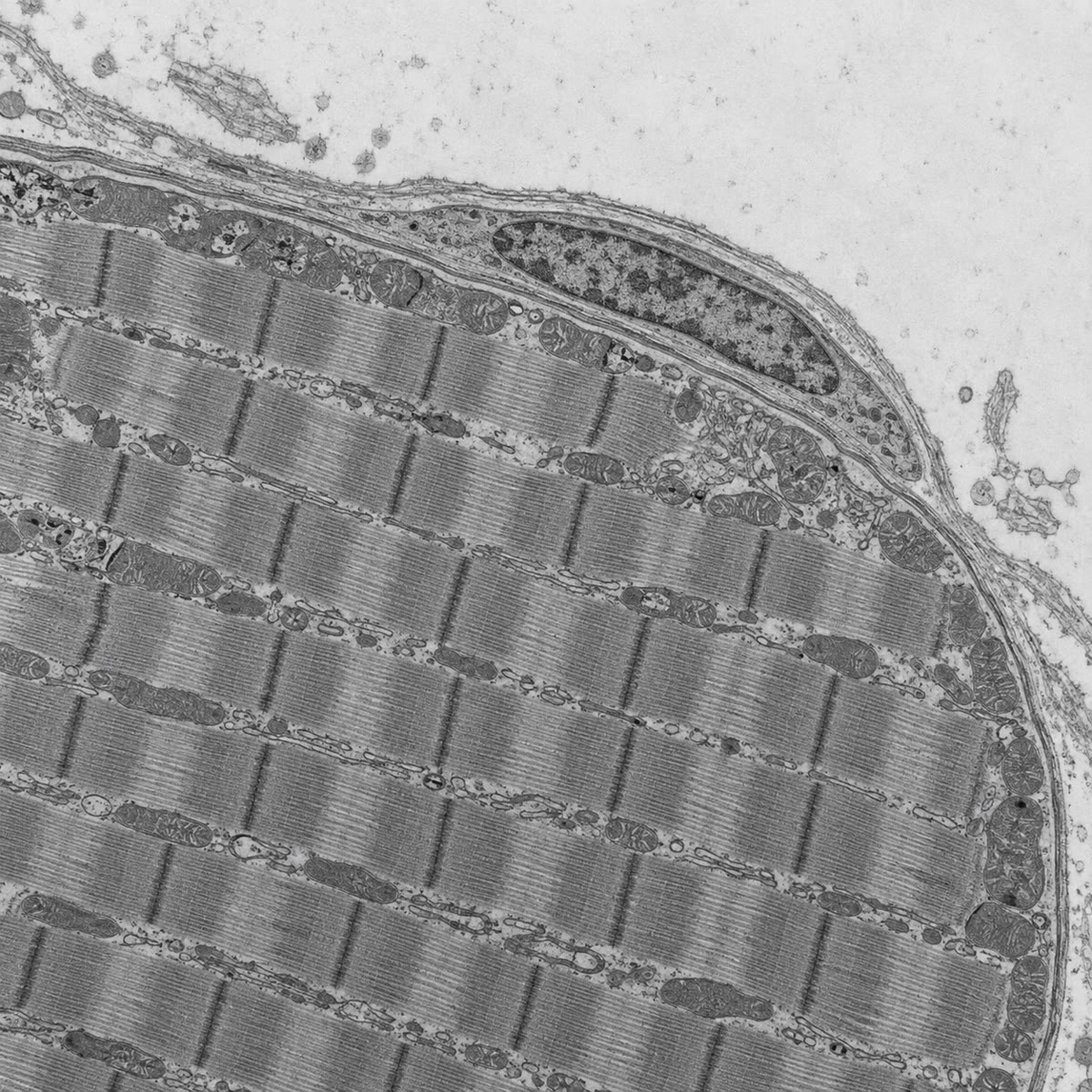
CoachA skeletal muscle fibre is a cylinder across and up to tens of centimetres long, bounded by the sarcolemma, with hundreds of nuclei just under it. Its interior is filled with parallel myofibrils, each a chain of sarcomeres long: between two Z discs, thin filaments of actin (with tropomyosin and troponin) anchored at the Z discs interdigitate with thick filaments of myosin held at the centre, and the overlap pattern gives the striations. Around each myofibril a lace of sarcoplasmic reticulum stores calcium; transverse tubules, invaginations of the sarcolemma, run between the myofibrils and carry the electrical signal from the surface to the interior. Mitochondria pack the spaces, and the cytoplasm holds glycogen, creatine phosphate and, in red fibres, myoglobin. The cell is built for one thing — to shorten on command — and the machinery of that command is Chapter 21.
Examples
Example 12.8 (Growth and repair)
A muscle grows in the adult not by adding fibres but by enlarging them: training adds myofibrils to each fibre, and satellite cells fuse into it to supply the extra nuclei a larger cytoplasm needs. After injury the same cells divide, re-express MyoD, fuse and rebuild the damaged segment within weeks — the embryonic programme run again in the adult. In muscular dystrophy the fibres lack dystrophin, a protein that ties the myofibrils to the membrane, and tear during contraction; the satellite cells repair them again and again until, after years, the stock is exhausted and fibrous tissue takes the muscle’s place.