University Biology — Year 2 · Bachelor Year 2
12Cell Differentiation: The Skeletal Muscle Cell
A fibre of your thigh muscle is a single cell thirty centimetres long, with hundreds of nuclei, packed from end to end with contractile rods so regular that it looks striped under the microscope. It began as a cluster of ordinary-looking cells that crawled out of a somite, multiplied, stopped dividing, lined up, fused, and switched on several hundred genes they had never used before. Every cell of the body has the same genome; a muscle fibre is what that genome does when one particular transcription factor is turned on. This chapter takes the skeletal muscle cell as the worked example of differentiation — how a cell acquires and keeps a specialised identity — and the muscle cell is a good one because the master switch is known, the steps can be watched in a dish, and the finished cell is a machine whose parts are visible.
12.1 What differentiation is
Definition 12.1 (Differentiation, determination, potency)
A cell is differentiated when it expresses the stable pattern of genes — and so the proteins, shape and function — of one cell type. It is determined (or committed) when its fate is fixed although its differentiation has not yet begun: a myoblast looks like a fibroblast but will make only muscle. Potency is the range of types a cell can still give: totipotent (the zygote and first blastomeres, which make everything including the placenta), pluripotent (the inner cell mass, which makes every tissue of the body), multipotent (a stem cell of the blood or of muscle, which makes the types of one tissue), unipotent. A stem cell is a cell that divides to give one daughter like itself and one that goes on to differentiate, which keeps the stock while supplying the tissue. Differentiation is in most cases not a loss of genes but a choice among them: the genome of a muscle nucleus is complete.
Evidence. Gurdon (1962) transplanted the nucleus of a differentiated intestinal cell of a tadpole into a frog egg whose own nucleus had been destroyed; a fraction of the eggs developed into normal, fertile frogs. The differentiated nucleus had lost none of the genes needed to make a whole animal; the egg’s cytoplasm had reset it. The same operation with a mammary cell nucleus in a sheep’s egg gave Dolly (1996), and the reprogramming of a skin fibroblast into a pluripotent stem cell by four transcription factors (Yamanaka, 2006) showed that the reset can be done with a handful of proteins. ∎
Proposition 12.2 (Differentiation is transcriptional)
The difference between two cell types lies in which genes are transcribed. A few transcription factors, expressed in response to the inductive signals of Chapter 10, bind the regulatory regions of hundreds of target genes and switch them on, while the genes of other fates are switched off; and because such a factor commonly activates its own gene, the state, once entered, sustains itself. For some tissues a single factor is a master regulator sufficient to impose the fate on a cell of another type: MyoD for skeletal muscle, Pax6 for the eye, Sox9 for cartilage. The state is stabilised further by changes to the chromatin — methylation of DNA, modification of histones — that keep the silenced genes silent through divisions (the epigenetics of the Year 3 volume). A differentiated cell therefore remembers what it is without any change to its DNA sequence, and the memory is written in the pattern of factors and chromatin, not in the genes.
12.2 The making of a muscle fibre
Definition 12.3 (Myogenesis)
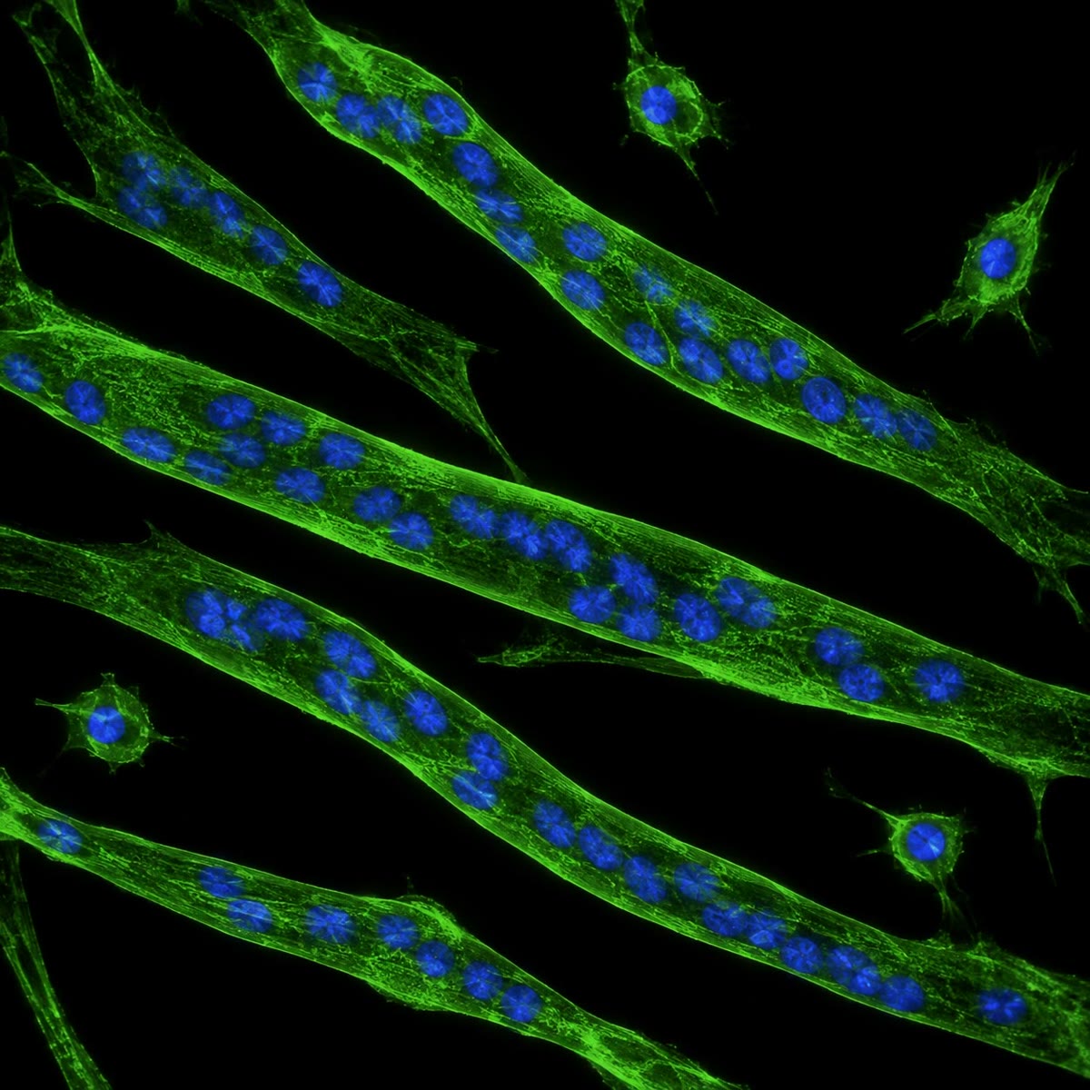
In the embryo, cells of the somite’s dermomyotome are induced by signals from the neural tube, notochord and surface ectoderm to express the myogenic factors MyoD and Myf5: they are now myoblasts, determined but still dividing and unremarkable to look at. Myoblasts migrate — into the limb buds, for instance — and multiply as long as growth factors (FGF) are present. When the factors run out, they withdraw from the cell cycle, express a second factor, myogenin, align end to end, and fuse: their membranes merge into a single long myotube with many nuclei in a row. The myotube switches on the muscle genes — actin, myosin, troponin, tropomyosin, creatine kinase, the acetylcholine receptor — assembles the contractile apparatus, and matures into a muscle fibre whose nuclei lie at its periphery. A fibre never divides again; a few myoblasts remain beside it, quiescent, as satellite cells, the stem cells that repair and grow the muscle for life.

Proposition 12.4 (MyoD: a master switch)
MyoD is a transcription factor of the helix-loop-helix family. It binds, as a dimer with a ubiquitous partner protein, a six-base sequence (the E-box, CANNTG) in the regulatory regions of the muscle genes, recruits chromatin-opening enzymes, and switches the genes on — including the genes for myogenin and for MyoD itself, which is how the state holds. Its activity is gated: in dividing myoblasts an inhibitor protein (Id), induced by growth factors, sequesters the partner and keeps MyoD idle; when growth factors are withdrawn Id disappears and MyoD acts. Because MyoD is sufficient to impose the muscle programme on a fibroblast, a fat cell or a nerve cell, the muscle fate is, in a sense, one gene deep: the difficulty of becoming a muscle is not in knowing how, but in being told to.
Evidence. Davis, Weintraub and Lassar (1987) transferred a single cDNA, isolated from myoblasts, into cultured fibroblasts: the fibroblasts stopped dividing, fused into myotubes and made muscle proteins. The gene was named MyoD. Its relatives Myf5, myogenin and MRF4 were found by homology; a mouse lacking both MyoD and Myf5 has no myoblasts and no skeletal muscle at all, while a mouse lacking myogenin has myoblasts that never fuse. ∎
Theorem 12.5 (A self-sustaining switch)
Let be the amount of a factor that activates its own gene with a cooperative (sigmoid) response and is degraded at rate :
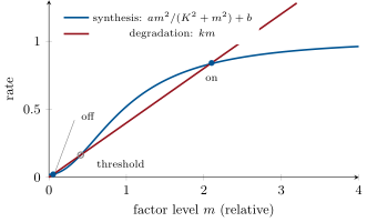
where is a small basal synthesis. For suitable , , and this equation has two stable steady states, a low one near and a high one near , separated by an unstable threshold: the cell is bistable. A transient inductive signal that pushes above the threshold sends the cell to the high state, where it stays after the signal is gone; below the threshold it returns to the low state. This is a memory built from a feedback loop, and it is why a cell, once committed, stays committed through divisions and in the absence of the inducer.
Proof. Steady states satisfy . The right-hand side is a sigmoid rising from to ; the left is a line through the origin of slope . For and small enough that the line cuts the steep part of the sigmoid, the two curves intersect three times: at , at an intermediate , and at . Stability: below synthesis exceeds decay and rises; between and decay exceeds synthesis and falls back; between and synthesis exceeds decay and climbs to ; above it, falls back to . Hence the outer two are stable, the middle one is unstable and is the threshold. ∎
12.3 The finished cell
Definition 12.6 (The muscle fibre)
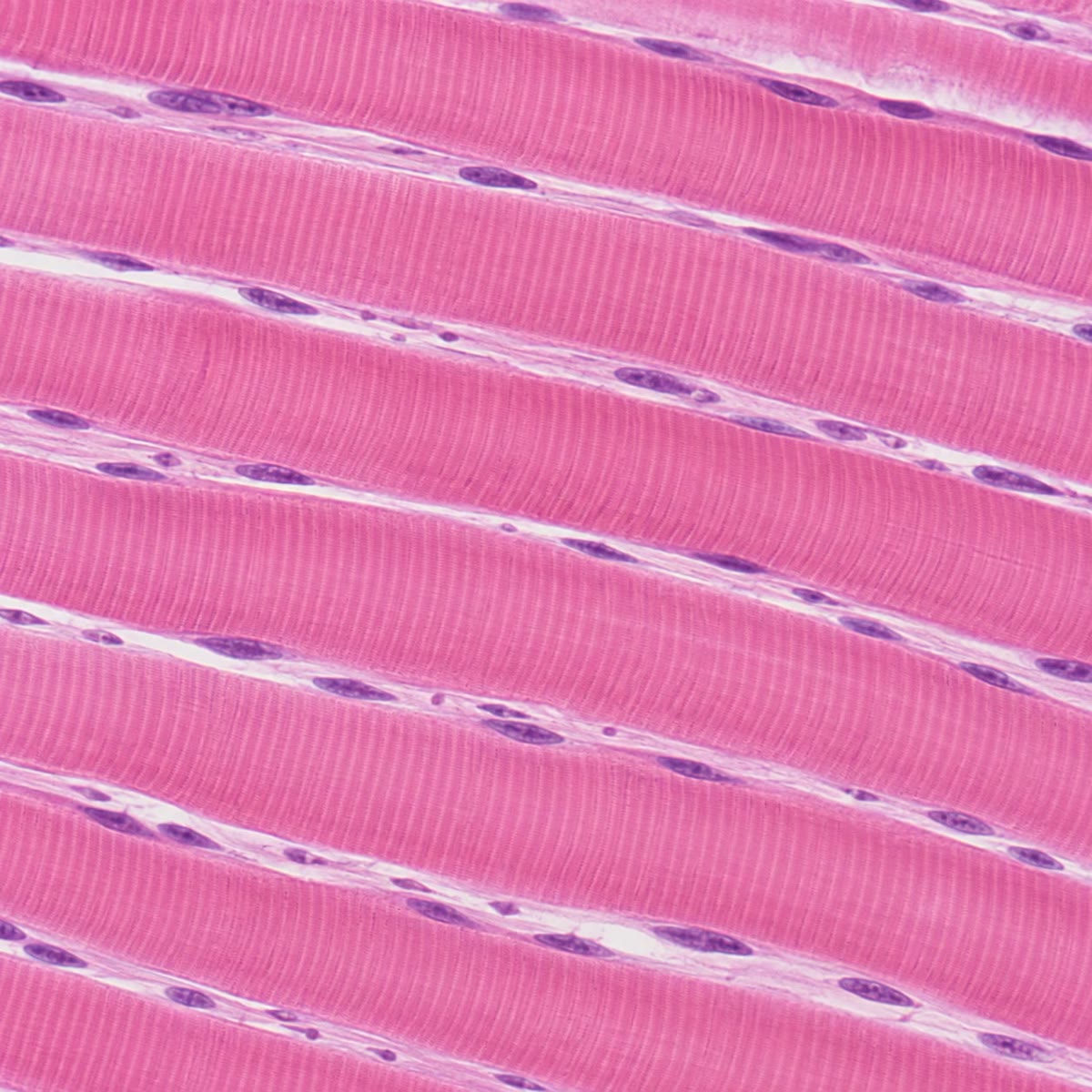
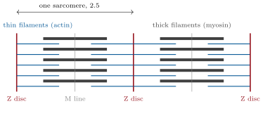
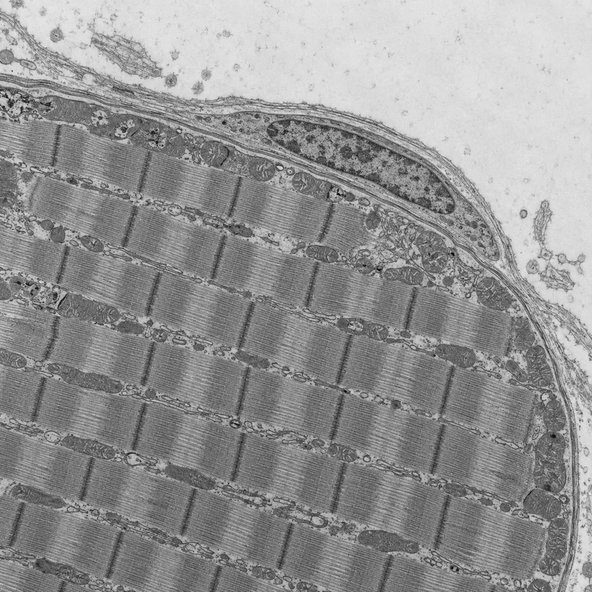
A skeletal muscle fibre is a cylinder across and up to tens of centimetres long, bounded by the sarcolemma, with hundreds of nuclei just under it. Its interior is filled with parallel myofibrils, each a chain of sarcomeres long: between two Z discs, thin filaments of actin (with tropomyosin and troponin) anchored at the Z discs interdigitate with thick filaments of myosin held at the centre, and the overlap pattern gives the striations. Around each myofibril a lace of sarcoplasmic reticulum stores calcium; transverse tubules, invaginations of the sarcolemma, run between the myofibrils and carry the electrical signal from the surface to the interior. Mitochondria pack the spaces, and the cytoplasm holds glycogen, creatine phosphate and, in red fibres, myoglobin. The cell is built for one thing — to shorten on command — and the machinery of that command is Chapter 21.
Proposition 12.7 (Fibre types)
A muscle is a mixture of fibre types, set partly by the myoblast lineage and partly by the nerve that innervates the fibre. Slow oxidative (type I) fibres have a slow myosin, many mitochondria and capillaries, and myoglobin — red, tireless, built for posture and endurance. Fast glycolytic (type IIx) fibres have a fast myosin, few mitochondria, much glycogen — white, powerful, exhausted in a minute. Fast oxidative (type IIa) fibres lie between. A sprinter’s calf is mostly fast fibres, a marathoner’s mostly slow, and the proportions are heritable; but the type is not fixed: cross-innervating a fast muscle with a slow nerve, or months of endurance training, converts fibres toward the slow type, because the pattern of nerve impulses switches the genes for the myosin isoforms and for mitochondrial biogenesis. A differentiated cell keeps its identity as a muscle fibre and yet revises its programme in response to use.
Example 12.8 (Growth and repair)
A muscle grows in the adult not by adding fibres but by enlarging them: training adds myofibrils to each fibre, and satellite cells fuse into it to supply the extra nuclei a larger cytoplasm needs. After injury the same cells divide, re-express MyoD, fuse and rebuild the damaged segment within weeks — the embryonic programme run again in the adult. In muscular dystrophy the fibres lack dystrophin, a protein that ties the myofibrils to the membrane, and tear during contraction; the satellite cells repair them again and again until, after years, the stock is exhausted and fibrous tissue takes the muscle’s place.
12.4 Exercises
Exercise 12.1 ★
Define differentiation, determination and potency, and place the zygote, an inner-cell-mass cell, a satellite cell and a muscle fibre on the scale of potency.
Solution
Solution of Exercise 12.1.
Differentiation: the stable expression of one cell type’s genes and functions. Determination: commitment to a fate before it is expressed. Potency: the range of fates still open. Zygote: totipotent; inner-cell-mass cell: pluripotent; satellite cell: unipotent (muscle only) — a stem cell nonetheless; muscle fibre: differentiated, post-mitotic, no potency.
Exercise 12.2 ★
List the steps of myogenesis from somite cell to muscle fibre, with the transcription factor expressed at each.
Solution
Solution of Exercise 12.2.
Dermomyotome cell (Pax3) induced myoblast (MyoD, Myf5), dividing while FGF lasts cycle exit, myogenin alignment and fusion into a myotube muscle genes on, myofibrils assembled (MRF4) mature fibre with peripheral nuclei, plus quiescent satellite cells (Pax7).
Exercise 12.3 ★
What did Gurdon’s nuclear transfer show, and what did it not show?
Solution
Solution of Exercise 12.3.
That the nucleus of a differentiated cell keeps every gene needed to make a whole animal, and that egg cytoplasm can reset its programme: differentiation is reversible and not a loss of DNA. It did not show that every differentiated nucleus can be reset (most transfers failed), nor how the reset works, nor that the differentiated cell itself could change fate in place.
Exercise 12.4 ★
Name the parts of a sarcomere and say which move and which do not when the fibre shortens.
Solution
Solution of Exercise 12.4.
Z discs bounding the sarcomere; thin actin filaments anchored to them; thick myosin filaments centred on the M line; the A band (thick filaments) and I band (thin only). On shortening the thin filaments slide toward the M line and the Z discs approach each other: the I band and the H zone narrow, the A band — the thick filament length — does not change, and neither filament shortens.
Exercise 12.5 ★★
A fibre long has sarcomeres of and a nucleus every along its length. How many sarcomeres in series, and how many nuclei? How many myoblasts fused to make it?
Solution
Solution of Exercise 12.5.
sarcomeres; nuclei; one myoblast per nucleus, so about myoblasts fused (satellite cells add more later).
Exercise 12.6 ★★
Explain why myoblasts do not fuse while FGF is present, using the inhibitor Id, and predict what happens to myoblasts engineered to make Id constitutively.
Solution
Solution of Exercise 12.6.
FGF induces Id, which binds the partner protein MyoD needs to form a DNA-binding dimer; MyoD is present but idle, and the cells keep dividing. When FGF is withdrawn Id decays, MyoD dimerises, myogenin is switched on, and the cells exit the cycle and fuse. Myoblasts making Id constitutively never fuse: they divide indefinitely and form no muscle.
Exercise 12.7 ★★
In the switch model with , , and , verify by substitution that is close to a steady state, find the low steady state approximately, and say what happens to a cell whose is briefly raised to and then left alone.
Solution
Solution of Exercise 12.7.
Condition . At : left , right : a steady state. Low state: for small , , so (left , right ). Raised to 1, which is above the threshold near , the cell climbs to and stays: it is committed.
Exercise 12.8 ★★
In the same model is raised to (the factor is degraded faster). Show graphically or numerically that the high state disappears. What does this predict for a cell in which MyoD is destabilised?
Solution
Solution of Exercise 12.8.
With : at , degradation exceeds synthesis ; at , ; at , : the line lies above the curve everywhere beyond the low state, which is the only steady state. A cell whose MyoD is degraded too fast cannot hold the on state: it relapses into the undifferentiated state whatever the induction — no muscle.
Exercise 12.9 ★★
Predict the phenotype of a mouse lacking (a) both MyoD and Myf5, (b) myogenin only, (c) MyoD only. Why is (c) nearly normal?
Solution
Solution of Exercise 12.9.
(a) No myoblasts, no skeletal muscle: the two factors are redundant for determination, so both must go. (b) Myoblasts form and align but never fuse or make muscle proteins: myogenin is required for differentiation. (c) Nearly normal, because Myf5 substitutes for MyoD in determination.
Exercise 12.10 ★★★
A fibroblast transfected with MyoD becomes a myotube; a liver cell transfected with MyoD does not. Propose an explanation involving chromatin and competence, and an experiment to test it.
Solution
Solution of Exercise 12.10.
In a fibroblast the muscle genes lie in chromatin that MyoD can open; in a liver cell they may be packed in heterochromatin, or the liver’s own master regulators may repress them, so the cell is not competent. Test: treat liver cells with a drug that loosens chromatin (a histone deacetylase inhibitor) before adding MyoD, or add MyoD together with a factor that displaces the liver programme, and score myosin expression.
Exercise 12.11 ★★★
Endurance training converts fast fibres toward slow ones without changing their genome. Explain in terms of gene expression how the nerve’s firing pattern can do this, and why the fibres nonetheless remain skeletal muscle and never become, say, cartilage.
Solution
Solution of Exercise 12.11.
Sustained low-frequency firing raises intracellular calcium for long periods; calcium-activated pathways switch on the genes of slow myosin, mitochondrial biogenesis, myoglobin and capillary growth, and switch off the fast isoforms. The fibre’s master programme (MyoD state, sarcomere architecture) is untouched: training changes which of the muscle genes are expressed, not the identity of the cell, and no signal from a nerve can reopen the cartilage programme that was closed at determination.
Exercise 12.12 ★★★
“A differentiated cell remembers what it is with a circuit, not with a mutation.” Discuss, with the bistable switch, chromatin, and the fact that Gurdon’s and Yamanaka’s resets are possible but inefficient.
Solution
Solution of Exercise 12.12.
The on state of a self-activating factor is a stable point of a feedback loop, kept by continuous synthesis, not by any change in the genes; chromatin marks add a second layer that keeps silenced genes closed. Resetting therefore requires driving the circuit below its threshold and reopening the chromatin at once, which egg cytoplasm or four factors can do only in a small fraction of cells: possible, because nothing is lost, but inefficient, because two independent locks must be picked together.
12.5 Problem: A Muscle Built and Rebuilt
Problem 12.1
Weekend problem — a muscle followed from somite to fibre and through injury: the numbers of myoblasts, fusions and nuclei, the bistable switch analysed, a repair timed, and training quantified, ending on the fibre’s nuclear count, the switch threshold and the repair time
A thigh muscle of an adult has fibres, each long and in diameter, with one nucleus per of length; sarcomeres are . Embryonic myoblasts divide every while FGF is present. Switch model: , , , . After an injury destroying of each fibre’s length, the satellite cells of the injured segment ( per millimetre of fibre) divide every for a time, then fuse.
Part I — The fibre.
- Compute the number of sarcomeres in series in one fibre and the number of nuclei.
- How many myoblasts fused to make one fibre? The whole muscle?
- Compute the volume of one fibre and of the muscle (in litres).
- Estimate the cytoplasmic volume served by one nucleus, and compare with an ordinary cell of .
- Why do the nuclei lie at the periphery of a mature fibre?
- The muscle’s myoblasts descended from somite cells. How many doublings did they need, and how many days, to reach the number of question 2?
Part II — The switch.
- Write the steady-state condition and show that satisfies it.
- Show that satisfies it.
- Show that there is a third solution near , and explain from the signs of on either side that it is unstable.
- A pulse of inducer raises to ; another to . What is the fate of each cell?
- The basal synthesis is raised to by a mutation. Show that the low state disappears. What does the cell do?
- Explain how this model accounts for commitment surviving cell division, and what must be true of at each division.
Part III — Repair.
- How many satellite cells does one fibre carry, and how many nuclei must be replaced after the injury?
- How many doublings must the satellite cells undergo to supply them, and how long does that take?
- Compare with the observed two to three weeks for repair, and say what else the time includes.
- Why must the satellite cells re-express MyoD before fusing, and why must they stop dividing first?
- Each round of repair uses up some satellite cells. If the pool falls by per injury, how many injuries before it is halved?
- How many satellite cells per fibre remain after twenty such injuries?
- Explain why a dystrophic muscle, whose fibres tear at every contraction, is replaced by fibrous tissue after years.
Part IV — Training.
- Training thickens each fibre to . By what factor does the fibre’s volume grow, and how many extra nuclei must satellite cells supply if the ratio of question 4 is kept?
- Compute the muscle’s new volume.
- Endurance training converts of the fast fibres to slow ones. What has changed in those fibres and what has not?
- Explain why an adult muscle enlarges by growth of fibres rather than by adding fibres, using the fact that fibres do not divide.
- A drug blocks the fusion of satellite cells. Predict the response of the muscle to training and to injury.
- State the result: the fibre’s nuclear count, the switch threshold, and the time for satellite cells to supply the nuclei of a repair.
Solution
Solution of Problem 12.1.
1. sarcomeres; nuclei. 2. About per fibre; for the muscle. 3. per fibre; , , for the muscle. 4. : 35 ordinary cells’ worth per nucleus. 5. The interior is packed with myofibrils that must run uninterrupted from end to end; nuclei at the edge leave the lattice continuous and sit near the membrane and its signals. 6. : 21 doublings, 10.5 days. 7. . At : left , right . 8. At : left , right . 9. At : left , right . Just below it synthesis is less than degradation, so falls toward ; just above, synthesis exceeds degradation and rises toward : any displacement grows — unstable. 10. : the first cell goes to the on state and differentiates; : the second relaxes to and does not. 11. With the synthesis curve starts at and stays above the line until : the low crossing is gone. The cell differentiates spontaneously, without induction. 12. Division halves ; the daughters inherit , above the threshold , and climb back to : the state is re-established in each daughter. Commitment survives as long as the halved level stays above the threshold. 13. per mm satellite cells per fibre; of nuclei, , must be replaced. 14. The injured segment holds satellite cells; , so 4 doublings (, giving : 800 fuse, 480 remain to restore the pool) — , three days. 15. Three days of division against two to three weeks observed: the rest is clearance of debris by macrophages, the activation lag, fusion, myofibril assembly, reinnervation and maturation of the new segment. 16. MyoD (and myogenin) must switch the muscle genes back on in the activated cells; division and differentiation are exclusive — Id must fall and the cycle stop before myogenin can act and fusion occur. 17. : , about fourteen injuries. 18. . 19. Each contraction tears fibres, each tear calls on the pool, and the pool shrinks by a few percent each time; after years it is exhausted, the torn fibres are not rebuilt, and fibroblasts fill the gaps with collagen. 20. ; nuclei : about extra from satellite cells. 21. . 22. Changed: the myosin isoform genes, mitochondrial and capillary density, myoglobin. Unchanged: the muscle identity (MyoD state), the sarcomere architecture, the nuclei and their genome. 23. Fibres are post-mitotic and cannot multiply; the only routes are thicker fibres and more nuclei per fibre from satellite cells. 24. Training: limited thickening, since the fibre cannot add nuclei; injury: no repair — the destroyed segments are replaced by fibrous tissue, as in dystrophy. 25. nuclei per fibre; threshold ; about three days of satellite-cell division for a repair.