 Coach
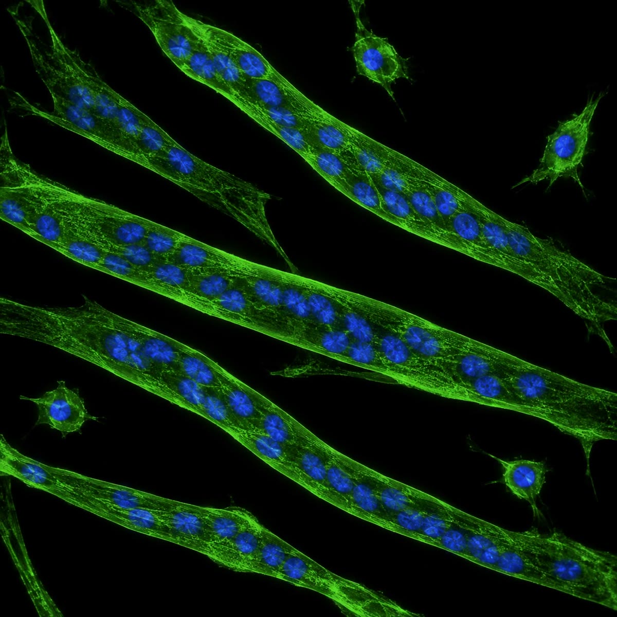
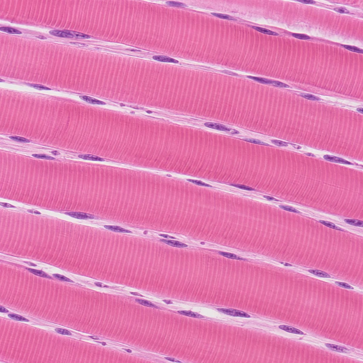
CoachIn the embryo, cells of the somite’s dermomyotome are induced by signals from the neural tube, notochord and surface ectoderm to express the myogenic factors MyoD and Myf5: they are now myoblasts, determined but still dividing and unremarkable to look at. Myoblasts migrate — into the limb buds, for instance — and multiply as long as growth factors (FGF) are present. When the factors run out, they withdraw from the cell cycle, express a second factor, myogenin, align end to end, and fuse: their membranes merge into a single long myotube with many nuclei in a row. The myotube switches on the muscle genes — actin, myosin, troponin, tropomyosin, creatine kinase, the acetylcholine receptor — assembles the contractile apparatus, and matures into a muscle fibre whose nuclei lie at its periphery. A fibre never divides again; a few myoblasts remain beside it, quiescent, as satellite cells, the stem cells that repair and grow the muscle for life.

Examples
Example 12.8 (Growth and repair)
A muscle grows in the adult not by adding fibres but by enlarging them: training adds myofibrils to each fibre, and satellite cells fuse into it to supply the extra nuclei a larger cytoplasm needs. After injury the same cells divide, re-express MyoD, fuse and rebuild the damaged segment within weeks — the embryonic programme run again in the adult. In muscular dystrophy the fibres lack dystrophin, a protein that ties the myofibrils to the membrane, and tear during contraction; the satellite cells repair them again and again until, after years, the stock is exhausted and fibrous tissue takes the muscle’s place.