Biology · Glossary

What is The retina?

Definition 18.3 University Biology — Year 3 · Chapter 18 — Sensory Systems

The retina is a sheet of brain at the back of the eye, three layers of cells with the photoreceptors, paradoxically, at the back, against the pigment epithelium. Rods10810^{8} per eye, one pigment, rhodopsin — serve dim light and saturate in daylight; cones6×1066\times 10^{6}, three pigments peaking in the blue, green and red, packed into the fovea — serve daylight, colour and acuity. Phototransduction is a cascade: a photon isomerises the retinal chromophore of one rhodopsin; the activated rhodopsin turns on hundreds of molecules of the G protein transducin, each of which activates a phosphodiesterase that destroys cyclic GMP; the fall in cGMP closes the cation channels that the cGMP held open in the dark, the inward “dark current” stops, and the cell hyperpolarises — light turns a photoreceptor off, and it releases less glutamate. In the dark the cell is depolarised and releasing continuously. The signal passes to bipolar cells (of ON and OFF types, which invert or preserve it) and on to the ganglion cells, a million per eye, whose axons form the optic nerve — a hundredfold compression. Horizontal and amacrine cells connect laterally, and their inhibition gives each ganglion cell a centre–surround receptive field: excited by light in a central disc and inhibited by light in the ring around it (or the reverse), so that the retina reports local contrast and edges rather than absolute light.
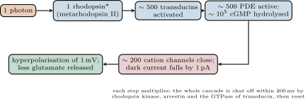
Phototransduction as an amplifier. One absorbed photon closes hundreds of channels through two catalytic stages, a gain of about a million in molecules — enough for a single photon to produce a measurable current in a rod.
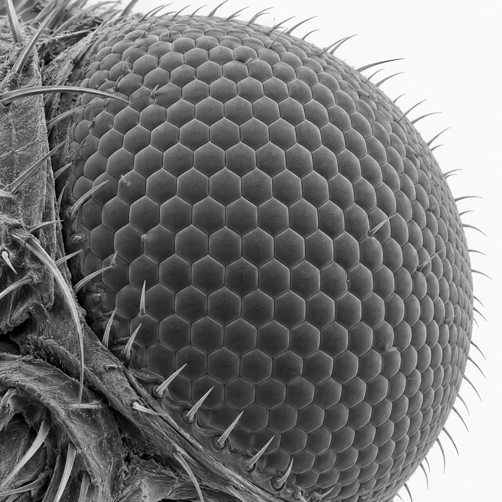
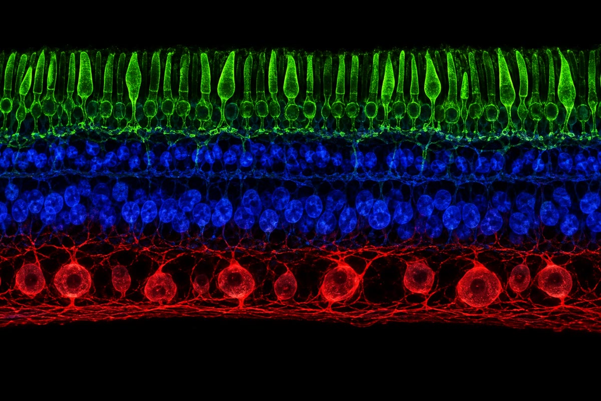
Left: a section of the retina, the photoreceptors (green) at the back, then the bipolar and horizontal cells, then the ganglion cells (red) whose axons leave for the brain. Right: a fly’s compound eye, hundreds of separate lenses each with its own receptors — a different solution to the same problem. Left: a section of the retina, the photoreceptors (green) at the back, then the bipolar and horizontal cells, then the ganglion cells (red) whose axons leave for the brain. Right: a fly’s compound eye, hundreds of separate lenses each with its own receptors — a different solution to the same problem.
Left: a section of the retina, the photoreceptors (green) at the back, then the bipolar and horizontal cells, then the ganglion cells (red) whose axons leave for the brain. Right: a fly’s compound eye, hundreds of separate lenses each with its own receptors — a different solution to the same problem.
Read in context →