University Biology — Year 3 · Bachelor Year 3
18Sensory Systems
On a dark night a human eye can detect a flash of a few photons — the smallest amount of light that physics allows to be detected at all. The ear hears a sound that moves the eardrum by less than the diameter of a hydrogen atom, and tells a tone from another that differs by a fifth of a per cent. A dog follows a trail of a few molecules per cubic centimetre; a shark finds a flatfish under the sand by the electric field of its heartbeat. Every sense begins with a cell that converts one kind of energy into a change of membrane potential, and every sense ends as spikes in a nerve; between the two lie the problems that this chapter treats: how a receptor amplifies a single molecule or photon into a signal a neuron can read, how a range of a trillionfold in intensity is squeezed into a rate of a few hundred spikes a second, how a mechanical wave is sorted by frequency, how ten thousand smells are told apart with four hundred receptors, and how the brain, which never receives anything but spikes, knows which sense they came from.
18.1 Principles common to every sense
Definition 18.1 (Transduction, coding and adaptation)
A sensory receptor cell contains a transduction mechanism — a protein or cascade — that converts a stimulus (a photon, a molecule, a displacement, heat, an electric field) into a change of membrane conductance and hence a receptor potential, graded with the stimulus; the cell, or the neuron it synapses on, turns this into a train of action potentials whose rate encodes intensity. The modality of a signal is given not by the spikes, which are identical in every nerve, but by the wire they travel on and where it ends in the brain: a labelled line. A sensory neuron responds to stimuli within a receptive field — a patch of skin, a region of the visual field, a band of frequencies — and neighbouring fields sharpen one another by lateral inhibition, each neuron suppressing its neighbours so that edges and contrasts are exaggerated. Most receptors adapt: their response to a maintained stimulus declines, so that they report change rather than level, and their working range shifts to sit around the prevailing background.
Theorem 18.2 (Weber, Fechner and Stevens)
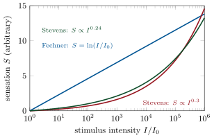
Weber’s observation (1834) is that the smallest detectable change in a stimulus of intensity is a fixed fraction of it: , with about for weight, for brightness, for loudness. If each just-noticeable difference is counted as one unit of sensation , then and
where is the threshold: sensation grows as the logarithm of the stimulus (Fechner, 1860), which is why intensities are measured in decibels and magnitudes, and why a candle added to a candle is noticed and a candle added to a floodlight is not. Direct scaling experiments, in which subjects assign numbers to sensations, give instead a power law (Stevens, 1957), with for loudness and brightness, for line length, and for electric shock; a logarithm and a small power are nearly indistinguishable over a few decades, and the two laws agree on the essential: the nervous system compresses.
Proof. From for one unit of , the increment of sensation per increment of stimulus is ; integrating from the threshold , where , gives . The power law is the alternative assumption that a fixed ratio of stimuli produces a fixed ratio of sensations, , whose integral is ; for with suitable scaling it tends to the logarithm. ∎
18.2 Vision
Definition 18.3 (The retina)
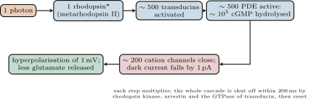
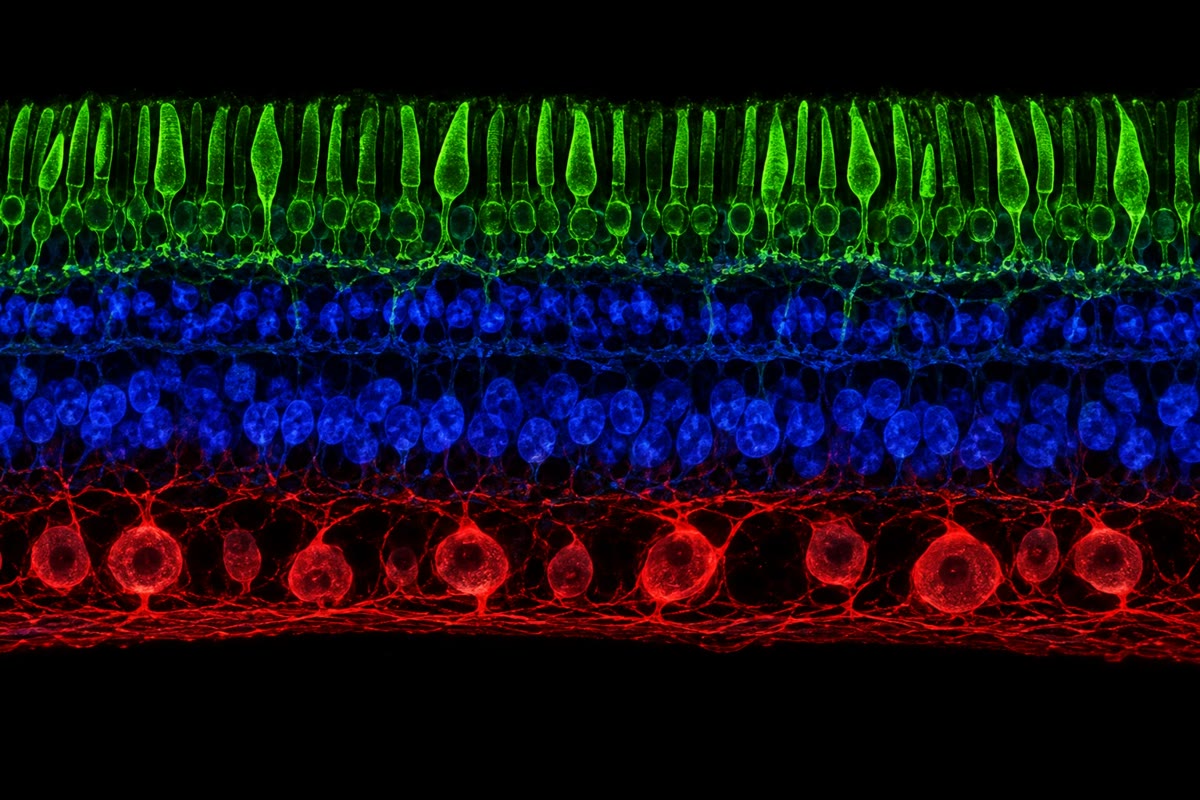
The retina is a sheet of brain at the back of the eye, three layers of cells with the photoreceptors, paradoxically, at the back, against the pigment epithelium. Rods — per eye, one pigment, rhodopsin — serve dim light and saturate in daylight; cones — , three pigments peaking in the blue, green and red, packed into the fovea — serve daylight, colour and acuity. Phototransduction is a cascade: a photon isomerises the retinal chromophore of one rhodopsin; the activated rhodopsin turns on hundreds of molecules of the G protein transducin, each of which activates a phosphodiesterase that destroys cyclic GMP; the fall in cGMP closes the cation channels that the cGMP held open in the dark, the inward “dark current” stops, and the cell hyperpolarises — light turns a photoreceptor off, and it releases less glutamate. In the dark the cell is depolarised and releasing continuously. The signal passes to bipolar cells (of ON and OFF types, which invert or preserve it) and on to the ganglion cells, a million per eye, whose axons form the optic nerve — a hundredfold compression. Horizontal and amacrine cells connect laterally, and their inhibition gives each ganglion cell a centre–surround receptive field: excited by light in a central disc and inhibited by light in the ring around it (or the reverse), so that the retina reports local contrast and edges rather than absolute light.
Theorem 18.4 (Counting photons: the frequency of seeing)
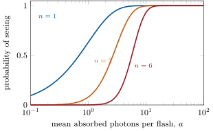
A flash that delivers on average absorbed photons to a patch of rods delivers, on any one presentation, a number that is Poisson distributed: . If the observer reports seeing the flash whenever at least photons are absorbed, the probability of seeing is
a curve that rises from to as increases, and whose steepness on a logarithmic scale of depends only on : the larger the threshold count, the steeper the curve. Fitting the measured frequency-of-seeing curve therefore gives without knowing how many of the delivered photons were actually absorbed.
Proof. Photons from a weak, steady source arrive independently at random, and the number absorbed in a short flash from a mean of is Poisson (the limit of a binomial with many photons each absorbed with small probability). Seeing requires , whose probability is one minus the sum of the first Poisson terms. Scaling the source by a factor multiplies by and shifts the curve along the axis without changing its shape, so the shape identifies : for , rises over two decades of ; for it rises over less than one. ∎
Evidence. Hecht, Shlaer and Pirenne (1942) sat observers in the dark for half an hour, then flashed a tiny green spot on the retina’s rod-rich periphery for a millisecond, hundreds of times at several intensities, and recorded the fraction of flashes seen. The threshold flash contained, at the cornea, some photons, of which (after reflection, absorption in the eye’s media and the chance of a photon that reaches a rod being absorbed by rhodopsin) about – were absorbed. The frequency-of-seeing curves had the steepness of to : the observer said “yes” when about six rods, out of the five hundred under the spot, had each caught a single photon. A rod detects one photon; the brain demands a few in coincidence, to keep the false alarms from the rods’ spontaneous isomerisations — one per rod per forty seconds — below the rate of the stars. ∎
18.3 Hearing and balance
Definition 18.5 (The cochlea)
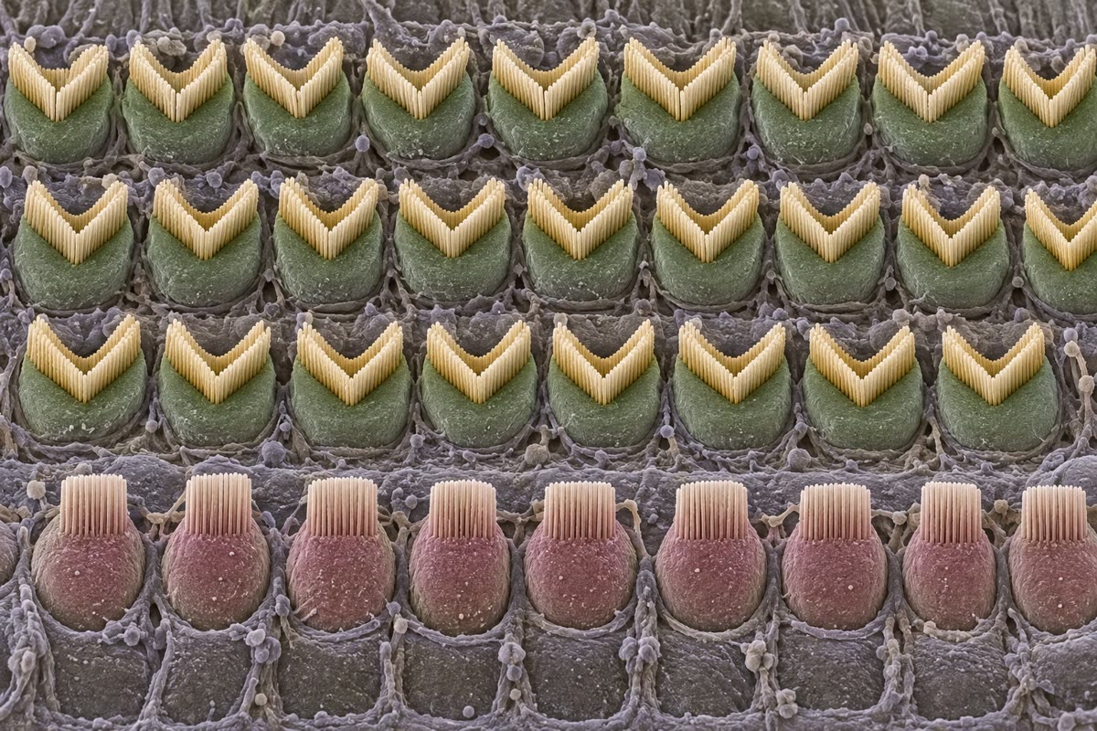
Sound is a pressure wave; the ear’s problem is to detect pressure changes of a few tens of micropascals in air and to sort them by frequency. The eardrum and the three middle-ear bones concentrate the force from the drum’s onto the of the oval window, raising the pressure some twentyfold — the impedance match between air and the fluid of the inner ear, without which most of the sound would reflect. Inside the cochlea, a coiled tube long, the pressure wave travels along the basilar membrane, which is narrow and stiff at the base and wide and floppy at the apex; each frequency makes the membrane vibrate most at one place — high frequencies near the base, low near the apex — a tonotopic map that the brain reads as pitch. On the membrane sit the hair cells: inner hair cells in a single row, each crowned with a bundle of stereocilia joined tip to tip by fine tip links; bending the bundle toward its tallest cilium stretches the links and pulls open cation channels within microseconds, without any second messenger, and potassium flows in from the endolymph, whose unusual composition and potential give a driving force of . The inner hair cells signal the auditory nerve; the outer hair cells, driven by the same motion, contract and lengthen with the sound through the motor protein prestin and pump energy back into the membrane’s vibration — an amplifier that sharpens tuning a hundredfold and is what fails in most deafness. Intensity is measured in decibels: , with the threshold of hearing; the ear works over , a trillionfold in intensity.
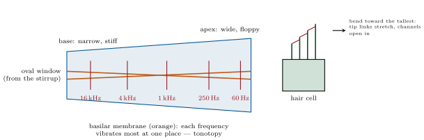
Evidence. Von Békésy (1940s) opened the cochleas of cadavers, sprinkled silver particles on the basilar membrane and watched under stroboscopic light as pure tones set up a travelling wave whose peak lay nearer the base the higher the tone — the place code, for which he received the Nobel prize in 1961. Hudspeth (1980s) pushed a single hair bundle with a glass fibre and recorded the current: it flowed within of the push, far too fast for any enzyme cascade, in proportion to how far the bundle moved toward its tallest row, and it vanished when the tip links were cut with a calcium chelator. Gating is mechanical — the link pulls the channel open — which is why hearing is the fastest of the senses and why its transducer is destroyed by the loud sounds that snap the links. ∎
Example 18.6 (The ear’s numbers)
At the threshold of hearing the basilar membrane moves about and the pressure amplitude is , a two-billionth of atmospheric; at the pressure is a million times greater and the membrane’s motion tens of nanometres. A young ear hears from to and separates tones apart, the discrimination of the place code sharpened by the outer hair cells. The auditory nerve’s fibres fire in step with the pressure wave up to about (phase locking), carrying timing to a precision of tens of microseconds, from which the brain computes a sound’s direction from the difference in arrival at the two ears — as little as . The balance organs use the same hair cells: in the semicircular canals the fluid’s inertia bends them when the head turns (angular acceleration), and in the otolith organs a layer of calcium carbonate crystals loads them with gravity and linear acceleration.
18.4 Chemical senses
Definition 18.7 (Olfaction and taste)
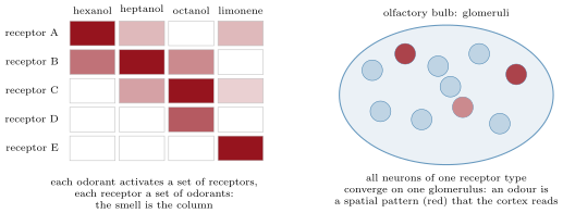
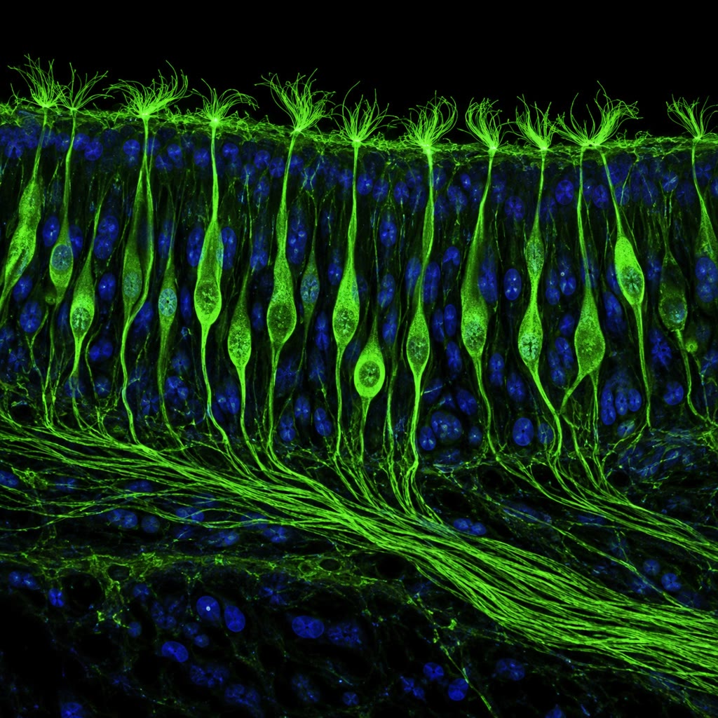
Smell begins in a patch of epithelium at the top of the nose, where some ten million olfactory sensory neurons each express one olfactory receptor gene out of about in humans ( in a mouse, the largest gene family in either genome) — G-protein- coupled receptors that bind odorant molecules in a pocket and, through cyclic AMP, open a channel. Each receptor binds several odorants and each odorant binds several receptors, so an odour is a combinatorial code, a pattern of activity across the receptor types, and a change of one carbon in a molecule changes the pattern and the smell. All the neurons expressing one receptor send their axons to the same one or two glomeruli in the olfactory bulb, so the bulb holds a map in which each odour is a spatial pattern of active glomeruli, read by the cortex without passing through the thalamus. Taste is simpler: five modalities, each a labelled line from its own receptor cells — sweet, umami and bitter through G-protein-coupled receptors (one sweet receptor, one umami, some twenty-five bitter), salt through a sodium channel, sour through a proton channel — and the rest of what we call flavour is smell, reaching the nose through the back of the mouth.
Evidence. Buck and Axel (1991) reasoned that odorant receptors would be G-protein-coupled receptors expressed only in the olfactory epithelium, and found by PCR with degenerate primers a family of hundreds of such genes, expressed there and nowhere else — the receptors for smell, unknown for a century. In-situ hybridisation showed one receptor per neuron and the convergence of like neurons on single glomeruli; Malnic, Buck and colleagues (1999) recorded single neurons and showed that each odorant activated several receptor types and each receptor several odorants — the combinatorial code. Bushdid and colleagues (2014) asked subjects to discriminate mixtures and estimated that humans can tell apart more than a trillion odours, far beyond the few thousand of common belief. ∎
Proposition 18.8 (The capacity of a combinatorial code)
With receptor types each either active or silent, an odour can be represented by any of patterns; with that is , and even if only patterns with exactly active types are considered there are . Discrimination is limited not by the code but by the noise of the receptors and by the brain’s reading: a labelled-line code with one receptor per odour would allow smells, a combinatorial one allows more than there are molecules to smell. The cost is that a single receptor tells the brain little on its own; the pattern must be read as a whole, which is what the glomerular map and the olfactory cortex do.
18.5 Touch, temperature, pain and the senses we lack
Definition 18.9 (Somatosensation)
The skin holds several kinds of mechanoreceptor, each an axon ending in its own capsule: Merkel cells for fine pressure and texture (slowly adapting, small fields), Meissner corpuscles for light touch and slip (rapidly adapting), Pacinian corpuscles deep in the skin for vibration (very rapidly adapting, huge fields), Ruffini endings for stretch. The transducer in most of them is Piezo2, a giant ion channel with a propeller of blades that opens when the membrane is stretched; without it touch and the sense of body position are lost, and the same channel in the lungs and bladder senses their filling. Temperature is sensed by channels of the TRP family tuned to different ranges — TRPV1, opened by heat above and by capsaicin, which is why chilli is “hot”; TRPM8, opened by cold and by menthol — and pain by free nerve endings, the nociceptors, that respond to damaging heat, pressure or chemicals (protons, ATP, bradykinin) and whose thresholds are lowered by inflammation (Chapter 15). Proprioception, the sense of where the limbs are, comes from the muscle spindles and tendon organs of Chapter 17 and from Piezo2 in joint capsules. The density of receptors sets acuity: two points apart are told apart on a fingertip, apart on the back.
Method 18.10 (Measuring a sense)
To characterise a sensory channel: (1) find the absolute threshold by presenting stimuli of graded intensity many times and fitting the fraction detected — the intensity detected half the time — as Hecht did; (2) find the difference threshold at several intensities and check Weber’s law; (3) map the spatial acuity with two stimuli at varying separation (the two-point test on skin, a grating on the retina); (4) map the temporal acuity with flicker or clicks (a flicker fuses at about in daylight vision); (5) in an animal, record from single afferent fibres while applying the same stimuli and match the neural to the behavioural thresholds — the psychophysics and the physiology should agree, and where they do not the difference is what the brain adds.
Remark 18.11 (Other animals, other senses)
Every sense here has a version an animal has pushed further, and there are senses we lack. Sharks and rays sense the microvolt fields of a prey’s heartbeat through jelly-filled canals; electric fish read distortions of their own field to see in the dark. Pit vipers image warm prey with infrared-sensitive pits; bats and dolphins echolocate, timing echoes of their own calls to a few microseconds and steering the call’s frequency to track a moth; bees see ultraviolet and the polarisation of skylight and navigate by it; migratory birds and turtles sense the Earth’s magnetic field by a mechanism still argued over. A dog’s nose has forty times our receptor neurons and a third of its brain to read them; an owl hears a mouse under snow. The principles do not change — a transducer, a labelled line, a code, compression, a map — and the variety is what evolution has done with them.
18.6 Exercises
Exercise 18.1 ★
Explain how the brain knows whether a train of spikes comes from the eye or the ear, given that the spikes are identical.
Solution
Solution of Exercise 18.1.
By the wire, not the message: each sensory nerve is a labelled line ending in its own region of the brain, and whatever arrives along the optic nerve is read as light — which is why pressure on the eyeball produces a flash and a blow to the ear a ring.
Exercise 18.2 ★
Why does light hyperpolarise a photoreceptor, and what is the dark current?
Solution
Solution of Exercise 18.2.
In the dark, cyclic GMP holds cation channels open and a steady inward “dark current” keeps the cell depolarised and releasing glutamate. Light activates rhodopsin, transducin and phosphodiesterase, which destroys cGMP; the channels close, the inward current stops, and the cell hyperpolarises and releases less — light is signalled by a decrease.
Exercise 18.3 ★
A whisper is , a conversation , a rock concert . Compute the intensity of each in W/m and the ratio between the loudest and the softest.
Solution
Solution of Exercise 18.3.
: , ; , ; , . Loudest to softest: .
Exercise 18.4 ★
State the five taste modalities and their receptor types, and explain why a cold blocks the “taste” of food.
Solution
Solution of Exercise 18.4.
Sweet, umami and bitter through G-protein-coupled receptors (one sweet, one umami, about twenty-five bitter); salt through the sodium channel ENaC; sour through a proton channel. Most of “flavour” is the smell of volatile molecules reaching the nose from the mouth; with the nose blocked only the five tastes remain and food seems bland.
Exercise 18.5 ★★
Weber’s fraction for weight is . Starting from a weight, how many just-noticeable steps lie between it and ? Use Fechner’s formula and check by counting steps of factor .
Solution
Solution of Exercise 18.5.
Fechner: steps. Counting: gives .
Exercise 18.6 ★★
Using Theorem 18.4, compute for , and absorbed photons with a threshold of . At what is the flash seen half the time? If of photons at the cornea are absorbed, how many must the flash deliver?
Solution
Solution of Exercise 18.6.
: : ; : ; : . Half the flashes are seen at ; with absorption the flash must deliver about photons at the cornea.
Exercise 18.7 ★★
The middle ear concentrates force from onto with a lever of . By what factor is the pressure raised, and in decibels? Why is the ear of a person with fused middle-ear bones (otosclerosis) duller by tens of decibels?
Exercise 18.8 ★★
Explain why the transduction current of a hair cell appears within microseconds while that of a photoreceptor takes tens of milliseconds, and what each sense gains from its design.
Solution
Solution of Exercise 18.8.
The hair cell’s channel is opened directly by the tip link pulling on it — no messenger, no enzyme — so it responds in microseconds; the photoreceptor amplifies one photon through two enzymatic stages, which take tens of milliseconds. Hearing gains the timing precision needed to locate sounds by interaural delays and to follow phase; vision gains the sensitivity to count single photons, and pays in speed.
Exercise 18.9 ★★
A mouse has receptor types and a human . If an odour activates about of the types, how many patterns of exactly that size are available to each (use with , and Stirling’s approximation with )?
Solution
Solution of Exercise 18.9.
. Human: , about patterns. Mouse: , about .
Exercise 18.10 ★★★
A rod isomerises a rhodopsin spontaneously once every in the dark. In a patch of rods observed for a window, what is the mean number of spontaneous events, and the probability that six or more occur by chance? Explain why the brain sets the threshold at about six and not at one.
Solution
Solution of Exercise 18.10.
Mean spontaneous events per window. , so . With a threshold of one, a spontaneous event would occur in most windows () and the dark would be full of flashes; at six, false alarms come once in five hundred windows. The threshold is set where the noise of the rods, not their sensitivity, dictates.
Exercise 18.11 ★★★
The auditory nerve locks its spikes to the sound’s phase up to , with a jitter of about . Sound travels at and the ears are apart. What is the largest interaural delay, what angular resolution does a discrimination give near the midline, and why does the place code, not timing, serve above ?
Solution
Solution of Exercise 18.11.
Largest delay (sound from one side). Near the midline , so corresponds to , about . Above the period () is shorter than the neurons can lock to, and a delay of several hundred microseconds spans more than one cycle, making the phase ambiguous; the brain then uses the level difference between the ears and the place code.
Exercise 18.12 ★★★
Lateral inhibition makes a uniform grey field next to a dark one look lighter at the border. Model a row of receptors each excited by its own light and inhibited by a fraction of each neighbour’s, , and compute the response across a step from to with . What does the retina gain and what does it lose by this?
Solution
Solution of Exercise 18.12.
Far from the edge, : on the dark side, on the bright. At the last dark receptor (, neighbours and ): ; at the first bright one: . The step is exaggerated into an undershoot and an overshoot — Mach bands. The retina gains contrast and edges, and a smaller dynamic range to transmit; it loses fidelity to absolute levels, and produces illusions of brightness.
18.7 Problem: A Candle Seen from Afar
Problem 18.1
Weekend problem — a candle’s photons counted into a distant eye, the flash’s detection computed from Poisson statistics and the rod’s amplifier, a concert’s decibels turned into eardrum motion and hair-cell current, and a nose’s code enumerated, ending on the distance at which the candle is seen, the pressure at the eardrum and the patterns a nose can form
Data: a candle emits about of visible light, at (photon energy J). A dark-adapted pupil is across; of photons entering the eye are absorbed by rhodopsin; the eye integrates over ; the threshold is absorbed photons. Each absorbed photon closes channels and reduces the dark current by for ; a rod’s dark current is . Sound: ; the eardrum has area ; a hair bundle of height at threshold deflects by . Olfaction: receptors; a hair cell’s transduction current is at saturation.
Part I — Photons.
- How many photons per second does the candle emit?
- At distance the photons are spread over a sphere of area . How many enter the pupil per second at ? At ?
- How many are absorbed in one integration window at each distance? Is the candle seen (threshold )?
- Find the distance at which the mean absorbed number in a window equals . With Poisson statistics, what fraction of windows exceed threshold there?
- At the distance of question 4, what is the probability that a given window contains no photon at all? Why does a faint star seem to flicker?
- Under a full moon the same rods absorb photons per window from the sky. Explain why the candle at is then invisible although it delivers the same photons.
Part II — The rod’s amplifier.
- One photon closes channels and reduces the current by . What current does one channel carry, and how many cations per second is that ()?
- What fraction of the dark current does one photon remove? How many simultaneous photons would shut the rod down completely (saturation)?
- The response lasts . How many cations does one photon prevent from entering? What is the gain in charge per photon?
- In daylight photons a second reach a rod. What happens to it, and why are cones, which adapt and saturate less, the daylight receptors?
- Cones need about photons to signal. Explain in terms of the trade between sensitivity and speed why cones respond in and rods in .
- A rhodopsin isomerises spontaneously once in . Over the rods of an eye, how many false photons per second does the retina generate, and why does that not blind the dark eye with noise? (Think of the threshold and the receptive field.)
Part III — Decibels.
- A concert at : intensity in W/m and power falling on one eardrum.
- How much acoustic energy does the eardrum receive in a two-hour concert? Compare with the energy of a falling raindrop ().
- The threshold of hearing at : power on the eardrum and energy per — compare with the energy of one visible photon.
- The hair bundle deflects at threshold on a height of . What angle is that, in degrees? Compare the deflection with the diameter of a hydrogen atom ().
- Prolonged exposure above damages hair cells, and hair cells do not regenerate in mammals. Two hours at deliver as much energy as how many hours at ?
- The range of hearing is . How many just-noticeable steps of loudness is that, if Weber’s fraction for intensity is about (one decibel)?
Part IV — The code of smell.
- With receptors each on or off, how many patterns? Write the answer as a power of ten.
- If an odour typically activates receptors, how many distinct such patterns are there (, use Stirling or logarithms)?
- Two odorants share of their receptors. Propose a measure of their distance in the code and explain why they smell alike.
- Humans discriminate about a trillion odours. What fraction of the patterns of question 20 is that, and what limits the number far below the code’s capacity?
- A mouse with receptors: how many more patterns of than a human, as a power of ten?
- A mutation deletes one receptor gene. Predict its effect on the ability to smell in general and on the perception of one odorant that binds that receptor strongly (specific anosmia).
- Summarise: the distance at which the candle is seen (question 4), the acoustic energy on the eardrum at threshold in (question 15), and the number of twenty-receptor patterns a human nose can form (question 20).
Solution
Solution of Problem 18.1.
1. photons a second. 2. Pupil area m. At : fraction , photons a second. At : a second. 3. In with absorbed: at , at — both above six; the candle is seen at ten kilometres (most of the time). 4. Six absorbed per window needs photons a second into the pupil: m, . There, : the candle is seen in about half the windows. 5. . At threshold the count fluctuates from window to window — — so the source seems to come and go: the scintillation of a faint star (to which the atmosphere adds its own). 6. The background’s fluctuation, photons per window, swamps the candle’s : detection requires the signal to exceed the noise of the background, not the threshold of the dark eye. 7. per channel; ions a second. 8. One thirtieth; about thirty simultaneous photons close every channel and saturate the rod. 9. C, cations: one photon controls over a million charges. 10. The rod saturates within milliseconds, every channel shut, and its rhodopsin is bleached faster than it is regenerated; cones, with lower gain and faster shut-off, keep responding and shift their range across six decades of light. 11. High gain needs long integration (each stage takes time and the response is prolonged to accumulate), so the rod is slow and sensitive; the cone’s smaller amplification and quicker shut-off give a response in , fast enough for movement, at the cost of needing a hundred photons. 12. spontaneous isomerisations a second across the retina — but only per window in any patch of rods, far below the threshold of six, so the noise is rejected patch by patch; the threshold and the pooling exist precisely for this. 13. ; on , . 14. — about four hundred raindrops’ worth over two hours. 15. W; in , J — about the energy of one or two visible photons. 16. rad, ; the deflection is about three hydrogen atoms. 17. : two hours at deliver the energy of hours at — nearly a month of working days. 18. About just-noticeable steps, one per decibel. 19. . 20. , less a Stirling correction of about : some patterns. 21. Count the receptors in one pattern but not the other: of (a Hamming distance); patterns that overlap by three quarters drive nearly the same glomeruli and are read as nearly the same smell. 22. of the patterns. The code is not the limit: receptors are noisy, many are tuned alike so their activities are correlated, real molecules produce only a small subset of patterns, and the brain’s discrimination and memory of patterns are finite. 23. , about — some times the human number of twenty-receptor patterns (and far more if the mouse uses more receptors per odour). 24. General smell hardly changes — the code is redundant and other receptors carry every odorant — but an odorant that depends on that receptor at low concentration becomes undetectable or changes its character: a specific anosmia, as with the many people who cannot smell androstenone. 25. The candle is seen to about ; threshold sound delivers J to the eardrum in , a photon or two; a human nose can form some twenty-receptor patterns.