Biology · Glossary

What is The testis and spermatogenesis?

Definition 8.1 University Biology — Year 2 · Chapter 8 — Sexual Reproduction of Mammals

The testis is a mass of seminiferous tubules, several hundred metres in all, whose walls make sperm; between the tubules lie Leydig cells, which make testosterone. In the tubule wall, spermatogonia at the outer edge divide by mitosis, keeping a stem population and sending cells inward; these grow into primary spermatocytes, which undergo meiosis I (to two secondary spermatocytes) and meiosis II (to four haploid spermatids); the spermatids shed most of their cytoplasm, condense their nucleus, grow a flagellum and cap the nucleus with the acrosome, a vesicle of enzymes, to become spermatozoa that are released into the lumen. Large Sertoli cells span the wall, nursing the germ cells and forming, by tight junctions between them, a blood–testis barrier that keeps the immune system away from cells that first appear at puberty. The whole sequence takes about 6464 days in a man and yields some 10810^{8} sperm a day from puberty to old age. Sperm leave the testis immotile and mature in the epididymis for two weeks.
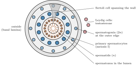
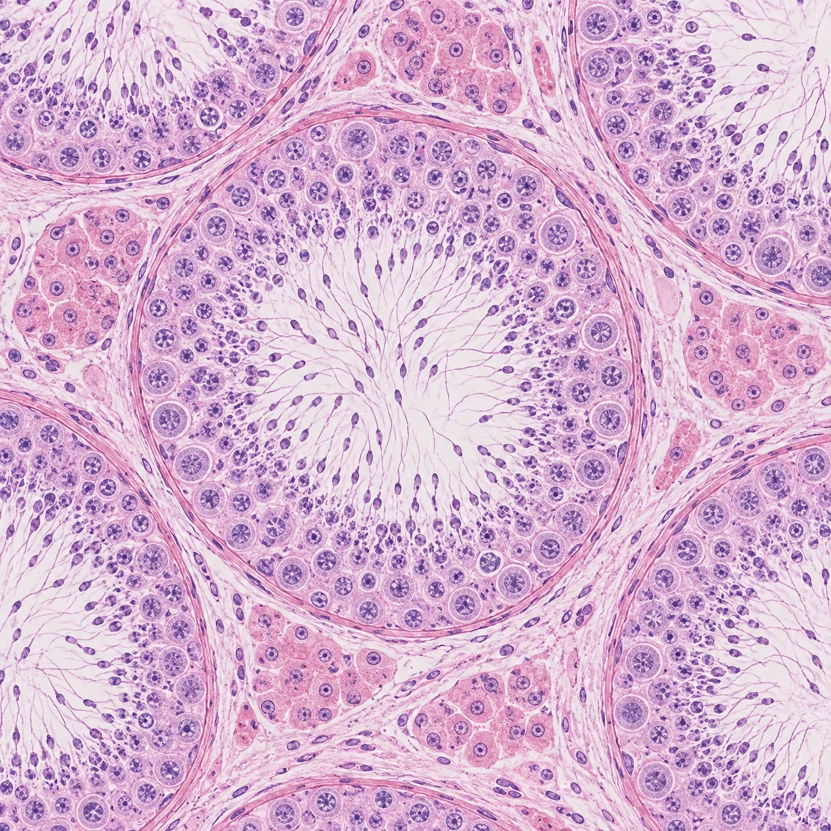
A seminiferous tubule in cross-section. Germ cells mature from the outer wall (spermatogonia) inward (spermatocytes, spermatids, spermatozoa in the lumen), supported by Sertoli cells; Leydig cells between the tubules make testosterone.
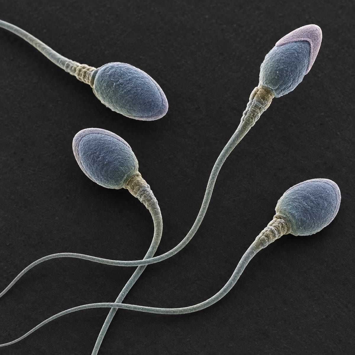
Left: seminiferous tubules in section, the germ cells layered from the wall to the lumen. Right: spermatozoa under the scanning electron microscope, the acrosomal cap visible on the heads. Left: seminiferous tubules in section, the germ cells layered from the wall to the lumen. Right: spermatozoa under the scanning electron microscope, the acrosomal cap visible on the heads.
Left: seminiferous tubules in section, the germ cells layered from the wall to the lumen. Right: spermatozoa under the scanning electron microscope, the acrosomal cap visible on the heads.
Read in context →