University Biology — Year 2 · Bachelor Year 2
8Sexual Reproduction of Mammals
A human egg is a tenth of a millimetre across, the largest cell in the body; a sperm cell is a nucleus with a propeller, one of three hundred million released at once, of which a few dozen will reach the egg and one will enter it. Within a week the fertilised egg has become a hollow ball that burrows into the wall of the uterus and builds, out of its own outer cells, an organ — the placenta — through which its mother will feed it for nine months. Then it is born and fed on milk. Every mammal does this; no other animals do all of it. This chapter follows the gametes to fertilisation, the embryo to implantation, and the organism through gestation, birth and lactation, and ends with how the two sexes are made.
8.1 Gonads and gametes
Definition 8.1 (The testis and spermatogenesis)
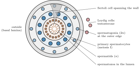
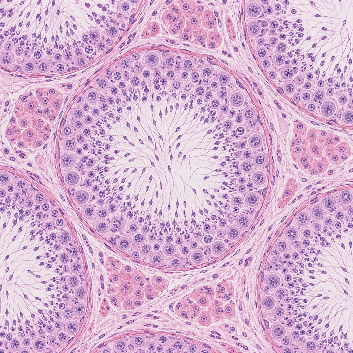
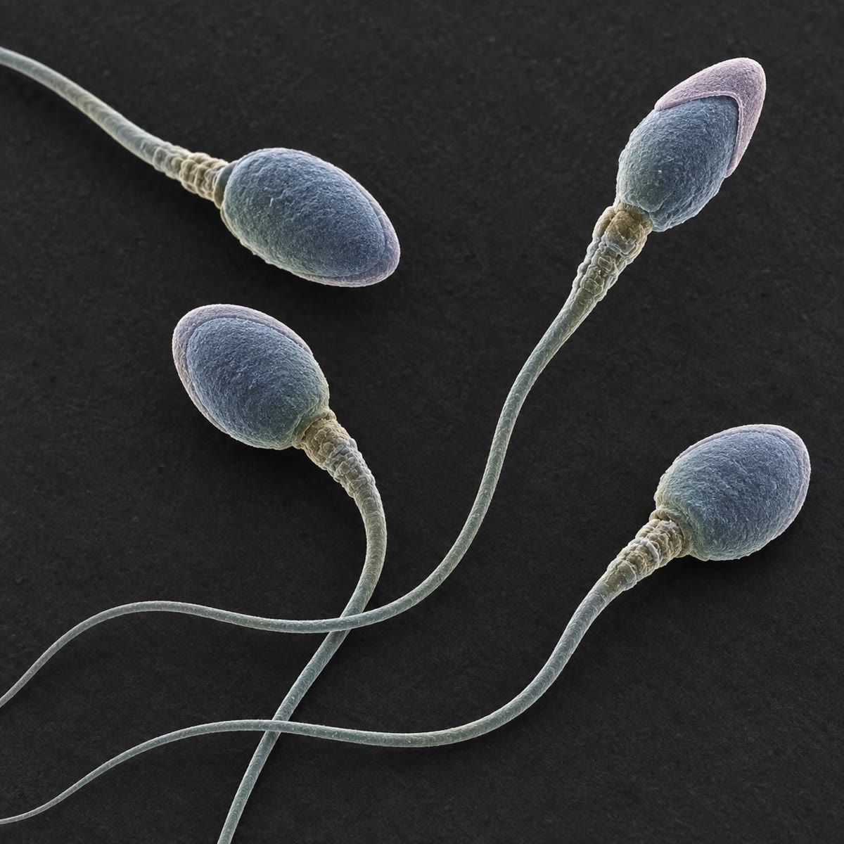
The testis is a mass of seminiferous tubules, several hundred metres in all, whose walls make sperm; between the tubules lie Leydig cells, which make testosterone. In the tubule wall, spermatogonia at the outer edge divide by mitosis, keeping a stem population and sending cells inward; these grow into primary spermatocytes, which undergo meiosis I (to two secondary spermatocytes) and meiosis II (to four haploid spermatids); the spermatids shed most of their cytoplasm, condense their nucleus, grow a flagellum and cap the nucleus with the acrosome, a vesicle of enzymes, to become spermatozoa that are released into the lumen. Large Sertoli cells span the wall, nursing the germ cells and forming, by tight junctions between them, a blood–testis barrier that keeps the immune system away from cells that first appear at puberty. The whole sequence takes about days in a man and yields some sperm a day from puberty to old age. Sperm leave the testis immotile and mature in the epididymis for two weeks.
Definition 8.2 (The ovary and oogenesis)
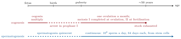
Oogenesis is the mirror image of spermatogenesis in almost every respect. The oogonia multiply only in the fetus: by birth a girl carries a stock of about a million primary oocytes, each arrested in prophase I of meiosis and wrapped in a single layer of cells — a primordial follicle — and no new ones are ever made. From puberty, each month a cohort of follicles grows: the oocyte enlarges, the surrounding granulosa cells multiply into layers, an outer theca forms, a fluid-filled cavity opens (the Graafian follicle, two centimetres across), and usually a single one reaches ovulation: the oocyte, only now completing meiosis I and arresting again in metaphase II, is expelled with its halo of granulosa cells into the oviduct. The emptied follicle becomes the corpus luteum, a temporary gland of progesterone. Meiosis produces one egg and three minute polar bodies — all the cytoplasm goes to one cell — and meiosis II is completed only if a sperm enters. Of the million oocytes, about are ovulated in a lifetime; the rest degenerate (atresia), and when the stock is exhausted, around fifty, cycles stop: the menopause.
8.2 Fertilisation
Proposition 8.3 (The steps of fertilisation)
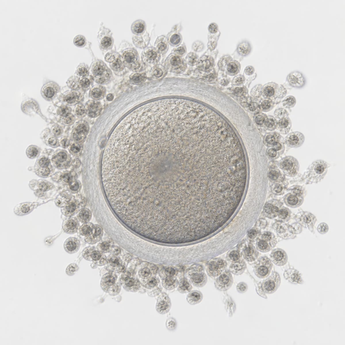
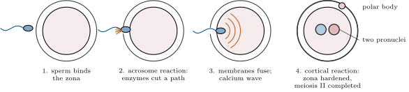
Sperm deposited in the vagina — some — cross the cervical mucus, are swept up the uterus by its contractions, and a few hundred reach the ampulla of the oviduct within an hour, where the egg waits for a day. On the way the sperm undergo capacitation: secretions of the female tract strip their membranes of coating proteins and make them hyperactive. At the egg a sperm pushes through the granulosa cells, binds a glycoprotein of the zona pellucida, and undergoes the acrosome reaction, releasing enzymes that digest a path through the zona; its membrane fuses with the egg’s, and the sperm nucleus enters. The egg responds within seconds: calcium sweeps across it, and cortical granules release enzymes that harden the zona and strip its sperm receptors — the cortical reaction, the block to polyspermy. The egg completes meiosis II, sheds the second polar body, and the two haploid pronuclei replicate their DNA, come together and enter the first mitosis: the zygote is a day old. Species specificity lies in the zona proteins, which is why a mouse sperm does not enter a human egg.
Evidence. Edwards and Steptoe (1978) fertilised a human oocyte, recovered from the ovary shortly before ovulation, with capacitated sperm in a dish, cultured the embryo to eight cells and returned it to the uterus, where it implanted and was born: in vitro fertilisation. Every step of the sequence above had first been worked out in the sea urchin and the mouse, where the fusion, the calcium wave and the cortical granule release can be watched; in the mouse, removing one zona glycoprotein (ZP2 or ZP3) by mutation leaves eggs that sperm cannot bind. ∎
Theorem 8.4 (Why one egg needs millions of sperm)
If, of sperm deposited, each reaches the egg’s surface independently with a small probability , the number arriving is Poisson with mean and the probability that at least one arrives is
With and , and ; with (the clinical threshold of low sperm count), and . The same arithmetic shows why the block to polyspermy is indispensable: when is high, several sperm arrive within seconds of one another, and an egg entered by two would be triploid and die.
Proof. Each sperm is an independent trial with success probability ; the number of successes is binomial, which for large and small is Poisson with mean . The probability of zero successes is , so of at least one, ; the probability of two or more is , about for . ∎
8.3 Implantation and the placenta
Definition 8.5 (Cleavage, blastocyst, implantation)
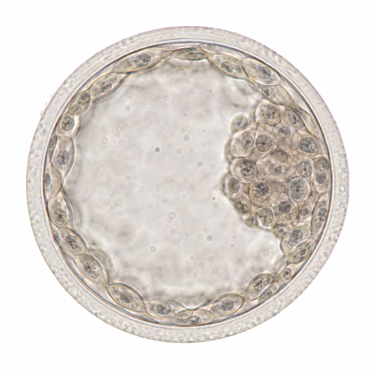
The zygote divides every twelve to twenty hours without growing (cleavage) as it is moved down the oviduct: two cells, four, eight, a compact ball of sixteen (morula), and by day five a blastocyst of about a hundred cells: an outer layer, the trophoblast, around a cavity, with a clump of cells at one side, the inner cell mass. The trophoblast will build the placenta and membranes; the inner cell mass will become the embryo — and, taken out at this stage, gives embryonic stem cells. On day six the blastocyst hatches from its zona, sticks to the lining of the uterus, and the trophoblast invades it: cells fuse into a multinucleate front that digests maternal tissue and opens maternal capillaries, so that by day twelve the embryo is buried in the wall and bathed in maternal blood. This is implantation. It succeeds only in a uterus prepared by progesterone, in a window of a few days; about half of all human conceptions fail before or at this step, usually from chromosomal errors of the egg.
Definition 8.6 (The placenta)
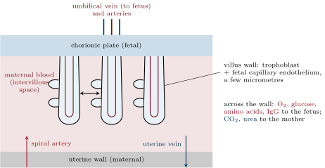
The placenta is an organ made jointly by the embryo (the chorion, from the trophoblast) and the mother (the uterine lining). Finger-like chorionic villi, branched into a surface of some at term, hang in lakes of maternal blood delivered by the uterine arteries at half a litre a minute; inside each villus run fetal capillaries, joined to the fetus through the two arteries and one vein of the umbilical cord. The two bloods never mix: they are separated by the villus wall, a few micrometres of trophoblast and capillary endothelium, across which oxygen, , water, urea and fat-soluble molecules diffuse, glucose and amino acids are carried by transporters, and maternal antibodies (IgG) are ferried by receptor, giving the newborn its first months of immunity. The placenta is also a gland: from the first days the trophoblast secretes human chorionic gonadotropin (hCG), which keeps the corpus luteum making progesterone, and from the third month the placenta itself makes the progesterone and oestrogens that maintain the pregnancy (Chapter 9). It is, in effect, a lung, a gut, a kidney and an endocrine gland grown for nine months and discarded at birth.
Theorem 8.7 (Exchange across the placenta)
A gas crossing a membrane of area and thickness moves at a rate , where is the difference in its partial pressure and the tissue’s permeability. For oxygen in tissue ; with , and (maternal in the intervillous space against in the umbilical artery), the placenta could pass about of oxygen a minute, some eight times the a term fetus consumes. Oxygen transfer is therefore limited not by diffusion but by blood flow; the fetus works at a low oxygen pressure and compensates with a haemoglobin of higher affinity and a higher red-cell count.
Proof. Fick’s law states that flux is proportional to the gradient and to the area; the constant absorbs the solubility and diffusion coefficient of the gas in tissue. Numerically: with in and in . The fetal demand is about per kilogram per minute for . ∎
8.4 Gestation, birth and lactation
Proposition 8.8 (Gestation and birth)
Gestation lasts days in a mouse, in a human, in an elephant — roughly as the quarter power of body mass, with primates slow for their size. It is maintained by progesterone, which keeps the uterine muscle quiet and the cervix closed. Birth (parturition) is triggered by the fetus itself: its adrenal cortisol, rising at term, shifts the placenta from progesterone to oestrogen, which fits the uterus with oxytocin receptors and gap junctions and starts prostaglandin release. Then a positive feedback takes over: the head presses on the cervix, sensory nerves signal the hypothalamus, oxytocin is released from the posterior pituitary, the uterus contracts, the head presses harder; the loop escalates until delivery, after which the stimulus ends and the loop stops — one of the few positive feedbacks in physiology, and one that must end in an event. The placenta is delivered minutes later.
Evidence. Ferguson (1941) showed in the rabbit that distending the cervix provoked uterine contractions and that cutting the spinal cord or removing the posterior pituitary abolished the response: a neuroendocrine reflex from cervix to pituitary to uterus. Oxytocin, isolated and synthesised by du Vigneaud in 1953, is now the standard drug for inducing labour. ∎
Proposition 8.9 (Lactation)
The mammary gland, a modified sweat gland, is built during pregnancy by oestrogen, progesterone and prolactin but held back from secreting by progesterone; when the placenta leaves, progesterone falls and, under prolactin from the anterior pituitary, the alveoli begin to make milk. Two reflexes run the process: suckling stimulates prolactin release (which maintains synthesis and suppresses ovulation) and oxytocin release (which contracts the cells around the alveoli and ejects the milk). Human milk carries about lactose, fat, protein and ; the first days’ colostrum is rich in antibodies (IgA) that coat the infant’s gut. A mother makes about a day, at a cost of some — a quarter of her food — for months; lactation is the most expensive part of mammalian reproduction and the one that names the class.
8.5 Making the two sexes
Proposition 8.10 (Sex determination and differentiation)
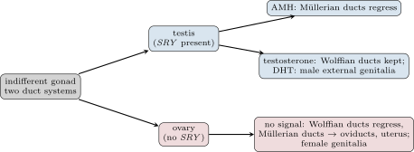
Until the sixth week a human embryo of either sex has the same equipment: an indifferent gonad, two pairs of ducts (Wolffian and Müllerian) and neutral external genitalia. The gene SRY on the Y chromosome switches on in the gonad and turns it into a testis; without it the gonad becomes an ovary. The fetal testis then does two things: its Sertoli cells secrete anti-Müllerian hormone, which makes the Müllerian ducts regress, and its Leydig cells secrete testosterone, which keeps the Wolffian ducts (the future epididymis, vas deferens, seminal vesicles) and, converted to dihydrotestosterone, moulds the external genitalia into penis and scrotum. In the absence of both signals the Wolffian ducts regress, the Müllerian ducts become the oviducts, uterus and upper vagina, and the genitalia develop as female. The female path is the one taken when nothing is said: the male path must be actively imposed by the testis, and every step at which the imposition fails — a mutant SRY, a missing hormone receptor — gives an XY individual with a female or intermediate body.
Evidence. Jost (1947) removed the gonads from rabbit fetuses in the uterus before their ducts had differentiated: castrated fetuses of either chromosomal sex developed a uterus and oviducts and female genitalia. A crystal of testosterone implanted in a castrated fetus kept the Wolffian ducts on that side but did not remove the Müllerian ducts; a grafted fetal testis did both, on its own side: the testis must make a second, then unknown, factor to suppress the female ducts, identified thirty years later as anti-Müllerian hormone. Later the sex-reversing effect of a small piece of the Y chromosome pinned the switch to SRY (1990), and a mouse made XX with the Sry gene alone developed as a male. ∎
8.6 Exercises
Exercise 8.1 ★
Compare spermatogenesis and oogenesis: when the stem cells divide, how many gametes one meiosis yields, how long a gamete takes to make, where the arrests are, and how many gametes a lifetime produces.
Solution
Solution of Exercise 8.1.
Spermatogonia divide from puberty for life; oogonia only in the fetus. One meiosis gives four sperm but one egg (and three polar bodies). A sperm takes about 64 days plus two weeks of maturation; an egg is arrested in prophase I from before birth until the month of its ovulation — decades — and again in metaphase II until fertilisation. A man makes sperm a day, some in a life; a woman ovulates about 400 eggs.
Exercise 8.2 ★
List the steps of fertilisation from the arrival of a sperm at the zona to the first mitosis, and say which step blocks polyspermy.
Solution
Solution of Exercise 8.2.
Binding to the zona pellucida; acrosome reaction and digestion of a path; fusion of the membranes and entry of the sperm nucleus; calcium wave and cortical reaction (hardening of the zona, loss of its receptors: the block to polyspermy); completion of meiosis II and extrusion of the second polar body; formation of the two pronuclei, DNA replication, first mitosis.
Exercise 8.3 ★
Name the two cell populations of the blastocyst and the fate of each. On what day does implantation begin, and what hormone must have prepared the uterus?
Solution
Solution of Exercise 8.3.
Trophoblast (outer layer) makes the placenta and membranes; inner cell mass makes the embryo. Implantation begins on day 6 to 7 after fertilisation, in a uterine lining prepared by progesterone from the corpus luteum.
Exercise 8.4 ★
For each of the following, say whether it crosses the placenta and how: oxygen, glucose, maternal red cells, IgG antibodies, urea, alcohol, most bacteria.
Solution
Solution of Exercise 8.4.
Oxygen: diffusion. Glucose: facilitated transport. Maternal red cells: do not cross (the bloods never mix). IgG: receptor-mediated transport. Urea: diffusion, from fetus to mother. Alcohol: diffuses freely. Bacteria: mostly do not cross (some, like syphilis and listeria, do).
Exercise 8.5 ★★
With per sperm, compute the probability of fertilisation for sperm counts of , , and . At which count does it drop below one half? What does this say about the shape of the dose–response of fertility to sperm count?
Solution
Solution of Exercise 8.5.
: . Below one half when , i.e. below sperm. The curve saturates: above about extra sperm add almost nothing, below it fertility falls nearly in proportion to the count.
Exercise 8.6 ★★
A girl is born with oocytes, has at puberty (age 13) and about at 50, having ovulated . Compute the mean rate of atresia (oocytes lost per day) before and after puberty, and compare with the rate of ovulation.
Exercise 8.7 ★★
Fetal haemoglobin has a half-saturation pressure , adult haemoglobin ; take the saturation as with . Compute both saturations at , the pressure in the umbilical vein, and explain the advantage.
Solution
Solution of Exercise 8.7.
Adult: , . Fetal: , . At the low pressures of the placenta the fetal blood loads a fifth more oxygen than adult blood would, and unloads it in tissues at pressures where adult haemoglobin would already be half empty.
Exercise 8.8 ★★
hCG appears in maternal blood at implantation (day 7 after fertilisation) at about and doubles every two days; a pregnancy test detects . On what day after fertilisation does the test turn positive? On what day after the last menstrual period (ovulation on day 14)?
Solution
Solution of Exercise 8.8.
: doublings, : day 16 to 17 after fertilisation; day 30 to 31 after the last period — about the day the period is missed.
Exercise 8.9 ★★
Recompute the placental oxygen transfer of the theorem for a placenta of with a villus wall of , as in some pathologies, and compare with the fetal demand. What happens to the fetus?
Solution
Solution of Exercise 8.9.
against a demand of : the margin falls from eight to one and a half; the fetus becomes hypoxic under any extra load, grows slowly and may need early delivery.
Exercise 8.10 ★★★
Predict the duct system and external genitalia of: (a) an XY fetus whose cells lack the testosterone receptor; (b) an XY fetus lacking anti-Müllerian hormone; (c) an XX fetus exposed to high testosterone from its adrenals; (d) an XX fetus carrying SRY on an X chromosome. Explain each from Jost’s results.
Solution
Solution of Exercise 8.10.
(a) Testes form and secrete both hormones; AMH removes the Müllerian ducts, but testosterone cannot act: the Wolffian ducts regress and the external genitalia are female — a woman with testes and no uterus. (b) Testosterone acts, AMH does not: a male with epididymis, vas deferens and male genitalia, plus a uterus and oviducts. (c) Ovaries and Müllerian derivatives (no testis, no AMH); the adrenal androgens virilise the external genitalia to varying degrees. (d) SRY makes testes, which impose the male path: a man with two X chromosomes, sterile because the Y genes of spermatogenesis are absent.
Exercise 8.11 ★★★
Identical twins arise when one embryo splits. If it splits before day 3 the twins have separate placentas and sacs; between days 4 and 8, one placenta and two sacs; between days 8 and 13, one placenta and one sac; later, they are conjoined. In a series of identical twins, have separate placentas, one placenta and two sacs, one sac. Deduce which stage of development most often splits, and explain in terms of the blastocyst which structures are shared in each case.
Solution
Solution of Exercise 8.11.
The commonest () is the day 4 to 8 split: after the trophoblast has formed (so one placenta) but before the amnion (so two sacs). Before day 3 each half makes its own trophoblast, hence two placentas (); after day 8 the amnion is already laid down and is shared (). Splitting the inner cell mass after the trophoblast has committed is evidently the easiest event.
Exercise 8.12 ★★★
Marsupials give birth after a few weeks to a bean-sized young that completes its growth on a teat in the pouch; placental mammals gestate long and give birth to a large young. Compare the two strategies in terms of maternal cost, risk, and the mother’s ability to abandon a litter in a bad season, and suggest why placentals have displaced marsupials on every continent but Australia.
Solution
Solution of Exercise 8.12.
A marsupial invests little before birth: a short gestation, a tiny young, and a lactation that can be cut short by dropping the pouch young if drought comes, so the mother’s risk per attempt is low and she can try again at once. A placental invests heavily in a long gestation with a large, well-developed young that survives better at birth, at the cost of a mother committed for months and vulnerable meanwhile. In productive, predictable environments the higher survival of placental young wins; Australia’s erratic climate rewards the marsupial’s option to abandon, and its isolation kept placentals out until recently.
8.7 Problem: A Pregnancy in Numbers
Problem 8.1
Weekend problem — a human pregnancy followed from the sperm to the milk, through the probabilities of fertilisation, the timing of implantation and its hormone, the exchange capacity of the placenta, and the energy cost of gestation and lactation, ending on the fertilisation probability, the day the test turns positive, the placenta’s oxygen margin and the cost of a day of milk
Data: sperm per ejaculate, each reaching the egg with probability ; sperm swim at ; the ampulla is from the cervix. hCG at implantation (day 7) , doubling every , test threshold . Placenta at term: , wall , , ; fetus consuming of oxygen per kilogram per minute; maternal placental blood flow carrying of oxygen per millilitre of blood. Glucose: fetus uses per kilogram per minute; maternal blood . Milk: at , made at efficiency; infant gains of tissue at deposited and spends on maintenance at .
Part I — Fertilisation.
- Compute the mean number of sperm reaching the egg and the probability of fertilisation.
- Compute the probability that two or more reach it, and say what would happen without the cortical reaction.
- How long would a sperm take to swim to the ampulla? Sperm are found there within : what carries them?
- Only a few hundred sperm reach the ampulla. What fraction of the ejaculate is that, and where are the others lost?
- An egg is fertilisable for about a day, sperm survive about three days in the tract. Over how many days of the cycle can intercourse lead to conception?
- A man’s count falls to . Recompute the probability of fertilisation per cycle, and the expected number of cycles to conception (geometric law).
- Explain why the egg, and not the sperm, provides the mitochondria of the embryo.
Part II — Implantation and its hormone.
- How many cleavage divisions have occurred by day 5 if they take each? How many cells?
- On what day after fertilisation does hCG reach the test threshold? After the last period (ovulation on day 14)?
- Why must hCG appear within days of implantation? What happens to the corpus luteum, and to the pregnancy, if it does not?
- hCG peaks near around week 10 and then falls. How many doublings from implantation is that, and why can it fall afterward without loss of the pregnancy?
- Half of conceptions are lost before or at implantation, mostly from aneuploid eggs. Explain why aneuploidy is more common in eggs than in sperm, using the timeline of oogenesis.
- Twins: an embryo that splits on day 6. State whether the twins share the placenta and the amniotic sac.
Part III — The placenta.
- Compute the maximal diffusive oxygen transfer.
- Compute the fetal oxygen demand and the diffusion margin.
- Compute the oxygen delivered per minute by maternal blood flow to the placenta. What fraction must be extracted to meet the demand?
- Compute the fetal glucose demand in grams per day, and the volume of maternal blood that contains it. Compare with the daily maternal placental flow.
- The fetus’s blood leaves the placenta at of oxygen. Why does it not suffocate at a pressure that would leave an adult unconscious?
- Name two things the placenta does that no other organ of the embryo could do at the time, and one thing it cannot do.
Part IV — Birth and milk.
- Describe the positive feedback of labour and explain why it does not run away before term.
- Compute the energy in a day’s milk and the energy the mother spends to make it.
- Compute the infant’s daily energy budget (growth plus maintenance) and compare with the milk supplied.
- Compute the mother’s total energy cost of nine months of lactation, and compare with the cost of the pregnancy itself.
- Explain why suckling delays the return of ovulation, and why this matters in populations without contraception.
- State the result: the probability of fertilisation, the day the test turns positive, the placenta’s oxygen margin, and the cost of a day’s milk.
Solution
Solution of Problem 8.1.
1. ; . 2. : without the block, four fertilisations in five would be polyspermic and the embryo triploid. 3. , fifty minutes; the contractions of the uterus and oviduct carry them. 4. : lost in the acid vagina, in the cervical mucus, to phagocytes in the uterus, and in the wrong oviduct. 5. From three days before ovulation to one day after: about four days. 6. , ; expected cycles . 7. The sperm’s few mitochondria, in the midpiece, are tagged and destroyed after entry; the egg carries a hundred thousand. 8. : seven divisions, cells — about a hundred. 9. doublings, : day 16 to 17; day 30 to 31 after the period. 10. The corpus luteum regresses about twelve days after ovulation unless hCG rescues it; without it progesterone falls, the lining sheds and the embryo is lost with the period. 11. doublings; by week 10 the placenta makes its own progesterone and the corpus luteum is no longer needed. 12. Eggs sit in prophase I for decades and the cohesins holding the bivalents decay, so nondisjunction at meiosis I rises steeply with maternal age; sperm are made fresh in two months. 13. After the trophoblast (day 5) and before the amnion (day 8): one placenta, two sacs. 14. . 15. : a margin of eight. 16. ; extraction . 17. ; in of maternal blood; daily flow : the fetus takes about of the glucose that passes. 18. Fetal haemoglobin is saturated at , the fetus has more red cells and a high cardiac output, and its tissues are adapted to work at low pressure — it lives, in effect, on the summit of Everest. 19. Gas exchange without a working lung, nutrition and excretion without a working gut or kidney, and the hormones that keep the pregnancy; it cannot keep out every toxin — alcohol, nicotine and some viruses cross. 20. Cervical stretch hypothalamus oxytocin contraction more stretch, until delivery removes the stimulus. Before term the progesterone-dominated uterus has few oxytocin receptors and no gap junctions, so contractions do not spread and the loop cannot close. 21. ; cost . 22. Growth , maintenance : , about two thirds of the milk’s energy; the rest goes to activity, warmth and losses. 23. , two and a half times the pregnancy. 24. Suckling raises prolactin and suppresses the pulsatile release of the hormone that drives ovulation, so ovulation returns only months later; without contraception this spaces births by two to three years. 25. ; test positive on day 17 after fertilisation (day 31 of the cycle); oxygen margin eight; a day’s milk costs the mother .