Biology · Glossary

What is Cilia and flagella?

Definition 9.10 University Biology — Year 3 · Chapter 9 — Cytoskeleton Dynamics and Cell Motility

A eukaryotic cilium or flagellum is a membrane-covered extension built on the axoneme: nine doublet microtubules in a ring around a central pair, held by nexin links and radial spokes, and grown from a basal body, a modified centriole. Axonemal dyneins attached to each doublet walk along the neighbouring doublet toward its minus end at the base; since the doublets are tied together they cannot slide freely, and the sliding is converted into bending, propagated along the axoneme as a wave by switching the active dyneins from one side of the ring to the other. A cilium is 5 to 10µm5\text{ to }10\,\text{µ}\mathrm{m} long and beats 10 to 2010\text{ to }20 times a second; a sperm flagellum is 50µm50\,\text{µ}\mathrm{m} and undulates. The proteins of a cilium are carried up from the cell body by kinesin-2 and back by dynein-2 along the doublets, intraflagellar transport, which is also how a non-motile primary cilium — present, one per cell, on most vertebrate cells — is built; that cilium is a sensory antenna, housing receptors for light (the rod outer segment), odours, flow and developmental signals, and its defects cause the ciliopathies: polycystic kidneys, retinal degeneration, obesity, extra digits.
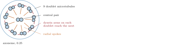
The axoneme in cross-section (left): nine doublets around a central pair, with dynein arms and radial spokes. Right: dyneins slide one doublet along the next, and because the doublets are tethered the sliding becomes a bend that travels along the cilium.
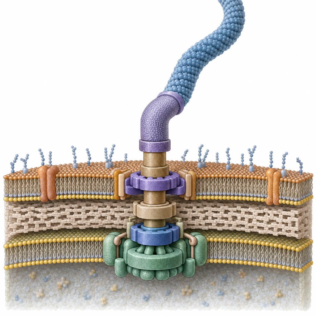
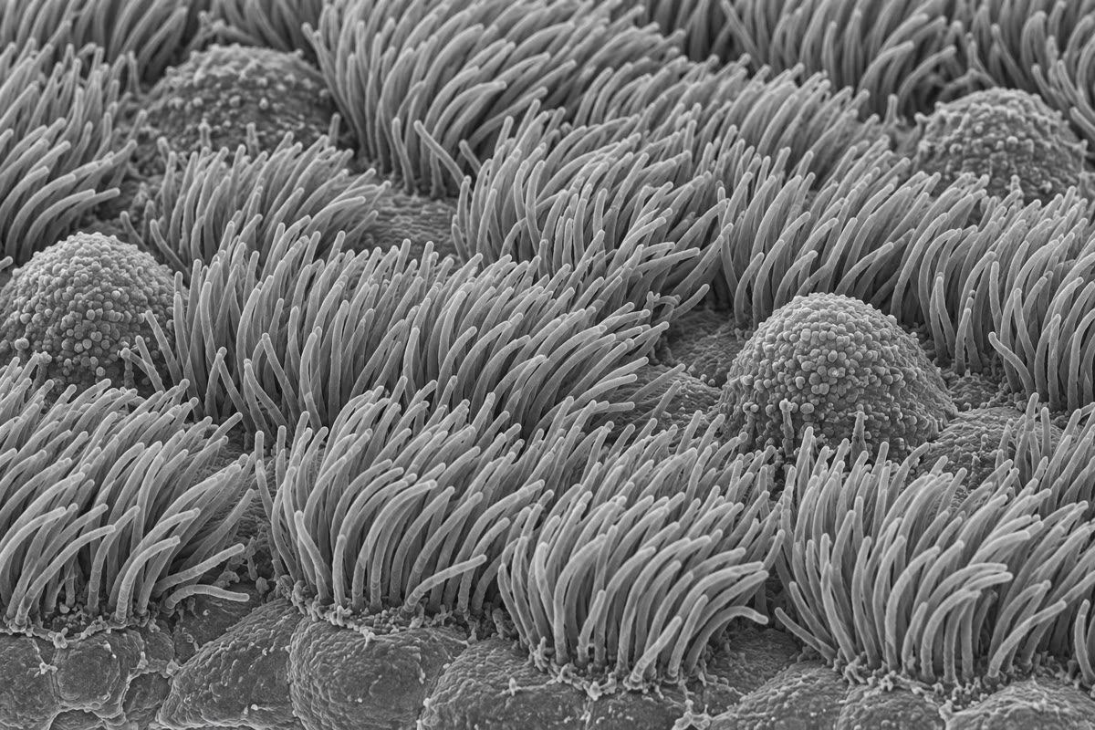
Left: the ciliated epithelium of the trachea in the scanning electron microscope, a lawn of cilia among dome-shaped mucus-secreting cells. Right: the bacterial flagellar motor, a rotary engine of rings in the cell envelope driven by the flow of protons. Left: the ciliated epithelium of the trachea in the scanning electron microscope, a lawn of cilia among dome-shaped mucus-secreting cells. Right: the bacterial flagellar motor, a rotary engine of rings in the cell envelope driven by the flow of protons.
Left: the ciliated epithelium of the trachea in the scanning electron microscope, a lawn of cilia among dome-shaped mucus-secreting cells. Right: the bacterial flagellar motor, a rotary engine of rings in the cell envelope driven by the flow of protons.
Read in context →