University Biology — Year 3 · Bachelor Year 3
9Cytoskeleton Dynamics and Cell Motility
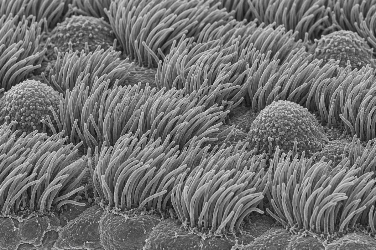
A neutrophil chasing a bacterium through a tissue crawls at ten micrometres a minute, changing direction within seconds when the scent shifts. A vesicle of neurotransmitter made in the cell body of a motor neuron travels a metre down the axon to the foot, hauled by a motor protein that takes eight-nanometre steps, a hundred a second, for a fortnight. The cilia lining the windpipe beat twelve times a second in coordinated waves that carry a day’s inhaled dust up to the throat. All of this is done by three kinds of protein filament that assemble and disassemble in seconds, and by motors that burn one molecule of ATP per step. The cytoskeleton is not a skeleton at all in the sense of a fixed frame; it is a set of polymers in constant turnover, whose dynamics are the mechanism of shape, division and movement. This chapter treats the polymers and their kinetics, the motors and the physics that limits them, the crawling of a cell, and the beating of cilia and flagella.
9.1 Three polymers
Definition 9.1 (Actin filaments, microtubules, intermediate filaments)
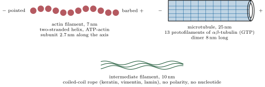
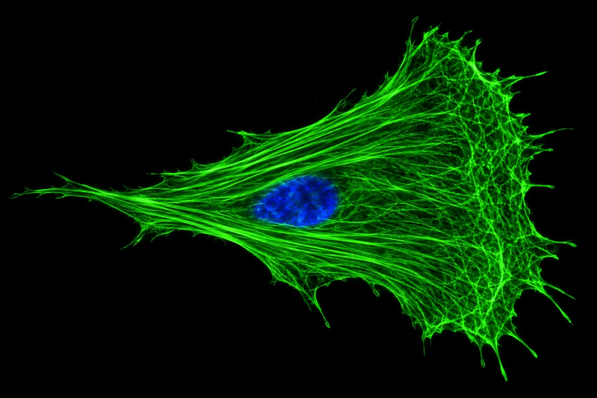
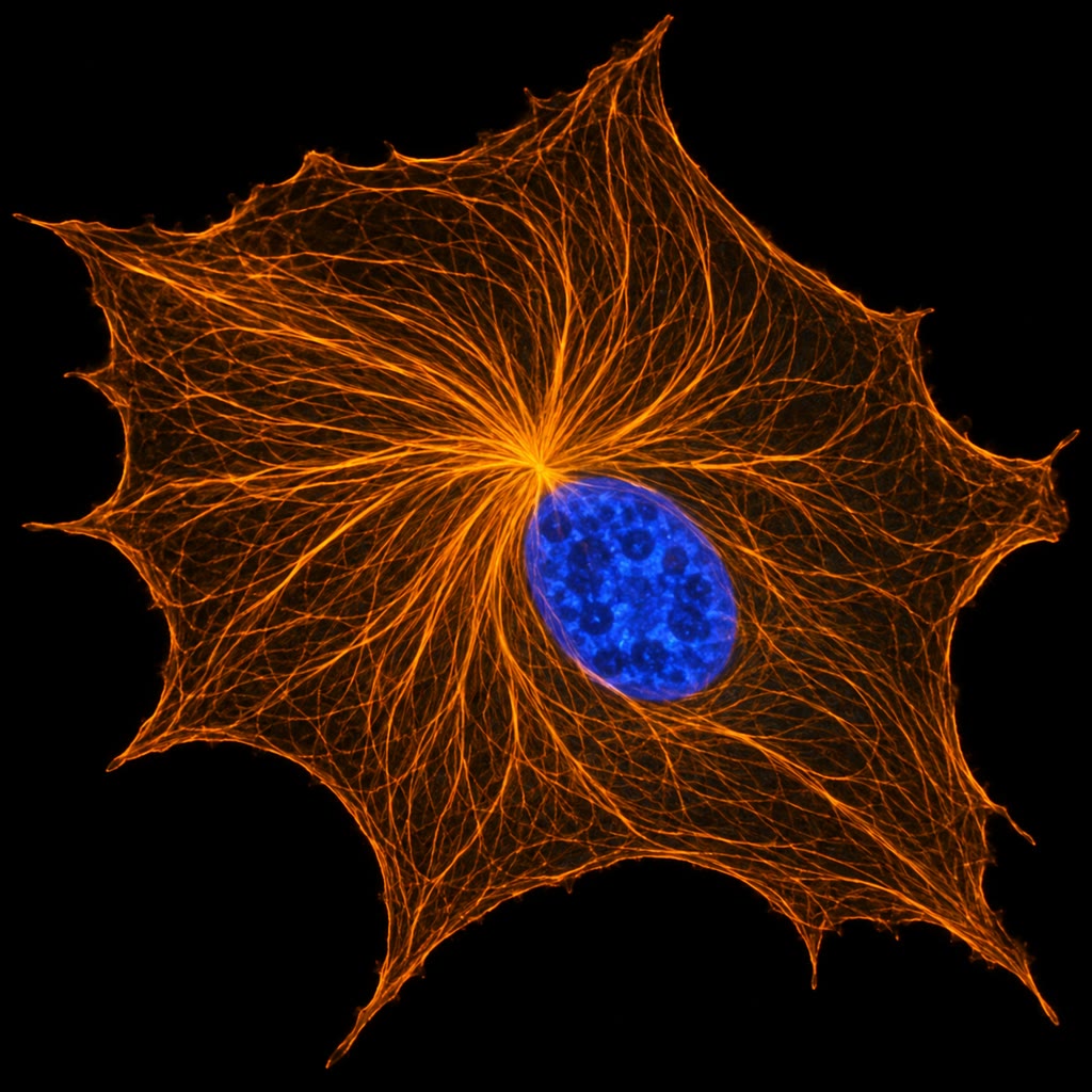
An actin filament is a two-stranded helical polymer of globular actin, thick, each subunit along the axis, polar: the barbed (plus) end grows faster than the pointed (minus) end, and each subunit carries an ATP that is hydrolysed after incorporation. A microtubule is a hollow tube of outer diameter built from thirteen protofilaments of -tubulin dimers ( per dimer), also polar, its plus end growing faster and its minus end usually anchored at the centrosome near the nucleus; the subunit hydrolyses its GTP after incorporation. An intermediate filament is a rope of made of coiled-coil proteins (keratins in epithelia, vimentin in mesenchyme, neurofilaments in axons, lamins under the nuclear envelope), apolar, without nucleotide, and far more stable — a tension-bearing cable rather than a dynamic track. Actin sits mostly under the plasma membrane and in protrusions; microtubules radiate from the centrosome and serve as the tracks of long-range transport and as the spindle; intermediate filaments give a cell and its nucleus their mechanical resilience.
9.2 The kinetics of a polymer
Theorem 9.2 (Critical concentration and treadmilling)
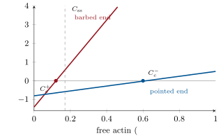
Let an end of a filament add subunits at rate , with the free monomer concentration, and lose them at rate . The end grows if exceeds its critical concentration and shrinks below it. If the two ends have different critical concentrations, , then at the steady state where the filament neither lengthens nor shortens the monomer concentration is
and the plus end grows while the minus end shrinks at the same rate : subunits flow through the filament from plus to minus — treadmilling — at the cost of one nucleotide hydrolysed per subunit.
Proof. The net rate at an end is , zero at . Constant length requires the sum of the two net rates to vanish, which gives ; it lies between the two critical concentrations because it is a weighted mean of them (). Above the plus end grows; below the minus end shrinks; the flux is the plus end’s net rate. Two ends of the same chemical polymer would have the same (the same equilibrium constant), so a difference requires that the subunit change after incorporation — the hydrolysis of ATP or GTP — which makes the ends differ and pays for the flow. ∎
Example 9.3 (Actin in the test tube)
For actin-ATP, , (); , (). Then and subunits per second: , imperceptible. In a cell the filaments treadmill a hundred times faster, because cofilin severs and strips the old ADP-actin from the pointed ends, profilin loads fresh ATP-actin onto barbed ends, and capping proteins decide which barbed ends may grow at all: the bare polymer supplies the mechanism, the regulators supply the speed.
Definition 9.4 (Dynamic instability)
Microtubules do something stranger than treadmilling. A growing plus end carries a GTP cap of freshly added dimers; behind it the GTP is hydrolysed, and GDP-tubulin, which prefers a curved conformation, is held straight in the lattice under strain. If the cap is lost — the end pauses, or growth slows — the strained protofilaments peel outward and the microtubule shortens at , twenty times its growth rate: a catastrophe, which ends when a GTP dimer lands and a new cap forms (a rescue). In one population, therefore, some microtubules grow while neighbours collapse: this is dynamic instability. It lets a cell explore its own volume with plus ends every few minutes and rebuild the whole array when the centrosome moves or the cell divides. The cell tunes it through proteins that track plus ends, stabilise (tau, MAP2), sever (katanin) or depolymerise (kinesin-13) microtubules; the drugs colchicine and the vinca alkaloids block assembly and taxol blocks disassembly, all of them poisons of the mitotic spindle used against cancer.
Evidence. Mitchison and Kirschner (1984) watched populations of microtubules grown from centrosomes in vitro. Diluting the tubulin below the critical concentration, they expected all the microtubules to shorten slowly; instead the number of microtubules fell while the survivors kept growing — some had disassembled completely and fast, others not at all. Individual microtubules filmed later by video microscopy switched abruptly between phases of steady growth and rapid shrinkage. The GTP cap explained both: only the capped ends grow, and the loss of the cap is a rare, all-or-nothing event. ∎
9.3 Motors
Definition 9.5 (Motor proteins)
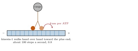
A motor protein converts the free energy of ATP hydrolysis into directed movement along a filament: myosins along actin (myosin II, the muscle and contractile motor, toward the barbed end; myosin V, a two-headed vesicle carrier stepping ), kinesins along microtubules toward the plus end (kinesin-1 steps , one tubulin dimer, per ATP, hand over hand, at about ), and dyneins toward the minus end (cytoplasmic dynein, with its adaptor dynactin, hauls cargo back to the cell centre; axonemal dyneins bend cilia). A motor is processive if it takes many steps before detaching: kinesin-1 walks about a micrometre, a hundred steps, alone; a single myosin II head takes one stroke and lets go, so muscle needs hundreds of heads working on one filament. Motors read the polarity of the track, so a cell’s geography — plus ends at the periphery, minus ends at the centre — is a map of where each motor will take its cargo.
Evidence. Vale, Reese and Sheetz (1985) found kinesin as the protein of squid axoplasm that made latex beads glide along microtubules in the plus direction, and in the following years the two-headed structure, the direction of each family and the retrograde partner dynein were sorted out. Svoboda, Schmidt, Schnapp and Block (1993) held a bead carrying a single kinesin in an optical trap and recorded its position with nanometre resolution: the bead advanced in discrete steps of , one per ATP at low ATP concentration, and stalled against a force of about . The molecular staircase was seen directly. ∎
Proposition 9.6 (What physics allows a motor)
The hydrolysis of one ATP in the cell releases about , that is J per molecule. A motor that advances per ATP against a force does work , so its stall force cannot exceed : for kinesin’s step, ; the measured means an efficiency near , higher than any engine. The thermal energy J, or , is only a twentieth of the ATP energy but is delivered to the motor constantly as Brownian kicks; a motor is a device that rectifies them, letting the head diffuse forward and binding when it lands, so that the chemical energy is spent on making the step irreversible rather than on pushing. Transport by motor beats diffusion over any distance that matters in a large cell: a vesicle with diffusion coefficient needs a time to wander a distance — half a second for a micrometre, but years for a metre of axon — whereas a motor at covers the metre in twelve days.
Proof. Admitted at this level. ∎
9.4 How a cell crawls
Definition 9.7 (Cell migration)
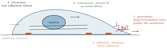
A crawling cell repeats a cycle of four steps. Protrusion: at the leading edge actin polymerises against the membrane in a sheet, the lamellipodium, whose branched network is built by the Arp2/3 complex, which nucleates a new filament from the side of an existing one at , while capping protein limits each filament’s growth and cofilin recycles the network a few micrometres back; finger-like filopodia of bundled filaments probe ahead. Adhesion: the new protrusion attaches to the substrate through integrins, transmembrane receptors that bind matrix proteins outside and, through adaptor proteins, actin inside, clustered in focal adhesions. Contraction: myosin II pulls on the stress fibres anchored at the adhesions, dragging the cell body forward. Retraction: the adhesions at the rear release and the tail is pulled in. The cycle is coordinated by three small GTPases of the Rho family — Rac drives lamellipodia, Cdc42 filopodia, Rho stress fibres and contraction — which are the targets of the receptors that read the direction of a chemical gradient (chemotaxis) or the stiffness and pattern of the substrate.
Example 9.8 (Listeria’s comet)
The bacterium Listeria monocytogenes, once inside a cell, displays on its surface a single protein, ActA, that recruits the host’s Arp2/3 complex. Actin polymerises at the bacterial surface and is left behind as a tail; the bacterium is pushed through the cytoplasm at up to and into neighbouring cells without ever leaving the cytosol. The tail is a lamellipodium turned inside out, with one protein where the cell uses dozens, and it showed that actin polymerisation alone — without any motor — generates the force of protrusion: each subunit that inserts between the network and the membrane, when a thermal fluctuation has opened a gap, ratchets the front forward by .
Method 9.9 (Watching the polymers turn over)
To measure the dynamics of a filament system in a living cell: (1) express the subunit fused to a fluorescent protein at a low level, so that it is incorporated into the endogenous polymer; (2) either bleach a small region with an intense laser pulse and film the return of fluorescence as unbleached subunits exchange in (fluorescence recovery after photobleaching, FRAP) — the half-time is the subunits’ residence time, and the fraction that never recovers is the immobile pool; or (3) express so little labelled subunit that the polymer appears as sparse speckles, and track them: their motion is the treadmilling flow, their appearance and disappearance the assembly and disassembly. In a lamellipodium the speckles flow backward at a micrometre a minute relative to the substrate while the edge advances: the network is built at the front and consumed a few micrometres behind it.
9.5 Cilia, flagella and a rotary motor
Definition 9.10 (Cilia and flagella)
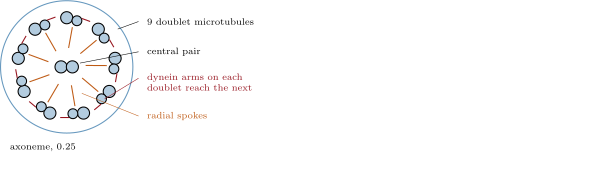
A eukaryotic cilium or flagellum is a membrane-covered extension built on the axoneme: nine doublet microtubules in a ring around a central pair, held by nexin links and radial spokes, and grown from a basal body, a modified centriole. Axonemal dyneins attached to each doublet walk along the neighbouring doublet toward its minus end at the base; since the doublets are tied together they cannot slide freely, and the sliding is converted into bending, propagated along the axoneme as a wave by switching the active dyneins from one side of the ring to the other. A cilium is long and beats times a second; a sperm flagellum is and undulates. The proteins of a cilium are carried up from the cell body by kinesin-2 and back by dynein-2 along the doublets, intraflagellar transport, which is also how a non-motile primary cilium — present, one per cell, on most vertebrate cells — is built; that cilium is a sensory antenna, housing receptors for light (the rod outer segment), odours, flow and developmental signals, and its defects cause the ciliopathies: polycystic kidneys, retinal degeneration, obesity, extra digits.
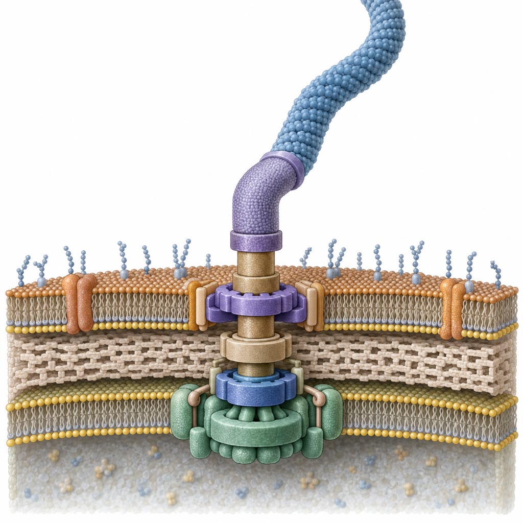
Proposition 9.11 (The bacterial flagellar motor)
The bacterial flagellum is unrelated to the eukaryotic one: a rigid helical filament of flagellin, thick and several micrometres long, turned by a rotary motor embedded in the cell envelope — a rotor of some forty-five proteins surrounded by a ring of stator units, each a channel through which protons flow down their gradient into the cell, about a thousand protons per revolution. The motor spins at up to , reverses direction in a millisecond, and develops a torque of some ; it is built from about twenty proteins whose genes are switched on in the order the parts are assembled, from the inside out. In E. coli, counterclockwise rotation bundles the several flagella into a propeller and the cell runs straight; a switch to clockwise flies the bundle apart and the cell tumbles to a new random direction. The chemotaxis pathway biases nothing but the frequency of tumbles — fewer when conditions improve — and that suffices to climb a gradient.
Remark 9.12 (The cytoskeleton is a verb)
Almost nothing described in this chapter is a structure in the sense that a bone is: the lamellipodium is a wave of polymerisation, the spindle of Chapter 10 a steady state of growing and collapsing microtubules, the cilium’s beat a switching pattern of motors. Turnover is not a cost the cell tolerates but the mechanism itself, which is why every one of these systems runs on nucleotide hydrolysis and stops within minutes of ATP depletion, and why the poisons that freeze the polymers in either state — taxol or colchicine, phalloidin or cytochalasin — are equally lethal.
9.6 Exercises
Exercise 9.1 ★
Compare actin filaments, microtubules and intermediate filaments in diameter, subunit, polarity, nucleotide and typical role.
Solution
Solution of Exercise 9.1.
Actin: , globular actin, polar (barbed/pointed), ATP; cortex, protrusion, contraction with myosin. Microtubule: , -tubulin dimer, polar (plus/minus), GTP; long-range transport, spindle, cilia. Intermediate filament: , coiled-coil proteins (keratin, vimentin, lamin), apolar, no nucleotide; mechanical strength of cell and nucleus.
Exercise 9.2 ★
Define critical concentration. Why do the two ends of an actin filament have different critical concentrations, and what would happen if they were equal?
Solution
Solution of Exercise 9.2.
The monomer concentration at which an end neither grows nor shrinks, . The two ends differ because the subunit hydrolyses its ATP after incorporation, so the ends present different chemical species (ATP-actin arriving at the barbed end, ADP-actin leaving the pointed end) with different affinities. With equal critical concentrations there would be a single equilibrium: no treadmilling, no flux, and no way to pay for directional turnover.
Exercise 9.3 ★
Name a motor for each: vesicle toward the cell periphery on microtubules; vesicle toward the centre; contraction of a stress fibre; bending of a cilium. Give the direction each takes on its track.
Solution
Solution of Exercise 9.3.
Periphery on microtubules: kinesin, toward the plus end. Centre: cytoplasmic dynein, toward the minus end. Stress fibre: myosin II, toward the barbed end of actin. Cilium: axonemal dynein, toward the minus end of the neighbouring doublet (the base).
Exercise 9.4 ★
List the four steps of the crawling cycle and the Rho-family GTPase that governs each of the first three.
Exercise 9.5 ★★
A polymer’s plus end has , , its minus end , . Find the two critical concentrations, the steady-state concentration and the treadmilling rate in subunits per second and, for subunits, in nanometres per second.
Solution
Solution of Exercise 9.5.
, ; ; subunits per second, .
Exercise 9.6 ★★
Kinesin walks at with steps, one ATP each. How many ATP per second per motor? A neuron’s axon is : how long is the trip, and how many ATP does one motor spend on it? Compare with the diffusion time from Proposition 9.6.
Solution
Solution of Exercise 9.6.
ATP per second. The metre takes s, about two weeks, and steps and ATP. Diffusion would take years: a thousand-million-fold difference.
Exercise 9.7 ★★
Myosin V steps per ATP. What is its maximal stall force, and why is it lower than kinesin’s? Which motor is better suited to carrying a large load, and which to moving fast?
Exercise 9.8 ★★
Predict the effect on a dividing cell, and on a crawling cell, of (a) taxol, (b) colchicine, (c) cytochalasin, (d) a Rac inhibitor, and explain each from the mechanism.
Solution
Solution of Exercise 9.8.
(a) Taxol freezes microtubules: the spindle cannot search, capture and correct, mitosis arrests at the checkpoint; the crawling cell keeps protruding but loses long-term polarity. (b) Colchicine removes microtubules: no spindle, mitotic arrest; crawling continues on actin but without persistent direction. (c) Cytochalasin caps barbed ends: no protrusion, no crawling; mitosis proceeds but cytokinesis fails, giving binucleate cells. (d) Rac inhibition: no lamellipodia, so no crawling; division largely unaffected.
Exercise 9.9 ★★
In a FRAP experiment on a lamellipodium, fluorescence recovers with a half-time of to of its initial value. What are the residence time of actin in the network and the immobile fraction? The speckles flow backward at while the edge advances at : at what rate is actin polymerising at the edge?
Solution
Solution of Exercise 9.9.
Residence time of the order of the half-time, (time constant ); immobile fraction . Polymerisation at the edge must supply both the advance and the retrograde flow: .
Exercise 9.10 ★★★
Explain dynamic instability in terms of the GTP cap, and show why a microtubule population held just above the critical concentration becomes bimodal in length rather than uniform. What does the cell gain from a behaviour that wastes GTP?
Solution
Solution of Exercise 9.10.
A growing end keeps a cap of GTP-tubulin; behind it hydrolysis produces strained GDP-tubulin. Loss of the cap — a random event whose probability rises as growth slows — releases the strain and the end shrinks fast until a new cap forms. Just above the critical concentration, each microtubule is at any moment either capped and growing or uncapped and collapsing, so lengths diverge into a long population and a vanishing one instead of clustering at a mean. The cell gains rapid exploration of its volume and the ability to stabilise selectively — a plus end that reaches a kinetochore or a cortical site is captured and kept, the others are recycled within minutes: search and capture.
Exercise 9.11 ★★★
A bacterium of swims at . In water at this scale viscous drag dominates and the cell stops within a nanometre when the motor stops. Explain why a reciprocal motion (an oar) cannot propel it and a rotating helix can, and estimate the number of protons the motor spends per second at .
Solution
Solution of Exercise 9.11.
At this scale inertia is negligible and the fluid equations are time-reversible: a motion that retraces itself (an oar going out and back) returns the body to where it started, whatever the speed of each stroke. A rotating helix never retraces itself — its motion is chiral and continuous — so it produces net thrust. At with a thousand protons per turn, protons per second per motor.
Exercise 9.12 ★★★
Kartagener syndrome — immotile cilia — gives chronic bronchitis, male infertility, and in half of patients a heart on the right side. Explain each from the biology of the axoneme, and say why the last affects only half.
Solution
Solution of Exercise 9.12.
Airway cilia cannot beat, mucus and bacteria are not cleared, and infections recur. Sperm flagella, built on the same axoneme, are immotile: infertility. In the embryo, motile cilia of the node drive a leftward flow that sets the left–right axis; without flow the side is chosen at random, so half the patients have their organs reversed.
9.7 Problem: A Neutrophil on the Move
Problem 9.1
Weekend problem — a neutrophil’s actin budget counted, its treadmilling computed, a vesicle’s journey down an axon timed against diffusion, a motor’s efficiency measured, and the crawling front’s polymerisation and ATP cost worked out, ending on the treadmilling rate, the time to cross an axon and the ATP a lamellipodium burns per second
Data: a neutrophil of volume holds actin at , half polymerised; a subunit adds to a filament. Actin rate constants as in Example 9.3. Kinesin: steps, , stall force ; ATP hydrolysis ; ; vesicle diffusion coefficient ; axon length . The lamellipodium is wide with filaments per micrometre of edge, advancing at ; the network is disassembled behind the edge.
Part I — The actin budget.
- How many actin molecules does the cell contain, and how many are in filaments?
- What total filament length is that? If the average filament is , how many filaments?
- The free actin is , far above the critical concentration of the barbed end. Why do the filaments not simply grow until the monomer is exhausted? (Two proteins.)
- Treadmilling in vitro: compute and from the rate constants, and the treadmilling speed in nanometres per second and micrometres per minute.
- In the cell the effective speed is . By what factor do the regulators accelerate the bare polymer?
- Each treadmilled subunit costs one ATP. If all filaments treadmilled at the cellular speed, how many ATP per second, and what fraction is that of a cell’s total turnover of about ATP per second?
Part II — A vesicle down the axon.
- How long does a kinesin-driven vesicle take to travel the axon, and how many steps and ATP does the motor spend?
- How long would diffusion take over ? Over , the width of a cell body? What does the comparison say about where cells can afford to rely on diffusion?
- Compute the energy per ATP in joules and in units of .
- The maximal force of a motor with steps, and kinesin’s efficiency at stall.
- A vesicle of radius in cytoplasm of viscosity (ten times water) moving at feels a drag . Compute it, and compare with the stall force: how many motors are needed?
- A neuron’s axon carries about vesicles at a time. What is the total ATP consumption of the transport, per second, and how does it compare with the neuron’s firing costs of about ATP per second? What does this imply for a neuron in which mitochondria fail?
Part III — The front.
- Convert the edge speed to nanometres per second, and compute the number of subunits per second each filament must add to keep up.
- How many filaments push the edge, and how many subunits per second does the whole edge consume?
- Each subunit added is one ATP hydrolysed and later recycled. ATP per second for protrusion; fraction of the cell’s per second.
- The network behind the edge is deep and disassembles there. What is the lifetime of a subunit in the network, and how many subunits are in the network at any time?
- The membrane resists protrusion with a force of about per filament. Compute the work done per subunit added () in and say whether the Brownian ratchet — a thermal fluctuation opening a gap of — is plausible.
- Why does a Listeria comet tail need no motor, and how fast can it push if it recruits the same machinery?
Part IV — Direction.
- The neutrophil senses a chemoattractant whose concentration rises by across its width. With receptors and equal to the mean concentration, how many more receptors are occupied on the front half than on the back half? (Occupancy ; use half the receptors per side.)
- The random fluctuation in the number occupied on one side is about the square root of the number. Is the difference of question 19 detectable in a single instant? How does averaging over a few seconds help?
- Which GTPase must be activated at the front and which at the rear for the cell to turn toward the source? What would a cell with constitutively active Rac everywhere do?
- The cell reaches the bacterium in from away. Check the speed against the data.
- A cell lacking Arp2/3 moves by blebbing — pressure-driven bulges of membrane — instead. Which step of the cycle has been replaced, and what does it say about the role of actin in the other steps?
- The whole crawling cycle takes minutes, yet the cell turns within seconds when the gradient shifts. Explain, using the lifetime of the network from question 16.
- Summarise: the treadmilling speed in vitro (question 4), the time for a vesicle to travel the axon (question 7), and the ATP per second spent on protrusion (question 15).
Solution
Solution of Problem 9.1.
1. L; mol/L mol molecules; in filaments. 2. of filament; at each, about filaments. 3. Capping protein blocks most barbed ends, and profilin and thymosin-4 bind the monomers so that the truly free ATP-actin is near the critical concentration; growth is confined to the ends the cell uncaps. 4. ; subunits per second; . 5. -fold. 6. subunits per second per filament; ATP per second, about of the cell’s turnover. 7. s days; steps and ATP. 8. : s years for a metre; s for . Diffusion serves within a cell body; anything longer needs motors. 9. J . 10. ; efficiency . 11. N , four hundred times below the stall force: one motor is ample against viscous drag; the real loads are obstacles and tethers. 12. ATP per second, of the neuron’s budget: transport is cheap. But motors stop the moment ATP falls, so a failure of mitochondria starves the synapse of everything made in the cell body — the axonal transport failure seen in neurodegenerative disease. 13. ; subunits per second per filament. 14. filaments; subunits per second. 15. ATP per second, of the budget. 16. ; subunits in the network. 17. : a fluctuation of that energy occurs with probability about — gaps of one subunit open constantly, and the ratchet is entirely plausible. 18. Polymerisation itself pushes, the bacterium being the “membrane” at the front of its own network; with the cell’s Arp2/3 machinery it reaches the lamellipodial speed and more — up to , . 19. At , and : a difference in gives ; with receptors per side, more occupied at the front. 20. Each side has about occupied, fluctuating by , so the difference of two sides fluctuates by about : the is lost in the noise at any instant. Receptors rebind about once a second; averaging over seconds shrinks the noise by — ten seconds bring it to and the gradient is read. 21. Rac (with Cdc42) at the front, Rho at the rear. With active Rac everywhere the cell protrudes all round, spreads, and goes nowhere. 22. : as given. 23. Protrusion is replaced by pressure-driven blebbing; adhesion and contraction still depend on actin and myosin, so actin remains essential for the rest of the cycle. 24. The front is rebuilt every : a shift in Rac activity redirects polymerisation within one network lifetime, the new front leads, and the slow steps at the rear follow. 25. Treadmilling in vitro ; about days to travel the axon; some ATP per second for protrusion.