Biology · Glossary

What is Magnification and resolution?

Also known as: magnification · resolution · resolving power

Definition 2.4 High School Biology · Chapter 2 — Cells: The Common Unit of Life

The magnification of an instrument is the ratio of the size of the image to the size of the object. Its resolution is the smallest distance between two points that it still shows as separate. Magnifying an image beyond what the resolution justifies enlarges the blur, not the detail.
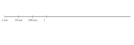
Sizes on a logarithmic scale: each step is ten times larger. The naked eye resolves about 0.1\, mm; the light microscope reaches 0.2\, µ m and sees whole cells and bacteria; the electron microscope resolves nanometres and shows the parts inside.
Sizes on a logarithmic scale: each step is ten times larger. The naked eye resolves about 0.1mm0.1\,\mathrm{mm}; the light microscope reaches 0.2µm0.2\,\text{µ}\mathrm{m} and sees whole cells and bacteria; the electron microscope resolves nanometres and shows the parts inside.

Examples

Example 2.7 (Cheek cells)

A cheek cell drawn 24mm24\,\mathrm{mm} wide from a ×400\times 400 image is 24/400=0.06mm=60µm24/400 = 0.06\,\mathrm{mm} = 60\,\text{µ}\mathrm{m} across, and its nucleus, drawn 3mm3\,\mathrm{mm}, is about 8µm8\,\text{µ}\mathrm{m}. A red blood cell is 7µm7\,\text{µ}\mathrm{m}; the bacteria of the mouth, 1µm1\,\text{µ}\mathrm{m} long, appear as barely resolved dots at the same magnification.
Read in context →
Definition 5.8 University Biology — Year 1 · Chapter 5 — The Cell: Unit of Life

A microscope magnifies (enlarges the image) and resolves (separates neighbouring points). Only the second limits what can be seen: the resolving power is the smallest distance dd between two points that still appear distinct,

d=0.61λNA,d = \frac{0.61\,\lambda}{\mathrm{NA}},

where λ\lambda is the wavelength of the illumination and NA\mathrm{NA} the numerical aperture of the objective (the sine of its half-angle of light collection times the refractive index of the medium, at most about 1.41.4 with oil). Magnifying beyond the resolving power enlarges the blur, not the detail.

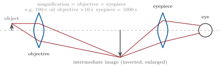
The compound light microscope: the objective forms an enlarged, inverted intermediate image, which the eyepiece magnifies again for the eye. The objective’s numerical aperture, not the total magnification, sets what can be resolved.
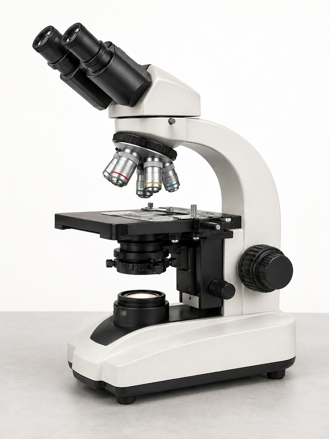
A compound light microscope: lamp and condenser below the stage, a turret of objectives above it, binocular eyepieces. Resolution 0.2\, µ m, magnification up to 1000\,, living specimens possible.
A compound light microscope: lamp and condenser below the stage, a turret of objectives above it, binocular eyepieces. Resolution 0.2µm0.2\,\text{µ}\mathrm{m}, magnification up to 10001000\,, living specimens possible.

Examples

Example 5.11 (A bacterium and a mitochondrion)

Under the light microscope at 1000×1000\times, an E. coli cell is a rod 2mm2\,\mathrm{mm} long on the retina’s scale, with no visible interior; a mitochondrion is a dot at the limit of resolution. In a TEM section the same bacterium shows its two membranes, its wall, its nucleoid as a pale fibrous region and its ribosomes as dark grains; the mitochondrion shows its double membrane and the folds of the inner one. Neither is alive.

Read in context →