 Coach
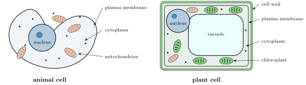
CoachAn organelle is a structure inside a cell, enclosed by its own membrane, that performs a specific task. The principal ones are:
- the nucleus, which contains the chromosomes — the DNA of Chapter 3 — and controls the cell’s activity;
- the mitochondria, some long, where organic molecules are broken down with oxygen to release usable energy (cellular respiration, Chapter 4);
- in plant cells, the chloroplasts, green with chlorophyll, where photosynthesis takes place, and a large water-filled vacuole that keeps the cell taut.
Outside the membrane, a plant cell is boxed in a rigid cell wall of cellulose; animal cells have none. The cytoplasm of every cell also contains millions of ribosomes, particles of about that assemble proteins, visible only with the electron microscope.
Examples
Example 2.7 (Cheek cells)
A cheek cell drawn wide from a image is across, and its nucleus, drawn , is about . A red blood cell is ; the bacteria of the mouth, long, appear as barely resolved dots at the same magnification.
Example 2.10 (Reading a bacterium)
An electron micrograph of the gut bacterium Escherichia coli shows a rod long and wide: a wall, a membrane just inside it, a granular cytoplasm packed with ribosomes, and a paler central region where the single circular DNA molecule lies. No nucleus, no mitochondria. Yet the cell feeds, grows and divides every twenty minutes in a rich broth: everything the cell theory demands, in a thousandth of the volume of a cheek cell.