University Biology — Year 1 · Bachelor Year 1
5The Cell: Unit of Life
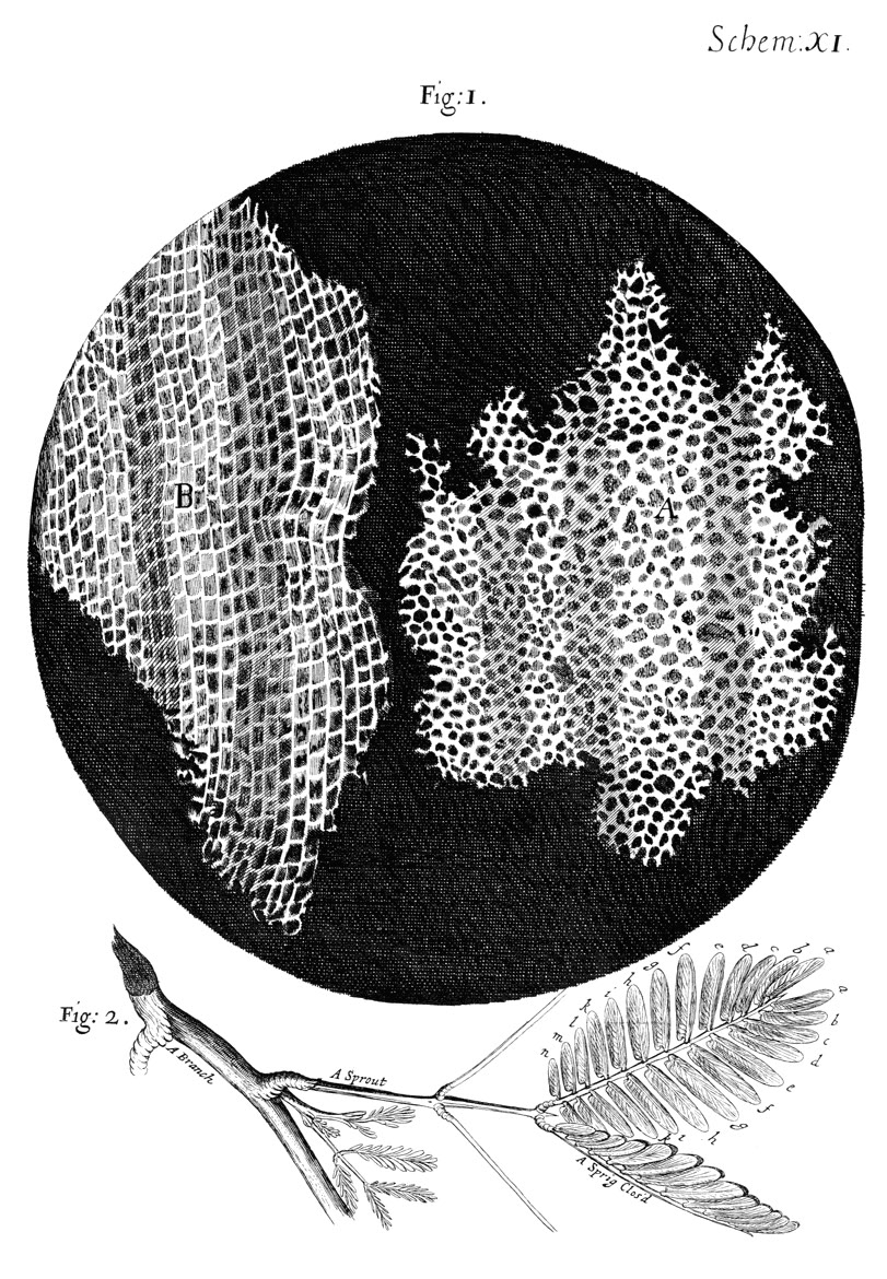
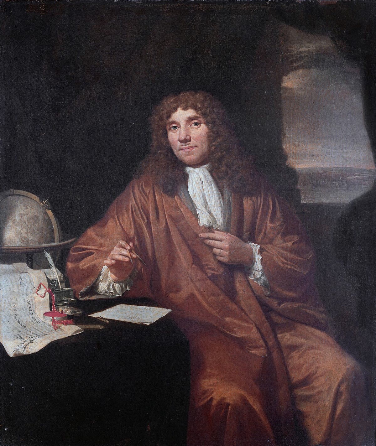
In 1665 Robert Hooke put a sliver of cork under a lens and saw it divided into tiny boxes he called cells, after the rooms of a monastery. Ten years later a Dutch draper, Antoni van Leeuwenhoek, grinding his own lenses to a power no one else could reach, saw living things swimming in a drop of pond water, in the scrapings of his teeth, in his own semen. It took another century and a half to state what the two had seen: that every organism is made of cells, and that every cell comes from a cell. This chapter states the cell theory and its evidence, contrasts the two kinds of cell, and describes the instruments — the microscopes, the centrifuge, the culture flask — that let us see cells, take them apart, and keep them alive outside an organism.
5.1 The cell theory
Definition 5.1 (Cell)
A cell is the smallest unit of living matter: a volume of aqueous cytoplasm bounded by a plasma membrane, containing the genetic material (DNA) and the machinery that expresses it, able to take in nutrients, transform energy, maintain its own organisation and, when conditions permit, divide into two cells.
Proposition 5.2 (The cell theory)
- Every organism is made of one or more cells, and the cell is the unit of structure and function of living things (Schleiden and Schwann, 1838–1839).
- Every cell arises from a pre-existing cell by division (Virchow, 1855: omnis cellula e cellula); there is no spontaneous generation.
- The hereditary material is passed from cell to cell at division, so that the continuity of life is the continuity of cells.
Evidence. Hooke’s cork (1665) and the plant sections of Grew and Malpighi showed plant tissues as arrays of chambers; Leeuwenhoek’s lenses (1670s) showed single-celled organisms, blood cells and sperm. Two centuries of improving lenses found cells in every tissue examined, animal as well as plant, once Schwann recognised nucleated cells in cartilage and embryos. Division was seen directly in algae and in embryos, and Virchow found no tissue, healthy or diseased, whose cells did not come from other cells. Pasteur’s swan-necked flasks (1861) — broths that stayed sterile for months when dust could not reach them, and teemed within days when it could — closed the question of spontaneous generation. The third statement is the work of the later nineteenth century, which followed the chromosomes through division (Chapter 18). ∎
Example 5.3 (One cell, many cells)
A bacterium, a yeast, an amoeba are whole organisms of one cell, doing everything — feeding, moving, dividing — within a few micrometres. A human is some cells of two hundred kinds, none of which could live alone in a pond, each of which descends by unbroken divisions from one fertilised egg. The rabbit of Chapter 2 and the oak of Chapter 3 are the same theory applied twice.
5.2 Two kinds of cell
Definition 5.4 (Prokaryotic and eukaryotic cells)
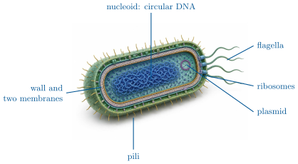
A prokaryotic cell (bacteria, archaea) has no nucleus: its DNA, usually one circular chromosome, lies in a region of the cytoplasm called the nucleoid, and it has no membrane-bounded compartments. It is small () and bounded by a plasma membrane and, outside it, a cell wall. A eukaryotic cell (protists, fungi, plants, animals) keeps its DNA, as several linear chromosomes, inside a nucleus bounded by a double membrane, and divides its cytoplasm into membrane-bounded organelles (Chapter 6). It is larger () and has an internal skeleton of protein filaments.
Proposition 5.5 (The bacterial cell)
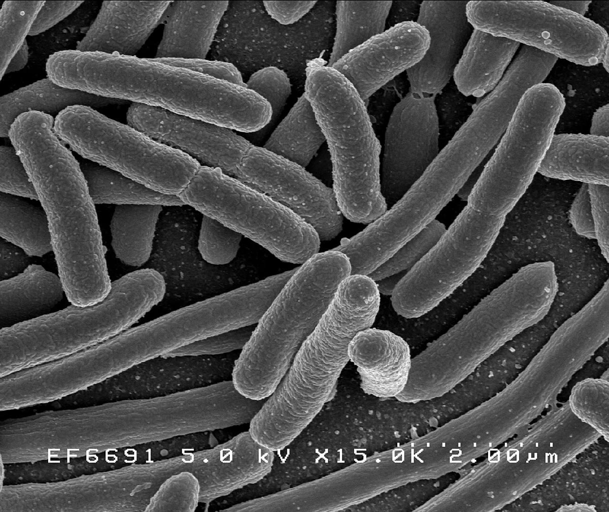
E. coli, the model bacterium, is a rod long and wide: a volume of about holding one chromosome of base pairs ( of DNA folded ten-thousand-fold), some ribosomes, a few million protein molecules of kinds, and often one or more plasmids — small circular DNA molecules carrying extra genes. Its envelope is, from inside out, the plasma membrane, a thin wall of peptidoglycan (a mesh of sugar chains cross-linked by short peptides, which resists the cell’s internal pressure), and an outer membrane. Bacteria with a thick peptidoglycan wall and no outer membrane retain the crystal-violet stain of the Gram stain (Gram-positive); those with a thin wall under an outer membrane lose it (Gram-negative, as E. coli). Flagella rotate like propellers; pili attach the cell to surfaces and to other cells.
Method 5.6 (The Gram stain)
- Fix a thin smear of bacteria to a slide by heat.
- Stain with crystal violet (all cells purple), then with iodine, which forms a large complex with the dye inside the cells.
- Rinse with alcohol: the thick peptidoglycan of Gram-positive cells shrinks and traps the complex; the thin wall of Gram-negative cells, once the alcohol has dissolved the outer membrane, lets it wash out.
- Counterstain with a pink dye (safranin): Gram-positive cells appear purple, Gram-negative pink. The result predicts the wall structure and, with it, the sensitivity to many antibiotics.
Example 5.7 (Why the eukaryotic cell is compartmented)
A cell of has a thousand times the volume of a bacterium but only a hundred times its membrane surface (Proposition 1.6). Membranes are where a cell carries its energy-transducing chains and its transport systems; the eukaryote recovers surface by folding membranes inside itself — the inner membrane of a mitochondrion, the sheets of the endoplasmic reticulum — and gains, besides, separate chambers in which incompatible chemistries (digestion in the lysosome, synthesis in the cytosol) can run side by side.
5.3 Seeing cells: microscopy
Definition 5.8 (Magnification and resolution)
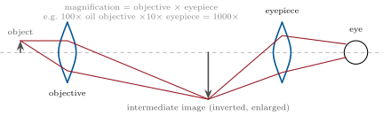
A microscope magnifies (enlarges the image) and resolves (separates neighbouring points). Only the second limits what can be seen: the resolving power is the smallest distance between two points that still appear distinct,
where is the wavelength of the illumination and the numerical aperture of the objective (the sine of its half-angle of light collection times the refractive index of the medium, at most about with oil). Magnifying beyond the resolving power enlarges the blur, not the detail.
Proposition 5.9 (Light and electron microscopes)
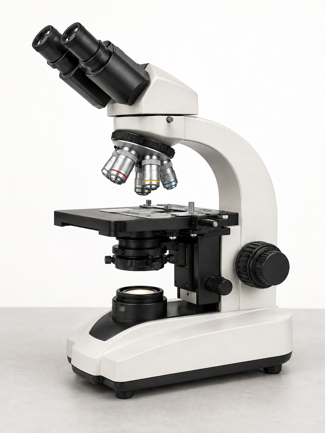
With visible light () and , the light microscope resolves : cells, nuclei, chloroplasts, mitochondria as dots, bacteria as rods — and living, moving specimens. Contrast in a transparent cell is obtained by staining (which usually kills), by phase contrast (converting differences of refractive index into differences of brightness), or by fluorescence (labelling one molecule with a dye or a fluorescent protein and imaging only its light). The electron microscope uses electrons of wavelength a few picometres; its practical resolution, limited by its lenses, is about in transmission (TEM: electrons pass through a section thick and stained with heavy metals, giving the internal structure of organelles) and a few nanometres in scanning (SEM: a beam swept over a metal-coated surface, giving its relief in depth). Electron microscopy requires a vacuum and dead, fixed, dehydrated specimens.
Method 5.10 (Choosing the instrument)
- To follow a living process (cell division, movement, a fluorescent protein moving between compartments): light microscopy, phase contrast or fluorescence, on unfixed cells.
- To count and identify cells in a tissue: a fixed, sectioned, stained preparation under the light microscope.
- To see the internal structure of an organelle, a membrane, a virus: transmission electron microscopy of thin sections, or of particles negatively stained.
- To see the surface relief of a cell or a tissue: scanning electron microscopy.
- To locate one protein: immunofluorescence (light) or immunogold labelling (electron), using an antibody against it.
Example 5.11 (A bacterium and a mitochondrion)
Under the light microscope at , an E. coli cell is a rod long on the retina’s scale, with no visible interior; a mitochondrion is a dot at the limit of resolution. In a TEM section the same bacterium shows its two membranes, its wall, its nucleoid as a pale fibrous region and its ribosomes as dark grains; the mitochondrion shows its double membrane and the folds of the inner one. Neither is alive.
5.4 Taking cells apart and keeping them alive
Definition 5.12 (Cell fractionation)
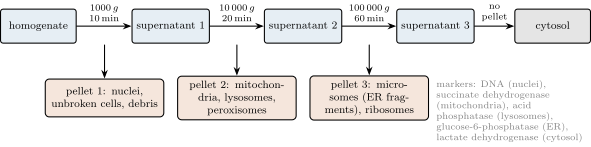
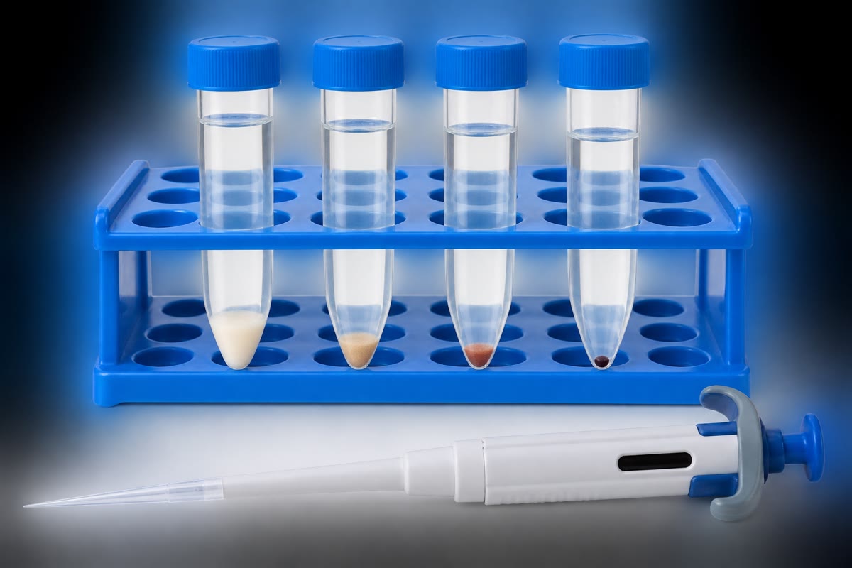
Cell fractionation separates the components of a cell for study. The tissue is homogenised — cells broken open in a cold, isotonic buffer by grinding or shearing, leaving the organelles intact — and the homogenate is subjected to differential centrifugation: a series of spins of increasing force and duration, each of which pellets the largest and densest particles remaining in suspension. Each fraction is identified by its appearance under the electron microscope and by marker enzymes, activities known to reside in one compartment only.
Proposition 5.13 (Sedimentation)
A spherical particle of radius and density in a fluid of density and viscosity , in a centrifugal field (a multiple of ), sediments at the steady speed
(Stokes’ law). Speed grows as the square of the radius: a nucleus of falls a hundred times faster than a mitochondrion of , which falls a thousand times faster than a ribosome of . This is why the same tube separates them at increasing speeds.
Proof. At steady speed the drag of Stokes, , balances the net weight of the particle in the fluid, ; solving for gives the formula. ∎
Definition 5.14 (Cell culture)
Cell culture keeps cells alive and dividing outside the organism, in a sterile medium supplying nutrients, salts, growth factors and, for animal cells, a surface to attach to, at the organism’s temperature. Bacteria and yeasts grow in simple media and double in minutes to hours; animal cells taken from a tissue (primary cultures) divide a limited number of times, while cell lines derived from tumours or transformed cells divide indefinitely. Culture makes the cell an experimental object: one type, one medium, one variable at a time.
Example 5.15 (A growth curve)
A flask of E. coli in a rich medium at : a lag of half an hour, then exponential growth — the count doubles every , so — until a nutrient runs out or wastes accumulate and the culture reaches a plateau near cells per millilitre. From a single cell, cells in ten hours. The same curve, with a doubling time of a day, describes a culture of human cells.
5.5 Exercises
Exercise 5.1 ★
State the three propositions of the cell theory and name a piece of evidence for each.
Solution
Solution of Exercise 5.1.
All organisms are made of cells (every tissue examined under the microscope, plant and animal, shows them); every cell comes from a cell (division observed in embryos and algae; Pasteur’s flasks exclude spontaneous generation); the hereditary material passes at division (chromosomes followed through mitosis).
Exercise 5.2 ★
List four differences between a prokaryotic and a eukaryotic cell.
Solution
Solution of Exercise 5.2.
Nucleus (absent / present), membrane-bounded organelles (absent / present), chromosome (one circular / several linear), size ( / ), cell wall of peptidoglycan (bacteria) / of cellulose, chitin or none, cytoskeleton (rudimentary / elaborate).
Exercise 5.3 ★
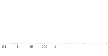
From the size-scale figure, how many bacteria laid end to end span a frog egg? How many animal cells?
Exercise 5.4 ★
Why does a light microscope show no more detail than a one?
Solution
Solution of Exercise 5.4.
Resolution depends on wavelength and numerical aperture, not on magnification; beyond about the image enlarges the blur of the diffraction limit () without separating anything new.
Exercise 5.5 ★★
Compute the resolving power of an objective of in green light (), and in blue light (). Can it resolve two bacteria apart? Two ribosomes apart?
Solution
Solution of Exercise 5.5.
; in blue, . Two bacteria apart: barely, in blue light; ribosomes apart: no, by a factor of fifteen.
Exercise 5.6 ★★
A Gram stain of a mixed culture shows purple cocci and pink rods. What does each colour say about the cell wall? Which of the two is E. coli?
Solution
Solution of Exercise 5.6.
Purple: thick peptidoglycan wall retaining the crystal violet–iodine complex, no outer membrane (Gram-positive). Pink: thin peptidoglycan under an outer membrane, dye washed out, counterstained (Gram-negative). E. coli is a Gram-negative rod: the pink rods.
Exercise 5.7 ★★
Using Stokes’ law with and , compute the sedimentation speed of a mitochondrion () at and the time to fall . Repeat for a nucleus () at .
Exercise 5.8 ★★
A homogenate’s marker activities are: succinate dehydrogenase in pellet 2, in pellet 1, in the supernatant; lactate dehydrogenase in the final supernatant. Interpret each number, including the in pellet 1.
Solution
Solution of Exercise 5.8.
Succinate dehydrogenase is mitochondrial: of the mitochondria are in pellet 2; the in pellet 1 are mitochondria trapped in unbroken cells and debris or dragged down with the nuclei; the in the supernatant come from mitochondria broken during homogenisation. Lactate dehydrogenase is cytosolic and soluble: it stays in the final supernatant.
Exercise 5.9 ★★
Which microscope would you use to (a) watch a white blood cell engulf a bacterium, (b) count the mitochondria in a section of muscle, (c) see the pores of a nuclear envelope, (d) see the shape of a pollen grain’s surface? Justify each choice.
Exercise 5.10 ★★★
Pasteur’s flasks: a broth boiled in a swan-necked flask stays clear indefinitely; tilted so that the broth touches the dust in the neck, it turns cloudy in two days. Explain what each observation excludes and what the pair proves, and say why boiling in a sealed flask alone had not settled the question.
Solution
Solution of Exercise 5.10.
The clear flask excludes that air itself, or boiled broth itself, generates life: air enters freely through the open neck. The cloudy flask, after contact with the dust, shows that what the neck had trapped — particles from the air — is the source of the organisms. Together they prove that microbes come from microbes carried by dust. A sealed boiled flask left open the objection that sealing had excluded the air a “vital force” needed; the swan neck admits air and excludes only dust.
Exercise 5.11 ★★★
A culture of E. coli starts at cells per millilitre and doubles every . When does it reach per millilitre? If each cell has a mass of , what is the bacterial mass in a litre at that point, and why does growth stop soon after even in an excess of glucose?
Solution
Solution of Exercise 5.11.
gives doublings, i.e. , about . per litre. Growth stops because oxygen (which the medium can supply at only a few milligrams per litre), a mineral, or the accumulation of acids and wastes becomes limiting, not the glucose.
Exercise 5.12 ★★★
“The eukaryotic cell is a bacterium that learned to fold.” Discuss in a paragraph, using surface-to-volume, the membranes of organelles, and what compartments allow.
Solution
Solution of Exercise 5.12.
A cell ten times larger in each dimension has a tenth of the membrane surface per unit volume; since membranes carry the energy-transducing and transport machinery, the large cell would starve. The eukaryote multiplies membrane by folding it inside itself (endoplasmic reticulum, inner mitochondrial membrane, thylakoids) and thereby also creates closed compartments in which incompatible processes — acid digestion, oxidative chemistry, DNA storage — run separately at their own conditions. The image is apt as far as membranes go, and incomplete: the eukaryote also gained a cytoskeleton and a nucleus, and its mitochondria are descendants of engulfed bacteria, not folds.
5.6 Problem: Fractionating a Liver
Problem 5.1
Weekend problem — ten grams of rat liver taken apart by centrifugation: pellets, markers, Stokes’ law and the balance sheet of an enzyme, ending on the purification factor of the mitochondrial fraction
of rat liver is homogenised in of cold isotonic sucrose and spun in three steps: for (pellet 1), for (pellet 2), for (pellet 3), leaving the final supernatant. The protein content and two enzyme activities are measured in the homogenate and in each fraction:
| fraction | protein (mg) | succinate dehydrogenase (U) | lactate dehydrogenase (U) |
|---|---|---|---|
| homogenate | 2000 | 100 | 400 |
| pellet 1 | 500 | 15 | 20 |
| pellet 2 | 200 | 60 | 12 |
| pellet 3 | 300 | 5 | 8 |
| final supernatant | 950 | 4 | 350 |
Take for the medium, a sedimentation path of , and .
Part I — What is where.
- Name the main organelle in each pellet.
- Why must the homogenisation medium be cold and isotonic?
- Succinate dehydrogenase is a marker of which compartment? Lactate dehydrogenase of which?
- Compute the recovery of protein: the sum over the fractions as a percentage of the homogenate. Comment.
- Compute the recovery of each enzyme activity in the same way.
- Compute the protein content per gram of liver and the fraction of the liver’s fresh mass that is protein.
Part II — Stokes’ law. Mitochondria: radius , density excess . Nuclei: radius , same excess. Ribosomes: radius , density excess .
- Compute the sedimentation speed of a mitochondrion at and the time to travel . Is enough?
- Compute the distance a mitochondrion travels during the first spin (, ). What fraction of the mitochondria, uniformly distributed at the start, would be pelleted in the first step?
- Compare with the of succinate dehydrogenase found in pellet 1. What else can drag mitochondria into pellet 1?
- Compute the time for a nucleus to travel at .
- Compute the time for a ribosome to travel at . Would a ribosome be pelleted at in ?
- Explain why the sequence of spins is in increasing force and why each step must be long enough but not too long.
Part III — Specific activity and purification. The specific activity of an enzyme in a fraction is its activity per milligram of protein. The purification factor of a fraction for an enzyme is its specific activity divided by that in the homogenate; the yield is the fraction’s activity divided by the homogenate’s.
- Compute the specific activity of succinate dehydrogenase in the homogenate and in each fraction.
- Compute the purification factor and the yield of succinate dehydrogenase in pellet 2.
- Compute the purification factor of succinate dehydrogenase in pellet 3 and interpret its value.
- Compute the specific activity of lactate dehydrogenase in the homogenate and in the final supernatant, and the corresponding purification factor and yield.
- Why is the purification factor of the cytosolic enzyme lower than that of the mitochondrial enzyme although its yield is higher?
- Pellet 2 contains of lactate dehydrogenase. Is the enzyme partly mitochondrial? Propose a better explanation and a way to test it.
- What fraction of the liver’s protein is mitochondrial, if all the succinate dehydrogenase is mitochondrial and pellet 2 is pure? (Hint: use the specific activity of pure mitochondria, deduced from pellet 2, and the total activity.)
Part IV — Checking the fractions.
- Pellet 2 is examined by electron microscopy: which instrument and preparation, and what should be seen?
- How would you check pellet 1 for unbroken cells, and what would you do if you found many?
- Pellet 2 is resuspended and layered on a sucrose density gradient, then spun until each particle stops where its density equals the medium’s. Mitochondria band at , lysosomes at , peroxisomes at . Explain why this separation works when differential centrifugation could not achieve it.
- Acid phosphatase activity in pellet 2 rises sharply when a detergent is added to the assay. Explain, and say what this tells about the state of the lysosomes in the pellet.
- A student homogenises in pure water instead of isotonic sucrose. Predict the effect on pellet 2 and on the distribution of succinate dehydrogenase and acid phosphatase.
- Summarise the result: the purification factor and the yield of the mitochondrial fraction, and what limits each.
Solution
Solution of Problem 5.1.
1. Pellet 1: nuclei (and unbroken cells, debris). Pellet 2: mitochondria (with lysosomes and peroxisomes). Pellet 3: microsomes (fragments of endoplasmic reticulum) and ribosomes. 2. Cold slows the enzymes (including proteases and phospholipases released by breakage) and preserves the organelles; isotonic so that organelles neither swell and burst nor shrink. 3. Mitochondria (inner membrane); cytosol. 4. , : losses on tube walls and in transfers. 5. SDH: , (some activity lost by damage to mitochondria). LDH: , . 6. : of the fresh mass (most of the rest is water). 7. ; in : is ample. 8. At , ; in , . Uniformly distributed over , the mitochondria within of the bottom reach it: , if nothing else intervened. 9. Only actually pellets: a real pellet does not trap everything that reaches it (the packed nuclei are resuspended gently and the loose upper layer stays in the supernatant), and the formula overestimates the speed of non-spherical, hydrated organelles. Mitochondria inside unbroken cells and adhering to nuclei also enter pellet 1. 10. ; . 11. ; in , . At the speed is : in , , so ribosomes stay in supernatant 2. 12. Speed goes as : at low force only the largest particles move appreciably, so each step pellets one class and leaves the smaller ones. Too short, the class is incompletely pelleted; too long, the next class begins to contaminate the pellet. 13. Homogenate ; pellet 1 ; pellet 2 ; pellet 3 ; supernatant . 14. Purification ; yield . 15. : pellet 3 is depleted in mitochondria; the little activity it holds is from fragments of mitochondrial membrane produced by homogenisation, which sediment with the microsomes. 16. LDH: homogenate ; supernatant ; purification ; yield . 17. The cytosol is half the cell’s protein, so even a pure cytosol can only double the specific activity; the mitochondria are a sixth or less, so isolating them concentrates their enzymes sixfold or more. The yield is high for the soluble enzyme because it is not lost with damaged organelles. 18. No: pellet 2 holds of protein and traps supernatant between its particles; is of the total, consistent with trapped cytosol. Test: wash the pellet (resuspend and re-spin): a trapped enzyme leaves, a bound one stays. 19. If pellet 2 is mitochondria, pure mitochondria have ; the liver’s then correspond to of mitochondrial protein, of the . 20. TEM of fixed, embedded, thinly sectioned, heavy-metal stained pellet: bean-shaped profiles with a double membrane and cristae, with some lysosomes (dense bodies) and peroxisomes among them. 21. Look at a drop of the resuspended pellet under the phase-contrast microscope and count intact cells among the free nuclei; if many, homogenise more thoroughly (more strokes, tighter pestle) and re-spin, since trapped organelles lower every yield. 22. Differential centrifugation separates by size (speed ) and the three organelles have similar sizes; in a density gradient a particle stops at its own density regardless of size, and the three densities differ. 23. Intact lysosomes keep the enzyme inside a membrane through which the substrate does not pass (latency); the detergent breaks the membrane and exposes it. The lysosomes in the pellet are therefore mostly intact. 24. Water is hypotonic: organelles swell and burst. Mitochondria and lysosomes rupture, so pellet 2 shrinks; succinate dehydrogenase (membrane-bound) partly goes to pellet 3 as membrane fragments; acid phosphatase (soluble inside the lysosome) is released into the final supernatant. 25. Purification factor , yield . The factor is limited by contamination (lysosomes, peroxisomes, trapped cytosol: pellet 2 is mitochondria); the yield by mitochondria lost to pellet 1 (trapped) and broken during homogenisation.