
Biology · Glossary

What is Cell?

Also known as: cytoplasm · plasma membrane

Definition 29.1 Primary & Middle School Biology · Chapter 29 — A First Look at Cells

A cell is the smallest living building block of a living thing. Almost all cells are far too small to see with the naked eye; a body is built of them as a wall is built of bricks. Your own body is made of tens of thousands of billions of cells.

Examples

Example 29.3 (How small is small?)

A typical cell of your body measures about a fiftieth of a millimetre. Line up 5050 of them and they span one millimetre — a pencil dot could cover the parade. That is why nobody suspected cells for most of human history: the building blocks of life are smaller than the eye’s smallest visible speck.

Example 29.5 (Two classics under the lens)

Two observations any school microscope can repeat: a shred of the thin skin inside an onion shows neat rows of brick-like cells, wall against wall; a gentle scrape from the inside of a cheek shows scattered, rounder cells — yours. Plant and animal, side by side, both cellular.

Example 29.8 (From one to trillions)

The fertilized egg divides into 22 cells, then 44, 88, 1616 … Repeated division, over months and years, built the tens of thousands of billions of cells reading this sentence. Every cell of you descends from that first one.
Read in context →
Definition 2.1 High School Biology · Chapter 2 — Cells: The Common Unit of Life

A cell is the smallest unit of a living thing that is itself alive: a volume of watery content, the cytoplasm, enclosed by a thin plasma membrane that separates it from its surroundings while letting selected substances cross. A cell takes in matter and energy, transforms them, grows, and divides into two cells.

Examples

Example 2.3 (One cell or many)

A yeast, a paramecium or a bacterium is a single cell that does everything — feeds, moves, divides. An oak or a human is a cooperative of cells of many kinds, each doing a share of the work: a human body contains some 3×10133 \times 10^{13} cells, of roughly two hundred kinds. Between the two lie colonies and simple multicellular organisms, but there is no living thing built of anything other than cells.

Example 2.7 (Cheek cells)

A cheek cell drawn 24mm24\,\mathrm{mm} wide from a ×400\times 400 image is 24/400=0.06mm=60µm24/400 = 0.06\,\mathrm{mm} = 60\,\text{µ}\mathrm{m} across, and its nucleus, drawn 3mm3\,\mathrm{mm}, is about 8µm8\,\text{µ}\mathrm{m}. A red blood cell is 7µm7\,\text{µ}\mathrm{m}; the bacteria of the mouth, 1µm1\,\text{µ}\mathrm{m} long, appear as barely resolved dots at the same magnification.

Example 2.10 (Reading a bacterium)

An electron micrograph of the gut bacterium Escherichia coli shows a rod 2µm2\,\text{µ}\mathrm{m} long and 0.5µm0.5\,\text{µ}\mathrm{m} wide: a wall, a membrane just inside it, a granular cytoplasm packed with ribosomes, and a paler central region where the single circular DNA molecule lies. No nucleus, no mitochondria. Yet the cell feeds, grows and divides every twenty minutes in a rich broth: everything the cell theory demands, in a thousandth of the volume of a cheek cell.

Read in context →
Definition 5.1 University Biology — Year 1 · Chapter 5 — The Cell: Unit of Life

A cell is the smallest unit of living matter: a volume of aqueous cytoplasm bounded by a plasma membrane, containing the genetic material (DNA) and the machinery that expresses it, able to take in nutrients, transform energy, maintain its own organisation and, when conditions permit, divide into two cells.

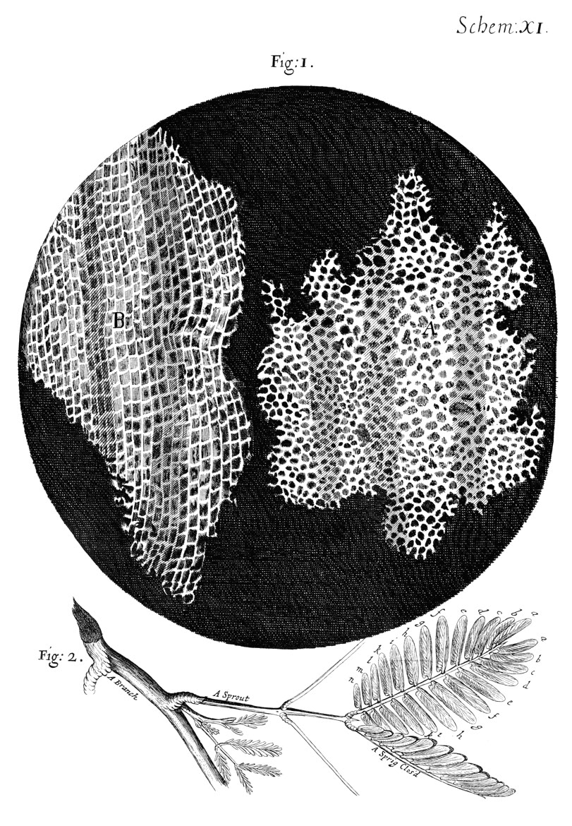
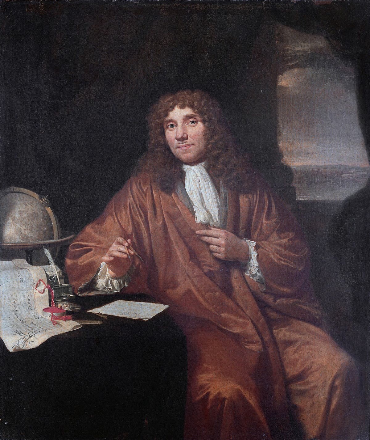
Left: Hooke’s drawing of cork in Micrographia (1665), the first cells ever figured — the empty walls of dead cells, seen in two orientations. Right: Antoni van Leeuwenhoek, painted by Jan Verkolje around 1680, whose single-lens microscopes first showed living cells.

Examples

Example 5.3 (One cell, many cells)

A bacterium, a yeast, an amoeba are whole organisms of one cell, doing everything — feeding, moving, dividing — within a few micrometres. A human is some 3×10133 \times 10^{13} cells of two hundred kinds, none of which could live alone in a pond, each of which descends by unbroken divisions from one fertilised egg. The rabbit of Chapter 2 and the oak of Chapter 3 are the same theory applied twice.

Example 5.7 (Why the eukaryotic cell is compartmented)

A cell of 20µm20\,\text{µ}\mathrm{m} has a thousand times the volume of a bacterium but only a hundred times its membrane surface (Proposition 1.6). Membranes are where a cell carries its energy-transducing chains and its transport systems; the eukaryote recovers surface by folding membranes inside itself — the inner membrane of a mitochondrion, the sheets of the endoplasmic reticulum — and gains, besides, separate chambers in which incompatible chemistries (digestion in the lysosome, synthesis in the cytosol) can run side by side.

Example 5.11 (A bacterium and a mitochondrion)

Under the light microscope at 1000×1000\times, an E. coli cell is a rod 2mm2\,\mathrm{mm} long on the retina’s scale, with no visible interior; a mitochondrion is a dot at the limit of resolution. In a TEM section the same bacterium shows its two membranes, its wall, its nucleoid as a pale fibrous region and its ribosomes as dark grains; the mitochondrion shows its double membrane and the folds of the inner one. Neither is alive.

Read in context →