High School Biology · Grades 10–12
2Cells: The Common Unit of Life
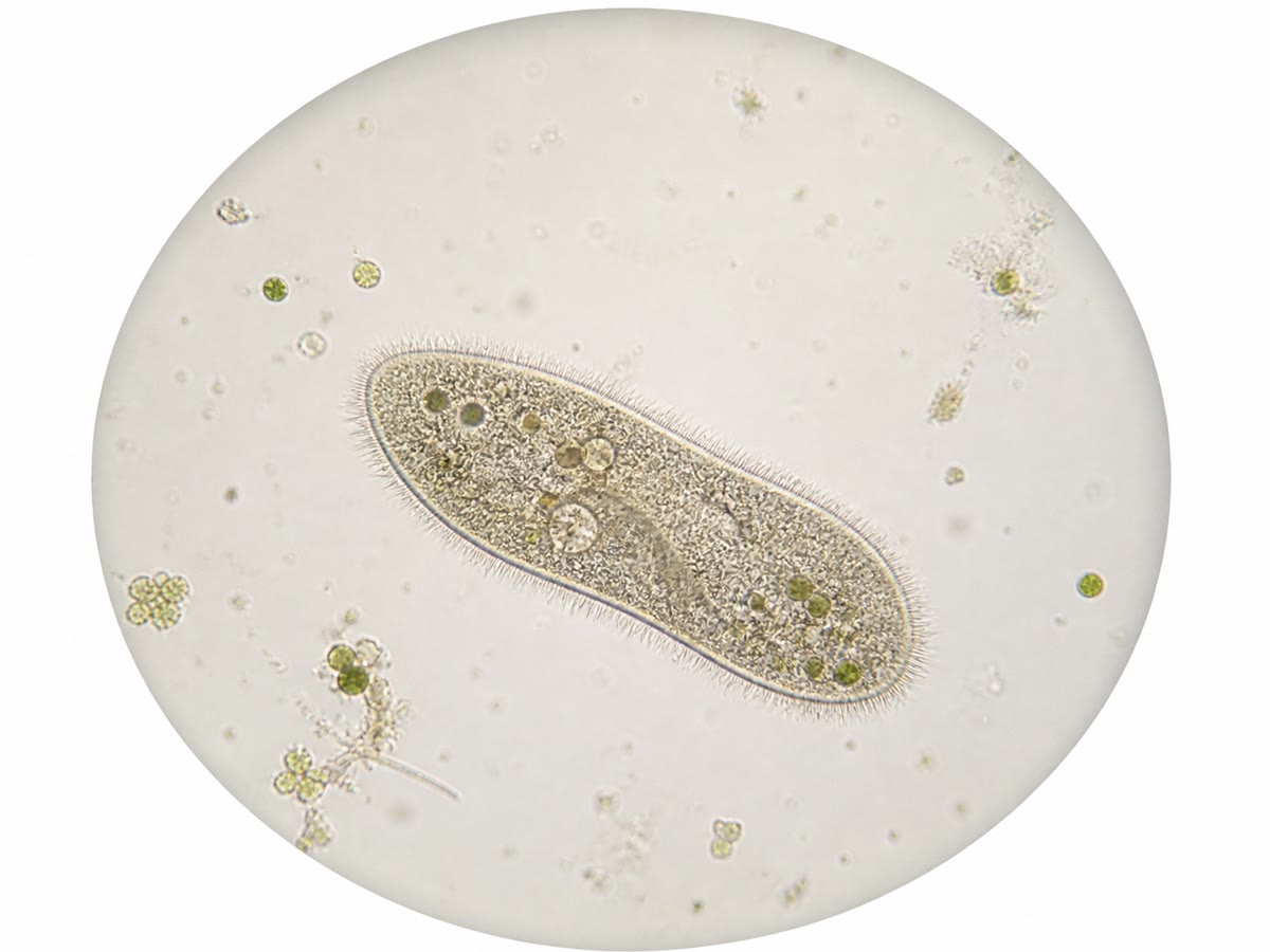
Scrape the inside of your cheek with a cotton bud, smear it on a slide, add a drop of blue dye, and look through a microscope at four hundred times: a scatter of flat, irregular tiles, each with a darker spot near its centre. Peel the thin skin from inside an onion scale and look again: neat rectangular bricks, each with its own spot. Pond water shows oval creatures rowing past with a fringe of hairs. The three samples have nothing in common to the naked eye. Under the lens they are built from the same object — the cell — and this chapter is about what that object is, how it is seen, and why every living thing is made of it.
2.1 The cell theory
Definition 2.1 (Cell)
A cell is the smallest unit of a living thing that is itself alive: a volume of watery content, the cytoplasm, enclosed by a thin plasma membrane that separates it from its surroundings while letting selected substances cross. A cell takes in matter and energy, transforms them, grows, and divides into two cells.
Proposition 2.2 (The cell theory)
Three statements, established by microscopy in the nineteenth century, form the cell theory:
Evidence. Statement 1 rests on the systematic examination of plants (Schleiden, 1838) and animals (Schwann, 1839) with improved microscopes: every tissue examined, however different in appearance, resolved into cells. Statement 3 was established by Virchow (1855) and by the observation of dividing cells in growing tissues; the alternative — cells forming spontaneously from fluid — was never observed, and Pasteur’s experiments on microbes (Chapter 18 returns to them) closed the question. Statement 2 follows from the first and from the fact that isolated cells, kept in a suitable liquid, go on living while any fragment of a cell does not. ∎
Example 2.3 (One cell or many)
A yeast, a paramecium or a bacterium is a single cell that does everything — feeds, moves, divides. An oak or a human is a cooperative of cells of many kinds, each doing a share of the work: a human body contains some cells, of roughly two hundred kinds. Between the two lie colonies and simple multicellular organisms, but there is no living thing built of anything other than cells.
2.2 Seeing cells: scales and microscopes
Definition 2.4 (Magnification and resolution)
The magnification of an instrument is the ratio of the size of the image to the size of the object. Its resolution is the smallest distance between two points that it still shows as separate. Magnifying an image beyond what the resolution justifies enlarges the blur, not the detail.
Proposition 2.5 (Two instruments, two scales)
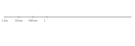
The light microscope magnifies up to about with a resolution of about : it shows cells, nuclei, chloroplasts and bacteria, in colour and alive. The electron microscope uses a beam of electrons instead of light: resolution below , magnification up to ; it reveals the inner structure of the cell — membranes, mitochondria, ribosomes, even large molecules — but on a sample that is dead, dried and cut into slices thinner than a micrometre.
Proof. Admitted at this level. ∎
Method 2.6 (Measuring an object on a micrograph)
A micrograph carries either a magnification or a scale bar.
- With a scale bar: measure the bar on the page (say for a bar labelled ) and the object (); the real size is .
- With a magnification: real size measured size divided by the magnification; a cell drawn long at is .
- Always state the unit, and convert: , .
Example 2.7 (Cheek cells)
A cheek cell drawn wide from a image is across, and its nucleus, drawn , is about . A red blood cell is ; the bacteria of the mouth, long, appear as barely resolved dots at the same magnification.
2.3 Inside the cell
Definition 2.8 (Organelle)
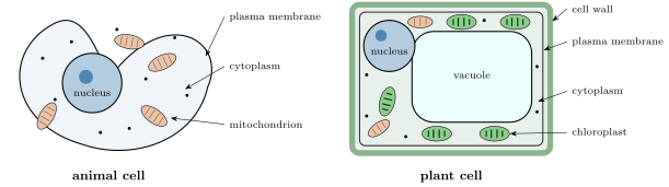
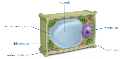
An organelle is a structure inside a cell, enclosed by its own membrane, that performs a specific task. The principal ones are:
- the nucleus, which contains the chromosomes — the DNA of Chapter 3 — and controls the cell’s activity;
- the mitochondria, some long, where organic molecules are broken down with oxygen to release usable energy (cellular respiration, Chapter 4);
- in plant cells, the chloroplasts, green with chlorophyll, where photosynthesis takes place, and a large water-filled vacuole that keeps the cell taut.
Outside the membrane, a plant cell is boxed in a rigid cell wall of cellulose; animal cells have none. The cytoplasm of every cell also contains millions of ribosomes, particles of about that assemble proteins, visible only with the electron microscope.
Definition 2.9 (Prokaryotes and eukaryotes)
A cell whose DNA is enclosed in a nucleus is a eukaryotic cell: animals, plants, fungi and many single-celled organisms are eukaryotes. A prokaryotic cell has no nucleus and no membrane-bound organelles: its DNA lies free in the cytoplasm, its size is around , and it is surrounded by a wall of its own kind. Bacteria are prokaryotes.
Example 2.10 (Reading a bacterium)
An electron micrograph of the gut bacterium Escherichia coli shows a rod long and wide: a wall, a membrane just inside it, a granular cytoplasm packed with ribosomes, and a paler central region where the single circular DNA molecule lies. No nucleus, no mitochondria. Yet the cell feeds, grows and divides every twenty minutes in a rich broth: everything the cell theory demands, in a thousandth of the volume of a cheek cell.
2.4 Many cells, many shapes: specialisation
Proposition 2.11 (Specialised cells)
In a multicellular organism the cells share the same basic plan and the same DNA, but each kind has a shape and an equipment matched to its task: a neuron extends a fibre up to a metre long to carry signals; a red blood cell has lost its nucleus and is a flattened disc packed with haemoglobin; a muscle fibre is a giant cell full of contractile proteins; a root hair is a thin tube that maximises the surface for absorbing water; a guard cell of a leaf changes shape to open or close a pore. Specialisation is a matter of which parts of the shared plan are developed.
Proof. Admitted at this level. ∎
Example 2.12 (Surface and volume)
Why are cells so small? Everything a cell takes in or gives out crosses its membrane, whose area grows with the square of the size, while the needs grow with the volume, the cube. A cubic cell of side has a surface of for a volume of : of membrane per cubic micrometre. At side the ratio drops to : ten times less membrane per unit of content. A large cell would starve at its centre; the cells that are large (a frog egg, a muscle fibre) are either mostly inert reserve or elongated so that no point is far from the surface.
Remark 2.13 (The cell as an open system)
A cell is not a sealed bag. Water, oxygen, glucose and ions cross its membrane continuously, and its wastes leave the same way; a cheek cell placed in pure water swells and bursts within minutes, and one placed in strong salt water shrivels. The membrane’s control of what enters and leaves is the subject of the Year 1 volume; for now, remember that a cell lives by exchanging with its surroundings, and dies when the exchanges stop.
2.5 Exercises
Exercise 2.1 ★
State the three statements of the cell theory.
Exercise 2.2 ★
Convert: to micrometres; to micrometres; to millimetres.
Solution
Solution of Exercise 2.2.
; ; .
Exercise 2.3 ★
A cell is drawn long from a image. What is its real length?
Solution
Solution of Exercise 2.3.
.
Exercise 2.4 ★
Name three structures found in a plant cell but not in an animal cell, and one structure found in every cell.
Solution
Solution of Exercise 2.4.
Plant only: cell wall, chloroplasts, large central vacuole. Every cell: a plasma membrane (also cytoplasm and ribosomes).
Exercise 2.5 ★
What is the difference between a prokaryotic and a eukaryotic cell? Give an example of each.
Solution
Solution of Exercise 2.5.
A eukaryotic cell keeps its DNA inside a nucleus and has membrane-bound organelles (a cheek cell, a yeast); a prokaryotic cell has neither, its DNA lies free in the cytoplasm, and it is about (a bacterium such as E. coli).
Exercise 2.6 ★★
On a micrograph a scale bar labelled measures ; a mitochondrion measures long. Compute the real length of the mitochondrion and the magnification of the image.
Exercise 2.7 ★★
Why can a light microscope not show ribosomes, whatever its magnification? Use the two words of Definition 2.4.
Solution
Solution of Exercise 2.7.
A ribosome () is ten times smaller than the light microscope’s resolution ( ): two neighbouring ribosomes, or a ribosome and its surroundings, cannot be told apart. More magnification only enlarges the blur.
Exercise 2.8 ★★
Iodine solution is added to a slice of potato under the microscope: the blue-black colour appears in oval grains inside the cells. Using Chapter 1, say what the grains are and what this shows about where a plant stores its reserves.
Exercise 2.9 ★★
Onion skin cells show a nucleus and a wall but no chloroplast, while the cells of a moss leaf are full of them. Explain the difference from where each tissue lives.
Solution
Solution of Exercise 2.9.
The onion scale grows underground, in the dark, and stores reserves: chloroplasts would be useless there. The moss leaf lives in the light and makes its food by photosynthesis, so its cells are packed with chloroplasts. Same plan, different equipment.
Exercise 2.10 ★★
Compute the surface-to-volume ratio of a cubic cell of side (a bacterium) and of side (a typical animal cell). Which one can rely on simple diffusion through its membrane alone?
Solution
Solution of Exercise 2.10.
Side : surface , volume , ratio 6 per micrometre. Side : surface , volume , ratio 0.3. The bacterium has twenty times more membrane per unit of content and lives by diffusion alone; the larger cell needs internal transport and organelles.
Exercise 2.11 ★★
A bacterium in rich broth divides every . Starting from one cell, how many are there after ? After ?
Exercise 2.12 ★★★
A red blood cell has no nucleus and no mitochondria. Does it satisfy Definition 2.1? Discuss, knowing that it lives about 120 days, cannot divide, and is produced by nucleated cells in the bone marrow.
Solution
Solution of Exercise 2.12.
It has a membrane, a cytoplasm and takes in and gives out substances, so it fits most of the definition, but it cannot divide and cannot renew its proteins: it is a cell that has given up part of the programme in exchange for room for haemoglobin, and it is made and replaced by complete cells. The definition describes the general case; the red blood cell is a specialised, terminal form of it.
Exercise 2.13 ★★★
In 1665 Hooke saw "cells" in a slice of cork — empty boxes with thick walls. Explain what he actually saw, in the vocabulary of this chapter, and why he could not have seen a nucleus in it.
Solution
Solution of Exercise 2.13.
Cork is dead tissue: Hooke saw the cellulose walls of cells whose living content had disappeared — empty boxes. A nucleus exists only in a living cell’s cytoplasm, and in any case his microscope’s resolution and the absence of stains would have hidden it.
Exercise 2.14 ★★★
Viruses are particles of to that contain nucleic acid and protein, cannot grow or divide on their own, and multiply only inside a cell. Are they cells? Are they alive? Argue from the cell theory.
Solution
Solution of Exercise 2.14.
A virus has no cytoplasm, no membrane of its own making, no metabolism, and cannot divide: it fails the definition of a cell. By the cell theory it is not alive on its own; it is a particle that borrows a cell’s machinery to make copies of itself. Whether to call it "alive" is a matter of definition; that it is not a cell is not.
Exercise 2.15 ★★★
A muscle fibre is a single cell long and wide, containing hundreds of nuclei. Compute its volume and compare with that of a cubic cell; then explain, from Example 2.12, why its shape and its many nuclei make such a size workable.
Solution
Solution of Exercise 2.15.
Volume , about 7000 times the of the cube. Being a thin cylinder, no point of the fibre is more than from the membrane, so exchanges remain fast; and each nucleus governs only its own stretch of the fibre, so no nucleus has to serve a volume larger than an ordinary cell’s.
2.6 Problem: Counting the Cells of a Body
Problem 2.1
Weekend problem — a microscopy session, from the onion skin on the slide to an estimate of the number of cells in a human body
A class spends an afternoon at the microscope. The objectives available give total magnifications of , and ; the field of view at is across.
Part I — The onion.
- At , about 9 onion cells fit side by side across the field of view. Estimate the width of one cell, in micrometres.
- At the field of view is four times narrower. How wide is it, and how many of these cells fit across it?
- A student draws one cell wide from the image. What is the magnification of the drawing relative to the real cell?
- Iodine stains the nucleus yellow-brown; the student measures it at on the drawing. Real diameter?
- Name the structures the student should label on the drawing, and one structure present that cannot be seen at this magnification.
Part II — The cheek and the pond.
- Cheek cells stained with methylene blue appear as flat polygons about across but only thick. Why are they flat, given where they come from?
- A drop of pond water shows a paramecium long. Can it be seen with the naked eye? At which magnification does it fill a quarter of the field?
- Tiny moving dots about long are visible near the paramecium at . What are they, most probably, and what would an electron microscope add?
- The paramecium is a single cell; the cheek cell is one of trillions. In one sentence each, say what "specialised" means for the cheek cell and why it cannot apply to the paramecium.
- A student adds a drop of salt water to the cheek cells and sees them shrivel. Explain with Remark 2.13.
Part III — The size of a cell.
- Take a typical human cell as a cube of side . Compute its volume in cubic micrometres, then in cubic millimetres ().
- Compute its surface area and the ratio surface/volume.
- Repeat for a bacterium taken as a cube of side . By what factor is its ratio larger?
- Cells take up a mass roughly equal to that of an equal volume of water ( per ). Estimate the mass of the cell in nanograms ().
- A red blood cell is a disc across and thick. Estimate its volume (treat it as a cylinder, ) and compare with the cube of question 11.
Part IV — The number of cells in a body.
- A student of mass has a volume of about . If the body were made only of cubic cells, how many would it contain?
- In fact about a third of the body’s volume is fluid between the cells and other non-cellular material. Correct the estimate.
- Red blood cells alone number about . Using the volume of question 15, what volume do they occupy? Is that consistent with a blood volume of about of which 45% is cells?
- Combine questions 17 and 18: give an order of magnitude for the total number of cells in the body, and say why a precise count is impossible.
- State the result: how many cells, roughly, make a human, and by what process — from which starting number — were they all produced?
Solution
Solution of Problem 2.1.
1. .
2. ; about two cells fit across it.
3. : the drawing is .
4. , about .
5. Cell wall, plasma membrane (pressed against the wall), cytoplasm, nucleus, vacuole. Present but invisible at : the ribosomes (and the mitochondria are at the limit).
6. They come from the surface of a lining that is several cells thick and constantly rubbed: the outermost cells are flattened and shed, which is why a cotton bud collects them.
7. is just above the naked eye’s limit: a barely visible speck. At it spans of the field, at more than half; no available objective gives exactly a quarter.
8. Bacteria. The electron microscope would show their wall, membrane, ribosomes and DNA region, and the absence of a nucleus.
9. The cheek cell does one job (protecting a surface) in a body where other cells feed it, oxygenate it and coordinate it; the paramecium has nobody else and must feed, move, sense and divide as a single cell.
10. The outside is now saltier than the cytoplasm, so water leaves the cell across the membrane; the cell loses volume and shrivels.
11. .
12. ; ratio per micrometre.
13. Surface , volume , ratio 6: twenty times larger.
14. , hence about .
15. , about a hundredth of the cube.
16. ; divided by 8000: about cells.
17. Two thirds of that: about .
18. . Blood cells: — consistent.
19. About large cells plus red cells: some , a few tens of trillions. Cell sizes span a factor of a hundred and the mix of kinds varies, so any count is an estimate to within a factor of two or three.
20. Roughly thirty trillion () cells, all produced by successive cell divisions from a single starting cell, the fertilised egg.