 Coach
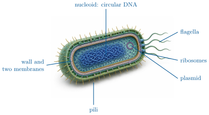
CoachA prokaryotic cell (bacteria, archaea) has no nucleus: its DNA, usually one circular chromosome, lies in a region of the cytoplasm called the nucleoid, and it has no membrane-bounded compartments. It is small () and bounded by a plasma membrane and, outside it, a cell wall. A eukaryotic cell (protists, fungi, plants, animals) keeps its DNA, as several linear chromosomes, inside a nucleus bounded by a double membrane, and divides its cytoplasm into membrane-bounded organelles (Chapter 6). It is larger () and has an internal skeleton of protein filaments.
Examples
Example 5.7 (Why the eukaryotic cell is compartmented)
A cell of has a thousand times the volume of a bacterium but only a hundred times its membrane surface (Proposition 1.6). Membranes are where a cell carries its energy-transducing chains and its transport systems; the eukaryote recovers surface by folding membranes inside itself — the inner membrane of a mitochondrion, the sheets of the endoplasmic reticulum — and gains, besides, separate chambers in which incompatible chemistries (digestion in the lysosome, synthesis in the cytosol) can run side by side.
Example 5.11 (A bacterium and a mitochondrion)
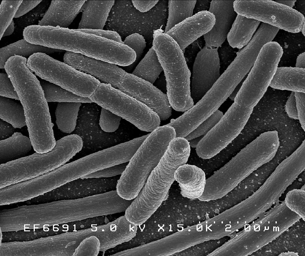
Under the light microscope at , an E. coli cell is a rod long on the retina’s scale, with no visible interior; a mitochondrion is a dot at the limit of resolution. In a TEM section the same bacterium shows its two membranes, its wall, its nucleoid as a pale fibrous region and its ribosomes as dark grains; the mitochondrion shows its double membrane and the folds of the inner one. Neither is alive.