University Biology — Year 2 · Bachelor Year 2
10Embryonic Development of Vertebrates
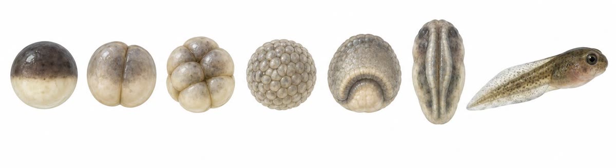
A frog’s egg is a sphere two millimetres across, dark on top and pale below. Within a day it has divided into thousands of cells; within two, a sheet of those cells has rolled inside through a groove to make a gut and lay a rod along the back; within three, the sheet above the rod has folded into a tube that will be the brain and spinal cord. Nothing has been added but water. The information that turns one cell into a swimming tadpole was in the egg and its genome, and the way it unfolds — cleavage, gastrulation, neurulation, the induction of each part by its neighbours — is recognisably the same in a fish, a chick and a mouse. This chapter follows a vertebrate embryo through those steps and describes the experiments that showed how each part of it is told what to become.
10.1 From egg to blastula
Definition 10.1 (The egg and its axes)
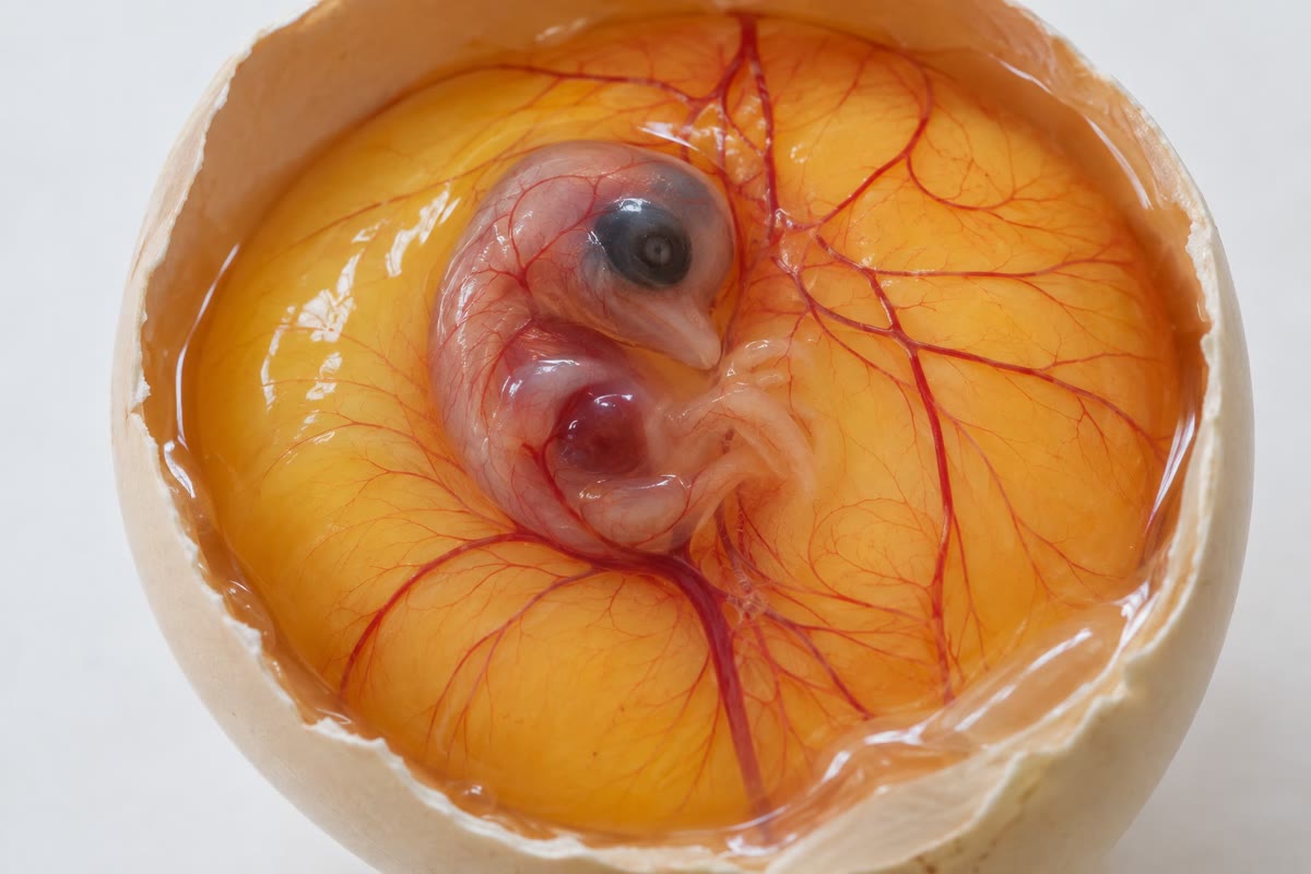
A vertebrate egg is polarised before fertilisation: the animal pole holds the nucleus and most of the cytoplasm, the vegetal pole the yolk, the store of protein and lipid on which the embryo will live until it feeds. The amount of yolk sets the pattern of everything that follows: a sea urchin or mammal egg has almost none and cleaves completely into equal cells; a frog egg has a moderate amount and cleaves completely but unequally (small cells above, large yolky cells below); a fish or bird egg is nearly all yolk and cleaves only in a disc of cytoplasm on top of it. In the frog, the point where the sperm enters fixes the second axis: the cortex rotates thirty degrees away from it, exposing a grey crescent opposite the entry point, and that side will become the back. Cleavage is a series of rapid mitoses with no growth — the cells halve in size at each — driven by maternal mRNAs and proteins stored in the egg, the embryo’s own genes staying silent until the mid-blastula transition, some twelve divisions in.
Theorem 10.2 (Cleavage arithmetic)
After synchronous cleavages an egg of volume is cells of mean volume and radius , and the total surface has grown by ; the ratio of nuclear to cytoplasmic volume, which starts near in a frog egg, rises by and reaches that of an ordinary cell () after about ten divisions. It is this rising ratio — the titration of a fixed maternal stock of some inhibitor by an exponentially growing amount of DNA — that triggers the mid-blastula transition: divisions slow, become asynchronous, and the zygotic genome switches on.
Proof. Volume is conserved in cleavage, so each of cells has and, as a sphere, radius ; their total area is . Each cell keeps one nucleus of fixed size, so the ratio scales as from : brings it to . The frog’s transition falls at the twelfth division, consistent with a threshold reached a little later. ∎
Evidence. Newport and Kirschner (1982) added extra DNA to frog eggs and found the transition came earlier by as many divisions as the DNA was in excess; removing cytoplasm had the same effect. The clock is not time or a count of divisions but a ratio. ∎
Definition 10.3 (Blastula)
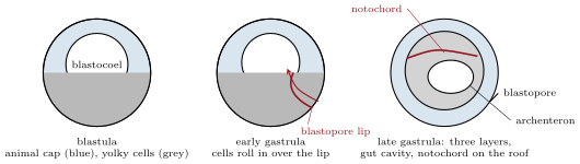
Cleavage ends in a blastula: a hollow ball (frog, sea urchin) or a disc (fish, bird) of a few thousand cells around a cavity, the blastocoel. Its cells are still unspecialised in appearance, but they are not equivalent: a fate map, made by staining patches of the surface with dyes and following them, shows that the animal cap will give skin and nervous system (ectoderm), the equatorial belt muscle, skeleton, kidney and blood (mesoderm), and the vegetal mass the gut and its organs (endoderm). These three germ layers are the same in every vertebrate, and the next step is to put them in their places: the mesoderm and endoderm are outside and must go in.
10.2 Gastrulation and neurulation
Proposition 10.4 (Gastrulation)
Gastrulation is the set of cell movements that turns the one-layered blastula into a three-layered gastrula. In the frog it begins below the grey crescent: cells there change shape, contracting their outer ends into bottles, and the surface buckles into a groove, the blastopore. Through it the mesoderm and endoderm stream inside (involution), rolling over the lip and moving forward along the roof of the cavity; the animal cap spreads down to cover the outside (epiboly); the blastopore closes to a ring around the last yolky cells. Inside, a new cavity lined with endoderm, the archenteron, is the future gut, and the mesoderm that rolled in first, along the midline, has become a stiff rod, the notochord, the axis of every vertebrate embryo and the structure that names the phylum. In birds and mammals the same movements happen through a slit, the primitive streak, in a flat disc; in fish the disc spreads over the yolk. The choreography differs with the yolk; the result — ectoderm outside, endoderm lining the gut, mesoderm between, notochord in the midline — is the same.
Proposition 10.5 (Neurulation and the body plan)
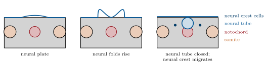
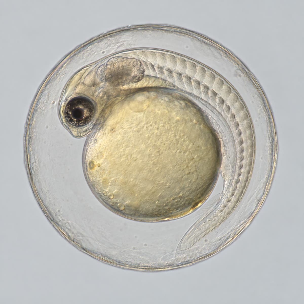
The ectoderm above the notochord thickens into a neural plate, whose edges rise as folds and meet in the midline to form the neural tube, the future brain and spinal cord, which sinks beneath the skin; cells from the crest of the folds, the neural crest, migrate away to make the peripheral nerves, the pigment cells, the adrenal medulla and much of the face. On each side of the notochord the mesoderm segments into blocks, the somites, one pair after another from head to tail, which give the vertebrae, the trunk muscles and the dermis; lateral mesoderm splits into the layers that line the body cavity and make the heart and limbs; the endoderm tube buds off the lungs, liver and pancreas. By the end of neurulation the embryo has the vertebrate body plan — a dorsal nerve cord over a notochord, a ventral gut, segmented muscle, a head — at a length of a few millimetres, and the rest of development is the elaboration of organs from this plan (Chapter 11).
10.3 Induction: how cells learn where they are
Definition 10.6 (Induction and competence)
Induction is the process by which one group of cells changes the fate of a neighbouring group by a signal; the responding cells must be competent — able to answer, which they are only for a limited time. Development is a chain of inductions: the vegetal cells induce mesoderm in the cells above them; the dorsal mesoderm — the organiser at the blastopore lip — induces the neural plate in the ectoderm above it and patterns the mesoderm on either side; the notochord induces the floor of the neural tube; the optic vesicle, a bud of the brain, induces a lens in the skin it touches; the lens induces the cornea. Each induced tissue becomes an inducer of the next. The signals are a handful of secreted protein families used over and over — and the same ones that pattern a limb (Chapter 11) and, in the adult, run the signalling of Chapter 19.
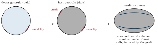
Evidence. Spemann and Mangold (1924) cut the dorsal blastopore lip from an early gastrula of one newt species and grafted it onto the ventral side of another, pigmented differently so that host and graft cells could be told apart. The graft did not simply become what it would have become in place: it rolled in, formed notochord, and induced the host’s own ventral ectoderm to make a second neural tube and, beside it, host somites — a complete second embryo, largely of host cells, joined belly to belly with the first. No other piece of the gastrula could do this. They called the lip the organiser; its signals (proteins that block the ventralising factor BMP) were identified seventy years later. ∎
Theorem 10.7 (A morphogen gradient)
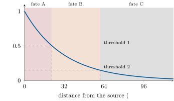
Many inductions work by a morphogen: a signal secreted by a source, spreading through the tissue and read by cells according to its local concentration — above one threshold one fate, above a higher one another. A morphogen produced at a plane source at rate per unit area, diffusing with coefficient and degraded at rate , has at steady state the exponential profile
so that the distance at which the concentration falls below a threshold is : a tissue can be patterned into bands of fixed width by fixed thresholds. With (a protein slowed by binding as it moves through tissue) and (a half-life of about twelve minutes), : a few cell diameters, the scale over which embryonic fields are patterned. Doubling the source doubles and shifts every boundary outward by .
Proof. At steady state diffusion balances degradation: , whose decaying solution is with . The flux leaving the source is , which gives . Setting gives ; changing to adds to it. ∎
Example 10.8 (Reading the gradient)
In the frog gastrula, cells of the marginal zone exposed to graded amounts of a signal of the activin family become, from high to low dose, notochord, muscle, kidney and blood; dissociated animal-cap cells put in a dish with a threshold dose switch fates within a factor of two of concentration. In the neural tube, a signal from the notochord and floor plate spreads upward and specifies, from the bottom, motor neurons and then successive classes of interneurons at thresholds a few cell diameters apart. The organism’s coordinates are written in concentrations.
10.4 Fate, commitment and the timing of decisions
Proposition 10.9 (When a cell’s fate is fixed)
A cell’s fate is what it will normally become; its potency is what it can become if moved; it is determined when its fate no longer changes with its surroundings. Early embryos regulate: each of the first two frog blastomeres, separated, makes a whole small tadpole, and the first cells of a mammal embryo are all totipotent — which is how identical twins arise. Determination comes gradually and at different times for different tissues: a piece of early gastrula ectoderm grafted to the belly becomes belly skin, the same piece taken from a late gastrula makes a neural plate wherever it is put; by the tailbud stage a limb field transplanted to the flank makes a limb there. In most invertebrates, by contrast, fate is fixed early by the cytoplasm each cell inherits (mosaic development) and a separated blastomere makes only its part. The vertebrate strategy — keep cells flexible and tell them what to do by induction — is what makes twinning, regeneration and the organiser experiment possible.
Evidence. Spemann (1901) tied a hair loop around a newt egg at the two-cell stage: if the loop lay in the plane of the grey crescent, so that each half received part of it, two complete embryos formed; if it lay perpendicular, so that one half had the whole crescent, that half made an embryo and the other a ball of belly tissue. The crescent — the future organiser — was the one part that could not be divided. ∎
Example 10.10 (The timetable of a frog)
At : fertilisation; first cleavage at , then every half hour; blastula of cells at ; mid-blastula transition at ; gastrulation to ; neural folds close at ; heart beats at ; hatching and feeding at , as a tadpole of . A zebrafish does the same in one day, a chick in three, a mouse in nine; a human embryo gastrulates in the third week and closes its neural tube by the fourth — before most women know they are pregnant, which is why folate, needed for neural tube closure, is prescribed before conception.
10.5 Exercises
Exercise 10.1 ★
Define cleavage, blastula, gastrulation and neurulation, and name the structure that each stage produces.
Solution
Solution of Exercise 10.1.
Cleavage: rapid mitoses without growth, producing the blastula. Blastula: a hollow ball or disc of small cells around the blastocoel. Gastrulation: the cell movements that bring mesoderm and endoderm inside, producing the three-layered gastrula with a gut cavity and notochord. Neurulation: the folding of the dorsal ectoderm into the neural tube, with the segmentation of the somites, producing the neurula with the vertebrate body plan.
Exercise 10.2 ★
Give the three germ layers and, for each, four adult tissues that derive from it.
Solution
Solution of Exercise 10.2.
Ectoderm: epidermis, brain and spinal cord, peripheral nerves (neural crest), lens and pigment cells. Mesoderm: notochord, muscle, bone and cartilage, kidney, heart and blood, dermis. Endoderm: the gut lining, liver, pancreas, lungs, thyroid.
Exercise 10.3 ★
How does the amount of yolk change the pattern of cleavage and of gastrulation? Compare a frog and a chick.
Solution
Solution of Exercise 10.3.
Little yolk: complete, nearly equal cleavage and gastrulation by involution through a round blastopore in a hollow ball (frog: complete but unequal, the yolky vegetal cells large and slow). Much yolk (chick): cleavage confined to a disc on top of the yolk, and gastrulation through a slit, the primitive streak, in the flat disc, the layers spreading over the yolk instead of enclosing a cavity.
Exercise 10.4 ★
What is induction? Give three inductions in the order they occur in a frog embryo, naming the inducer and the responding tissue.
Solution
Solution of Exercise 10.4.
Induction is the change of a competent tissue’s fate by a signal from a neighbouring tissue. Vegetal cells induce mesoderm in the overlying marginal zone; the organiser (dorsal lip mesoderm) induces the neural plate in the dorsal ectoderm; the optic vesicle induces the lens in the head ectoderm.
Exercise 10.5 ★★
A frog egg of radius cleaves twelve times. Compute the number of cells, their mean radius, and the factor by which the total cell surface has grown. Why does the embryo need the extra surface?
Solution
Solution of Exercise 10.5.
cells of radius ; surface multiplied by . The extra surface is the membrane of the epithelia that gastrulation will fold into skin and gut, and the area through which the blastula exchanges with its surroundings.
Exercise 10.6 ★★
The nuclear-to-cytoplasmic ratio starts at and the mid-blastula transition occurs when it reaches . After how many divisions? If the egg is injected with three times its own amount of DNA, at which division does the transition come?
Solution
Solution of Exercise 10.6.
: , . With four times the DNA the ratio starts at and reaches at : the tenth division, two earlier.
Exercise 10.7 ★★
A morphogen has and a half-life of . Compute and . Two thresholds are at and of the source concentration: give the widths of the three fate bands.
Solution
Solution of Exercise 10.7.
; . Thresholds at and : bands of 29, 67 and the remainder.
Exercise 10.8 ★★
In the gradient of the previous exercise the source is doubled by a mutation. By how much does each boundary move? What if the degradation rate is doubled instead?
Solution
Solution of Exercise 10.8.
Doubling the source shifts both boundaries outward by : the first band doubles, the second keeps its width. Doubling divides by () and, since , divides by as well: the boundaries move to and — both bands shrink.
Exercise 10.9 ★★
Describe the Spemann–Mangold experiment, say why the two species had to differ in pigmentation, and state what the result excluded.
Solution
Solution of Exercise 10.9.
A dorsal lip from an unpigmented newt gastrula was grafted to the ventral side of a pigmented one; it rolled in and the host formed a second embryo — neural tube, somites, gut — joined to the first. The pigment difference showed that the second axis was made of host cells, so the graft had induced them rather than built the axis itself. It excluded self-differentiation of the graft as the explanation, and showed that a small region carries the signal that organises the whole axis.
Exercise 10.10 ★★★
Predict the outcome of: (a) a piece of early-gastrula prospective neural plate grafted to the belly; (b) the same piece from a late gastrula; (c) a piece of belly ectoderm placed over a notochord in a late gastrula; (d) the whole organiser removed from an early gastrula. Explain each with the notions of competence and determination.
Solution
Solution of Exercise 10.10.
(a) Belly skin: the early ectoderm is competent but not yet induced, so it follows its new surroundings. (b) Neural tissue: by the late gastrula it has been induced and is determined. (c) Neural plate: late-gastrula belly ectoderm is still competent, and the notochord induces it. (d) No dorsal structures: a ball of belly tissue, since nothing induces the mesoderm to dorsal fates or the ectoderm to neural ones.
Exercise 10.11 ★★★
The frog’s first cleavage takes and the next eleven each, at ; at every step is 2.5 times slower. At what time is the mid-blastula transition reached at each temperature? Explain why the transition comes at the same cell number at both temperatures, and what that says about the clock.
Solution
Solution of Exercise 10.11.
At : , 7 hours. At : . The transition falls at the twelfth division in both cases because what triggers it is the ratio of DNA to cytoplasm, which depends on the number of divisions and not on how long they took: the clock counts doublings, not hours.
Exercise 10.12 ★★★
“A vertebrate embryo is built by conversation, an invertebrate one by inheritance.” Discuss the contrast between regulative and mosaic development, its consequences for twinning and regeneration, and why it is a difference of degree.
Solution
Solution of Exercise 10.12.
In regulative development cells acquire their fates by induction from neighbours, so the embryo can rebuild after loss (twinning from a split egg, regeneration of a grafted region); in mosaic development fates are inherited with localised cytoplasmic determinants, so each blastomere makes only its part and a split embryo makes two half-larvae. The difference is one of degree: the frog’s grey crescent is a determinant, and mosaic embryos use inductions later; every embryo mixes inheritance and conversation, vertebrates leaning on conversation for longer.
10.6 Problem: A Frog Embryo Timed and Patterned
Problem 10.1
Weekend problem — a frog embryo followed through cleavage, the mid-blastula transition, gastrulation and induction, with the morphogen gradient computed and the classical grafts predicted, ending on the cell count and time of the transition and the widths of the fate bands
A frog egg has radius and a nucleus of radius whose DNA content is ; each cleavage takes after a first one of . The mid-blastula transition occurs when total DNA reaches . Morphogen: , half-life , source concentration , thresholds at and .
Part I — Cleavage.
- Compute the volume of the egg and of its nucleus, and the initial nuclear-to-cytoplasmic ratio.
- After how many cleavages does the DNA reach ? How many cells?
- At what time after fertilisation?
- What is the mean cell radius then, and the ratio of nuclear to cell volume if nuclei keep their size? Comment.
- By what factor has the total cell surface grown?
- Each cleavage needs the genome copied: how much DNA has been synthesised in total, in units of , and why does the embryo not need to transcribe its genes to do it?
- What changes at the transition, and what experiment shows that it is triggered by a ratio and not by a count?
Part II — Gastrulation.
- Where does the blastopore form relative to the sperm entry point, and what fixed that position?
- List, in order, the tissues that roll in through the blastopore and their positions afterward.
- Gastrulation takes and the involuting sheet travels . Compute the mean cell speed in micrometres per minute and compare with a crawling fibroblast ().
- Why is gastrulation impossible before the mid-blastula transition?
- Explain in one sentence why the notochord, and not the neural tube, is the defining structure of the chordates.
- A drug that blocks cell-shape change is applied at the start of gastrulation. Predict the embryo.
Part III — The gradient.
- Compute from the half-life, and .
- Compute the positions of the two thresholds.
- Give the widths of the three fate bands in micrometres and in cells of .
- The source is halved. Recompute the two positions and state which band shrinks most.
- A cell at from the source is moved to before the gradient is read; then after. What does it become in each case, and why?
- Explain why an exponential profile gives boundaries whose positions depend on the logarithm of the source strength, and why this makes patterning robust.
Part IV — Grafts.
- Predict the result of grafting a dorsal lip to the ventral side of an early gastrula, and of grafting ventral marginal zone to the dorsal side.
- Predict the result of grafting a dorsal lip to the ventral side of a neurula, and explain the difference.
- A frog embryo at the two-cell stage is split along the plane of the grey crescent; another perpendicular to it. Predict both.
- An animal cap is cut from a blastula and cultured alone; the same cap is cultured next to vegetal cells. Predict the tissues formed.
- Why did Spemann and Mangold’s result require host and graft to be distinguishable, and what would a graft of the wrong species have shown?
- State the result: the cleavage number and time of the transition, its cell count, and the three band widths.
Solution
Solution of Problem 10.1.
1. Egg ; nucleus ; ratio . 2. : , 4096 cells. 3. , about 6.75 hours. 4. Radius ; ratio — that of an ordinary cell: the nuclei have caught up with the cytoplasm. 5. . 6. About of DNA; the egg holds enough stored histones, polymerases and nucleotides for twelve divisions, so no transcription is needed. 7. Zygotic genes switch on, divisions slow and lose synchrony, cells become motile. Injecting extra DNA brings the transition forward by as many divisions as the DNA is in excess, which a counter of divisions or of time could not explain. 8. Opposite the sperm entry point, at the grey crescent fixed by the rotation of the egg cortex after fertilisation. 9. Dorsal mesoderm first (prechordal plate, then notochord) onto the roof of the archenteron in the midline; lateral mesoderm (somites, lateral plate) beside it; the endoderm lines the archenteron; the ectoderm spreads over the outside by epiboly. 10. : three times a fibroblast — a coordinated sheet moves faster than a lone cell. 11. The cell movements need zygotic gene products (new adhesion molecules, motility) and slowed cycles; before the transition the cells can only divide. 12. Every chordate has a notochord at some stage, and vertebrates build the vertebral column around it; the neural tube is induced by it and is the consequence, not the origin, of the plan. 13. Without bottle-cell contraction no blastopore forms, nothing rolls in, and the embryo stays a ball with its layers in place: no gut, no notochord, no axis. 14. ; . 15. ; . 16. Bands of (2.5 cells), (4.5 cells), and the remainder. 17. ; : both boundaries move inward by ; the first band shrinks from 38 to 9, the middle band keeps its width, the outer band grows. 18. Moved before reading, it becomes fate A — it reads the concentration where it now is; moved after, it keeps fate B — it was determined. 19. : a change of source by a factor moves every boundary by , so a twofold error shifts the pattern by only , a fraction of a field that spans several — the pattern is nearly insensitive to the exact amount of morphogen. 20. Dorsal lip to ventral: a second axis. Ventral marginal zone to dorsal: it is re-specified by its surroundings and makes dorsal tissue; nothing extra. 21. In a neurula the ectoderm is no longer competent: the graft makes notochord but induces no second neural tube. 22. Split along the crescent’s plane: two complete embryos; perpendicular: one embryo and a ball of belly tissue. 23. Alone: an epidermal ball. With vegetal cells: mesoderm — muscle, notochord — induced in the cap. 24. To prove that the second axis was induced in host cells and not built by the graft; had the graft been indistinguishable, self-differentiation could not have been excluded. A graft from a different species works (they used two newt species), which is what made the cells distinguishable. 25. Twelfth cleavage, 4096 cells, at about 6.75 hours; bands of 38 and , and everything beyond .