 Coach
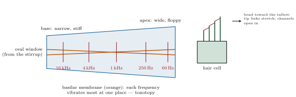
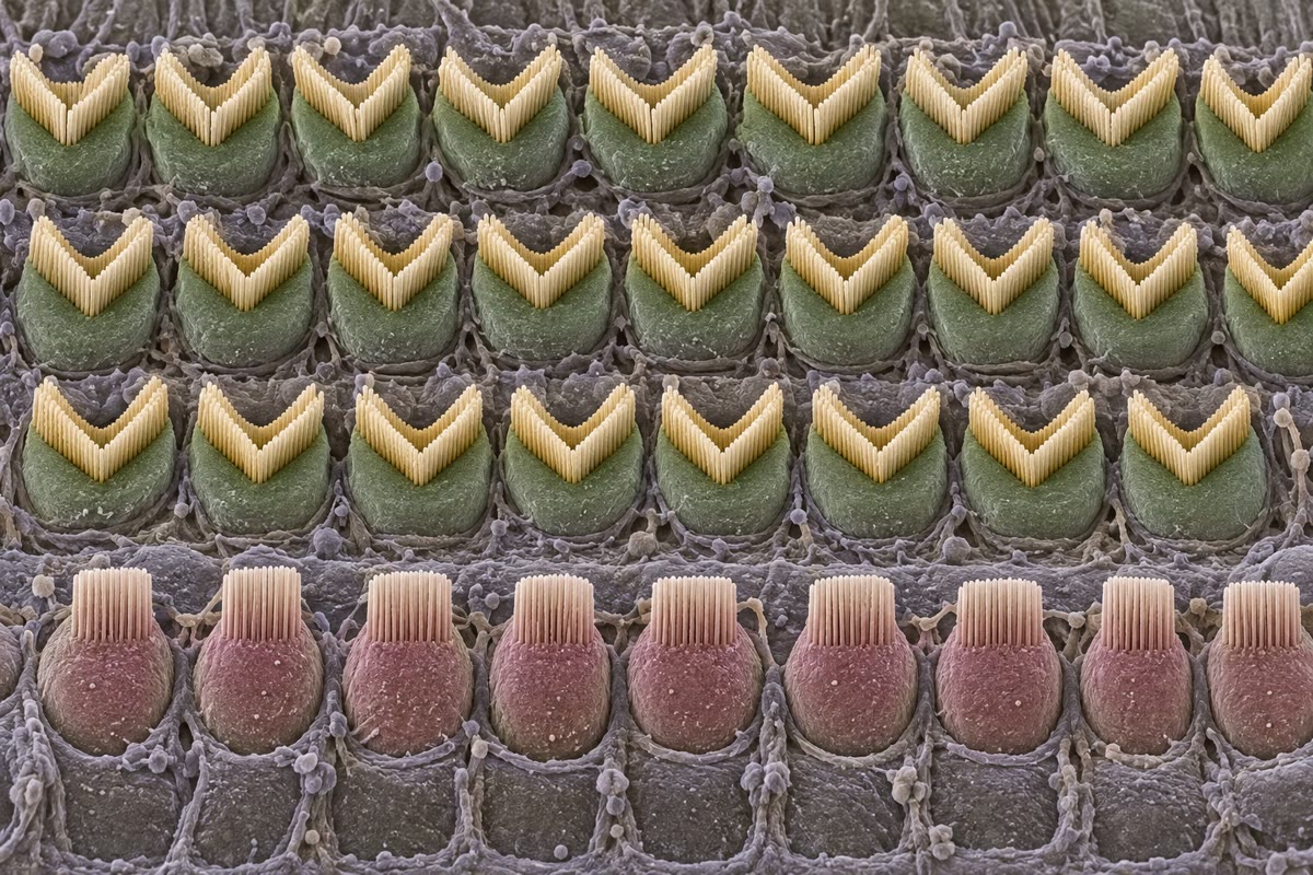
CoachSound is a pressure wave; the ear’s problem is to detect pressure changes of a few tens of micropascals in air and to sort them by frequency. The eardrum and the three middle-ear bones concentrate the force from the drum’s onto the of the oval window, raising the pressure some twentyfold — the impedance match between air and the fluid of the inner ear, without which most of the sound would reflect. Inside the cochlea, a coiled tube long, the pressure wave travels along the basilar membrane, which is narrow and stiff at the base and wide and floppy at the apex; each frequency makes the membrane vibrate most at one place — high frequencies near the base, low near the apex — a tonotopic map that the brain reads as pitch. On the membrane sit the hair cells: inner hair cells in a single row, each crowned with a bundle of stereocilia joined tip to tip by fine tip links; bending the bundle toward its tallest cilium stretches the links and pulls open cation channels within microseconds, without any second messenger, and potassium flows in from the endolymph, whose unusual composition and potential give a driving force of . The inner hair cells signal the auditory nerve; the outer hair cells, driven by the same motion, contract and lengthen with the sound through the motor protein prestin and pump energy back into the membrane’s vibration — an amplifier that sharpens tuning a hundredfold and is what fails in most deafness. Intensity is measured in decibels: , with the threshold of hearing; the ear works over , a trillionfold in intensity.
Examples
Example 18.6 (The ear’s numbers)
At the threshold of hearing the basilar membrane moves about and the pressure amplitude is , a two-billionth of atmospheric; at the pressure is a million times greater and the membrane’s motion tens of nanometres. A young ear hears from to and separates tones apart, the discrimination of the place code sharpened by the outer hair cells. The auditory nerve’s fibres fire in step with the pressure wave up to about (phase locking), carrying timing to a precision of tens of microseconds, from which the brain computes a sound’s direction from the difference in arrival at the two ears — as little as . The balance organs use the same hair cells: in the semicircular canals the fluid’s inertia bends them when the head turns (angular acceleration), and in the otolith organs a layer of calcium carbonate crystals loads them with gravity and linear acceleration.