 Coach
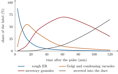
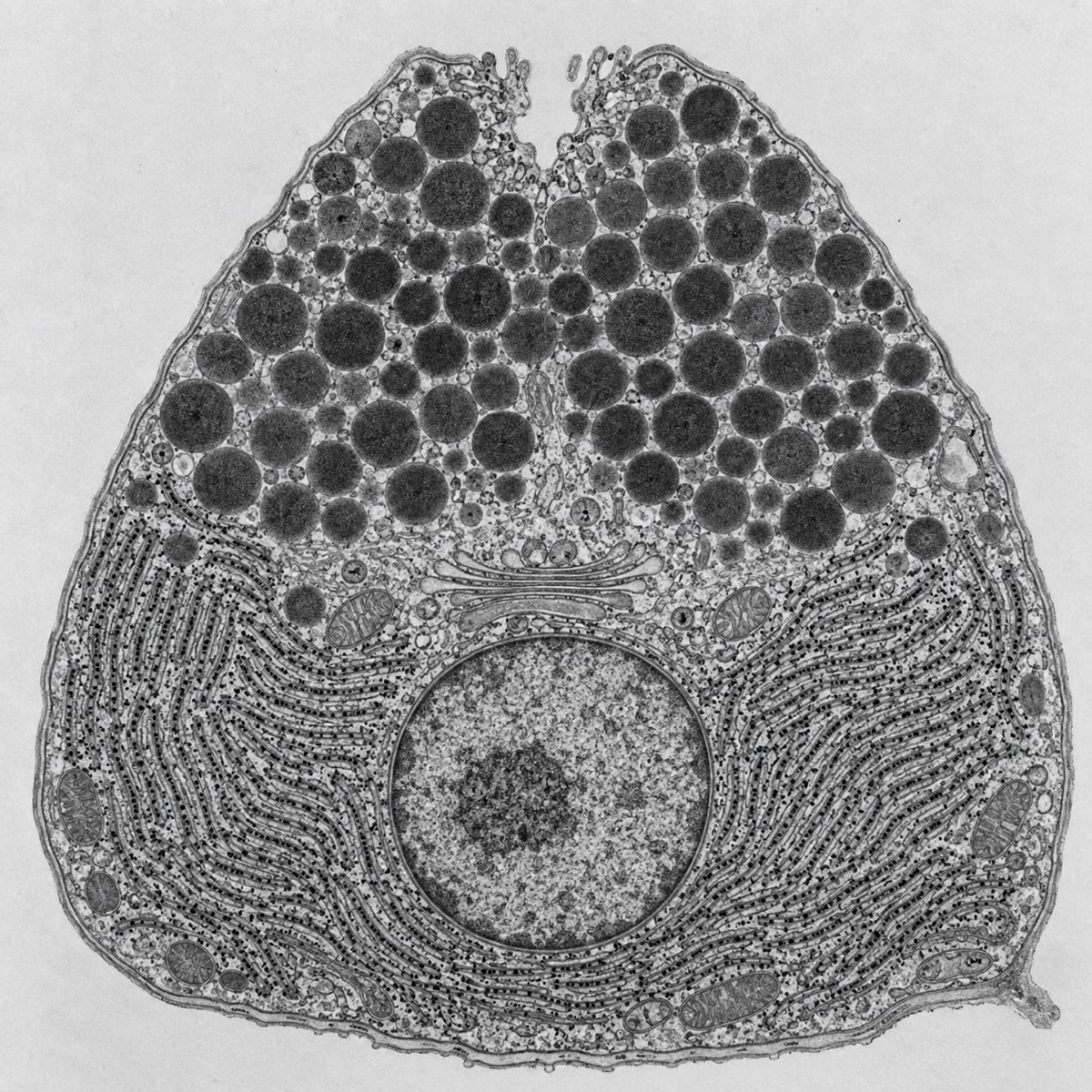
CoachThe endoplasmic reticulum (ER) is a network of membrane sheets and tubes enclosing one continuous space, the lumen, and continuous with the nuclear envelope. Its rough part is studded with ribosomes that inject the proteins they make into the lumen or into the membrane; its smooth part makes lipids, stores calcium and detoxifies. The Golgi apparatus is a stack of flattened sacs (cisternae), with a receiving cis face near the ER and a shipping trans face, in which proteins from the ER are modified (sugars trimmed and added), sorted, and packed into vesicles for their destinations. Lysosomes are acidic vesicles (pH ) full of hydrolytic enzymes that digest material brought in from outside or worn-out organelles. Together with the vesicles that move between them and the plasma membrane, these form the endomembrane system: one connected set of compartments whose lumens are topologically the outside of the cell.
Examples
Example 6.13 (Moving a vesicle)
A secretory vesicle leaving the Golgi is picked up by kinesin, a motor that walks along a microtubule toward the cell’s periphery at about , spending one ATP per step; in a neuron the same motor carries vesicles the length of an axon a metre long — a journey of ten days. Near the surface, the vesicle passes to the actin cortex and to myosin for the last micrometre. Diffusion would take a vesicle of about a minute to cross a cell of , and years to travel a metre: organelles are carried, not left to wander.