University Biology — Year 1 · Bachelor Year 1
6Functional Organization of the Eukaryotic Cell
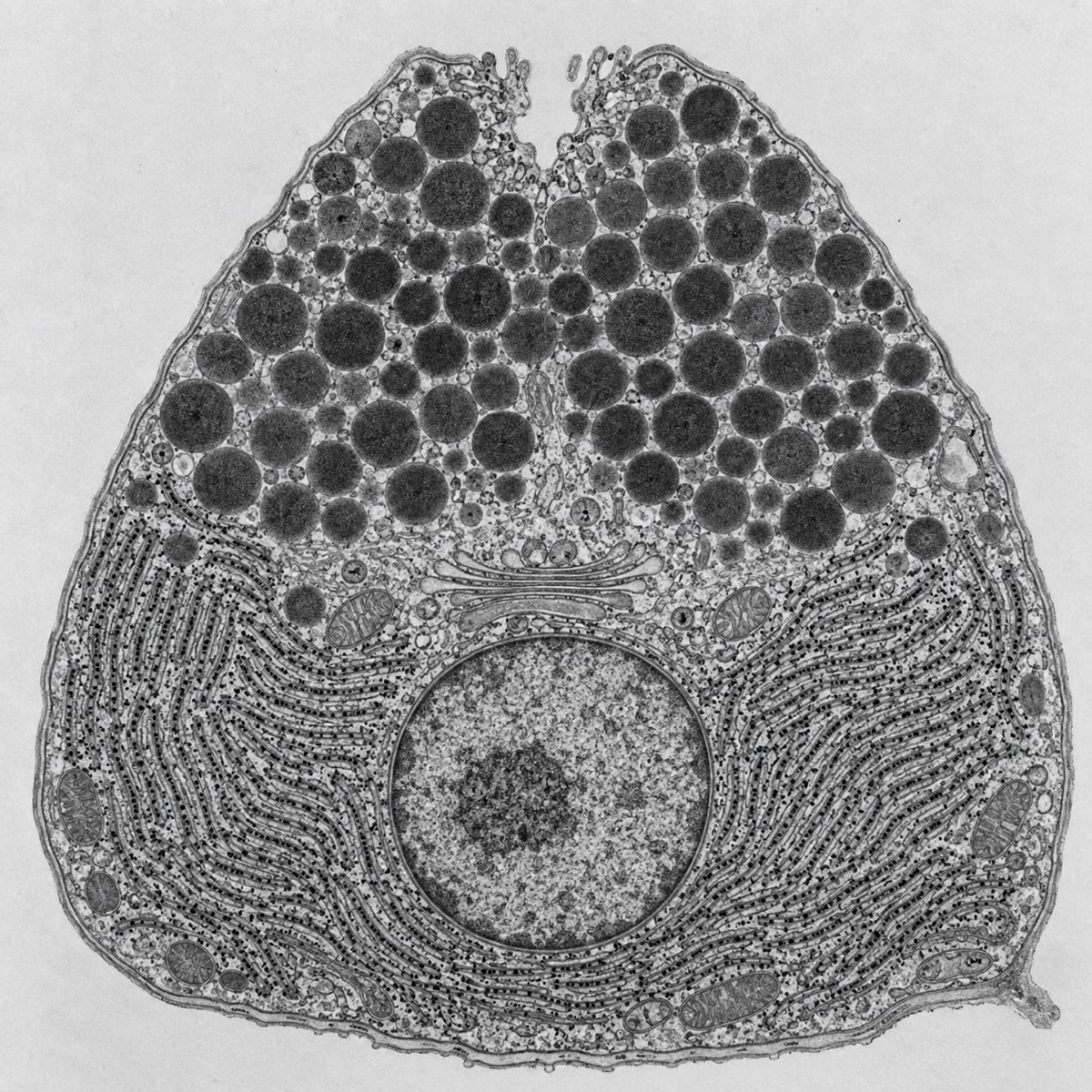
A cell of the pancreas makes, every day, about its own mass of digestive enzymes and pours them into a duct. The enzymes are proteins that would digest the cell that made them; so the cell builds them inside a sealed sheet of membrane, ships them through a second compartment where they are sorted and concentrated, stores them in granules, and releases them only at the signal of a meal. Every step happens in a different membrane-bounded space, and the traffic between the spaces is carried by vesicles. This chapter describes the compartments of the eukaryotic cell, the endomembrane system through which proteins flow, the organelles that transform energy, the skeleton that holds the cell’s shape and moves its parts, and the features that distinguish a plant cell from an animal one.
6.1 Compartments
Definition 6.1 (Organelle, cytosol)
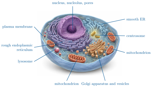
An organelle is a structure inside a cell with a definite composition and function; the membrane-bounded organelles are compartments whose interior differs in composition from the cytosol, the aqueous solution of the cytoplasm outside them. Membrane-bounded compartments: the nucleus, the endoplasmic reticulum, the Golgi apparatus, lysosomes, peroxisomes, mitochondria, and in plants the plastids and the vacuole. Organelles without a membrane: ribosomes, the centrosome, the cytoskeleton.
Definition 6.2 (The nucleus)
The nucleus holds the chromosomes as chromatin, DNA wound on histone proteins (Chapter 17). It is bounded by the nuclear envelope, two membranes, the outer one continuous with the endoplasmic reticulum, pierced by nuclear pores through which RNA and proteins pass in both directions under control. The nucleolus is the region where ribosomal RNA is made and ribosomes are assembled. The nucleus separates transcription (inside) from translation (outside), a separation prokaryotes do not have.
Proposition 6.3 (The compartments of a liver cell)
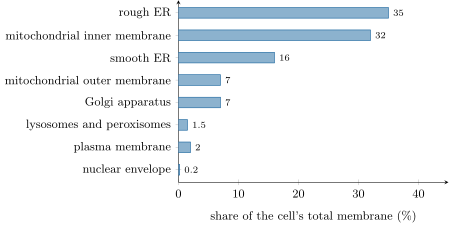
In a hepatocyte of , the cytosol takes about half the volume, the mitochondria a fifth, the endoplasmic reticulum a sixth, the nucleus a sixteenth, the Golgi, lysosomes and peroxisomes a few percent together. The membranes tell a different story: of the of membrane in the cell, half is endoplasmic reticulum, a third the inner membrane of the mitochondria, and only the plasma membrane. Most of a cell’s membrane is inside it.
6.2 The endomembrane system
Definition 6.4 (Endoplasmic reticulum, Golgi apparatus, lysosome)
The endoplasmic reticulum (ER) is a network of membrane sheets and tubes enclosing one continuous space, the lumen, and continuous with the nuclear envelope. Its rough part is studded with ribosomes that inject the proteins they make into the lumen or into the membrane; its smooth part makes lipids, stores calcium and detoxifies. The Golgi apparatus is a stack of flattened sacs (cisternae), with a receiving cis face near the ER and a shipping trans face, in which proteins from the ER are modified (sugars trimmed and added), sorted, and packed into vesicles for their destinations. Lysosomes are acidic vesicles (pH ) full of hydrolytic enzymes that digest material brought in from outside or worn-out organelles. Together with the vesicles that move between them and the plasma membrane, these form the endomembrane system: one connected set of compartments whose lumens are topologically the outside of the cell.
Proposition 6.5 (The secretory pathway)
A protein destined for secretion, for the plasma membrane, or for a lysosome is synthesised by ribosomes bound to the rough ER, enters the ER lumen as it is made, folds there, travels in vesicles to the cis face of the Golgi, crosses the stack while being modified, and leaves the trans face in a vesicle addressed to its destination: a secretory granule that fuses with the plasma membrane on a signal (regulated exocytosis), a vesicle that fuses continuously (constitutive exocytosis, which also delivers new membrane), or a lysosome. Cytosolic and nuclear proteins are made on free ribosomes and never enter the system. What decides is a signal sequence at the start of the protein (Chapter 19).
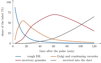
Evidence. Palade (1960s) fed slices of guinea-pig pancreas radioactive amino acids for three minutes (the pulse), then unlabelled ones (the chase), and located the radioactivity by autoradiography of electron micrographs at intervals. At the silver grains lay over the rough ER; at over the Golgi; at over condensing vacuoles and new granules; at in the duct lumen. Cell fractionation gave the same sequence chemically. Blobel (1970s) showed that secretory proteins made in a test tube without membranes are twenty residues longer than the secreted form and are not protected from added protease, whereas made with ER membranes present they are cleaved and protected: the extra residues are the signal that directs the growing chain into the ER. ∎
Method 6.6 (Following a molecule through the cell)
- Label a short cohort: expose cells to a radioactive or fluorescent precursor for a few minutes (the pulse), then replace it by excess unlabelled precursor (the chase), so that only molecules made during the pulse carry the label.
- Sample at intervals, fix, and locate the label: by autoradiography of sections, by fluorescence microscopy, or by fractionation and counting.
- The compartment whose label peaks first is upstream; the sequence of peaks is the route; the intervals are the transit times.
- Block a step (cold, an inhibitor of energy, a mutant) and see where the label accumulates: the compartment before the block.
Definition 6.7 (Endocytosis)
Endocytosis brings material in: the plasma membrane invaginates around it and pinches off a vesicle, which generally fuses with a lysosome. Phagocytosis engulfs particles (a bacterium, a dead cell); pinocytosis takes in droplets of fluid; receptor-mediated endocytosis concentrates a particular molecule (cholesterol-carrying particles, iron-carrying transferrin) on receptors in coated pits before taking it in. Exocytosis adds membrane to the cell surface and endocytosis removes it; in a secreting cell the two balance so that the surface stays constant while whole areas of membrane cycle through it every hour.
6.3 Mitochondria and plastids
Definition 6.8 (Mitochondrion)
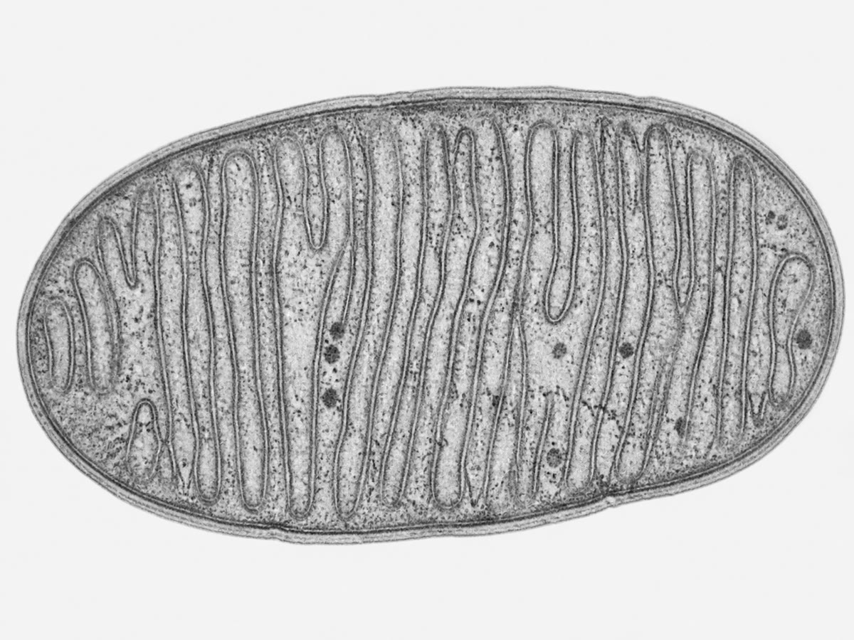
A mitochondrion is an organelle across and a few micrometres long, bounded by two membranes: a smooth outer membrane permeable to small molecules, and an inner membrane folded into cristae that is impermeable to ions and carries the respiratory chain and the ATP synthase (Chapter 15). Inside is the matrix, holding the enzymes of the Krebs cycle, ribosomes of the bacterial type and several copies of a small circular DNA. Mitochondria divide by fission, move along the cytoskeleton, and number from a few hundred to a few thousand per cell.
Definition 6.9 (Plastids)
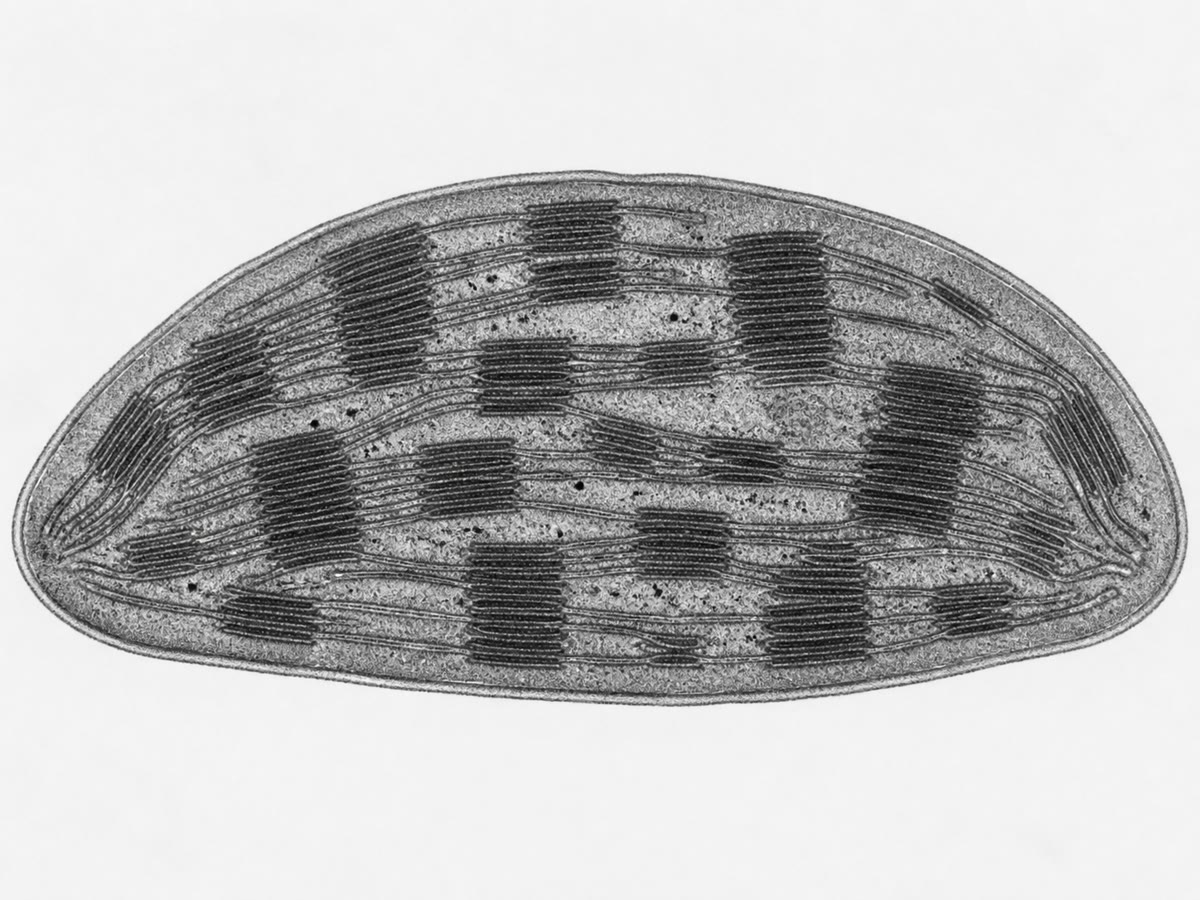
Plastids are the double-membraned organelles of plant cells: chloroplasts, in which a third system of membranes, the thylakoids, stacked in grana and bathed in the stroma, carries out photosynthesis (Chapter 14); amyloplasts, which store starch; chromoplasts, which hold the pigments of fruits and petals. All derive from proplastids and, like mitochondria, contain their own circular DNA and bacterial-type ribosomes and divide by fission.
Proposition 6.10 (Endosymbiotic origin)
Mitochondria and plastids descend from free-living bacteria engulfed by an ancestral eukaryotic cell and retained as symbionts: mitochondria from an aerobic bacterium, plastids from a cyanobacterium.
Evidence. Both organelles have two membranes, the inner of bacterial composition and the outer resembling the host’s; a circular chromosome without histones; ribosomes of the bacterial size and sensitivity to antibiotics; a genetic code with bacterial variants; division by binary fission independent of the cell’s; and their genes, when sequenced, group with those of specific bacterial lineages (alphaproteobacteria, cyanobacteria) rather than with the nuclear genes of the cell that houses them. Most of their original genes have since moved to the nucleus, and the organelles can no longer live alone. ∎
Definition 6.11 (Peroxisome)
A peroxisome is a small single-membraned organelle containing oxidative enzymes that transfer hydrogen from substrates (fatty acids, alcohol) to oxygen, producing hydrogen peroxide, and catalase, which destroys the peroxide on the spot. It confines a dangerous chemistry.
6.4 The cytoskeleton
Definition 6.12 (Cytoskeleton)
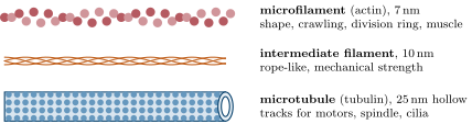
The cytoskeleton is a network of protein filaments that gives the cell its shape, anchors its organelles, and moves them and the cell itself. Three kinds: microfilaments of actin (, two twisted strands of globular subunits), concentrated under the plasma membrane, responsible for cell shape, crawling, the contractile ring of division and muscle contraction with myosin; microtubules ( hollow tubes of tubulin), radiating from the centrosome, tracks along which motor proteins (kinesin outward, dynein inward) carry vesicles and organelles, the spindle of division, and the core of cilia and flagella; intermediate filaments (, rope-like, of keratins and related proteins), the mechanically resistant fibres that stiffen cells and anchor at desmosomes. Microfilaments and microtubules assemble and disassemble continuously from their subunit pools.
Example 6.13 (Moving a vesicle)
A secretory vesicle leaving the Golgi is picked up by kinesin, a motor that walks along a microtubule toward the cell’s periphery at about , spending one ATP per step; in a neuron the same motor carries vesicles the length of an axon a metre long — a journey of ten days. Near the surface, the vesicle passes to the actin cortex and to myosin for the last micrometre. Diffusion would take a vesicle of about a minute to cross a cell of , and years to travel a metre: organelles are carried, not left to wander.
6.5 Plant and animal cells
Definition 6.14 (Cell wall, vacuole, plasmodesmata)
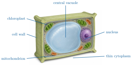
A plant cell adds three structures to the common plan. The cell wall, outside the plasma membrane, is a composite of cellulose microfibrils in a matrix of other polysaccharides (Chapter 10); it fixes the cell’s shape, resists the pressure inside, and cements neighbouring cells together through a shared middle lamella. The vacuole, a single-membraned compartment holding up to of the cell’s volume, stores water, ions, sugars, pigments and wastes; its osmotic pressure pushes the membrane against the wall, and this turgor is what holds a leaf up. Plasmodesmata are channels through the walls, lined by plasma membrane, through which the cytosols of neighbouring cells are continuous, so that a plant tissue is in part one connected cytoplasm (the symplast).
Proposition 6.15 (Plant and animal cell compared)
Both have a nucleus, ER, Golgi, mitochondria, peroxisomes, ribosomes and a cytoskeleton. The plant cell has a cellulose wall, a large vacuole, plastids and plasmodesmata, and no centrioles or lysosomes (the vacuole digests); it does not crawl or engulf, and divides by building a new wall across the middle. The animal cell has no wall (and so can change shape, crawl and phagocytose), lysosomes, a centrosome with centrioles, and communicates through junctions rather than plasmodesmata; it divides by pinching in two.
Example 6.16 (Turgor)
A leaf cell whose vacuole holds of solutes draws water in until the wall pushes back with a pressure of about , seven atmospheres; the cell is then turgid and the leaf firm. Deprived of water, the vacuole loses volume, the pressure falls to zero, and the leaf wilts — the wall unchanged, the pressure gone (Chapter 7). An animal cell in the same solution, having no wall, would simply burst.
6.6 Exercises
Exercise 6.1 ★
List the membrane-bounded organelles of an animal cell and give the main function of each in a few words.
Solution
Solution of Exercise 6.1.
Nucleus (chromosomes, transcription); rough ER (secretory and membrane proteins); smooth ER (lipids, calcium, detoxification); Golgi (sorting, modification, packing); lysosomes (digestion); peroxisomes (oxidations, catalase); mitochondria (respiration, ATP).
Exercise 6.2 ★
Trace the path of a digestive enzyme from its synthesis to the duct, naming each compartment and how the protein passes between them.
Solution
Solution of Exercise 6.2.
Synthesised by ribosomes bound to the rough ER and threaded into its lumen (signal sequence); vesicles bud from the ER and fuse with the cis Golgi; the protein crosses the stack, is modified and sorted; it leaves the trans face in a condensing vacuole that matures into a secretory granule; the granule fuses with the apical membrane on a signal and empties into the duct.
Exercise 6.3 ★
From the membrane-budget figure, what fraction of the cell’s membrane is mitochondrial (outer plus inner)? Why is the inner membrane so much larger than the outer?
Solution
Solution of Exercise 6.3.
. The inner membrane is folded into cristae because it carries the respiratory chain and ATP synthase, whose throughput is proportional to its area; the outer is a simple envelope.
Exercise 6.4 ★
Give three structures a plant cell has that an animal cell lacks, and two the animal cell has that the plant cell lacks.
Solution
Solution of Exercise 6.4.
Plant: cellulose wall, large vacuole, plastids (plasmodesmata too). Animal: lysosomes, centrioles (and the ability to crawl and phagocytose).
Exercise 6.5 ★★
From the pulse-chase figure, at what time does the label peak in the ER, in the Golgi, and in the granules? Estimate the transit time from ER to granule and the time before half the label has been secreted.
Solution
Solution of Exercise 6.5.
ER at (already declining), Golgi at about , granules at about . Transit ER to granule about ; half secreted at about .
Exercise 6.6 ★★
A cell is treated with a drug that depolymerises microtubules. Predict the effects on the position of the Golgi, on vesicle traffic, on cell division and on the beating of cilia.
Solution
Solution of Exercise 6.6.
The Golgi, held near the centrosome by dynein on microtubules, fragments and disperses; vesicle traffic slows to diffusion and transport to the periphery stops; the spindle cannot form and cells arrest in mitosis; cilia, whose core is microtubules, stop (existing cilia are stable, but they are not renewed).
Exercise 6.7 ★★
Give four pieces of evidence for the bacterial origin of mitochondria, and explain why a mitochondrion nevertheless cannot live outside a cell.
Solution
Solution of Exercise 6.7.
Two membranes with a bacterial-type inner one; circular DNA without histones; bacterial-size ribosomes sensitive to antibiotics; division by fission; gene sequences grouping with alphaproteobacteria. It cannot live alone because most of its genes have moved to the nucleus: it imports most of its proteins from the cytosol.
Exercise 6.8 ★★
The lumen of the ER is “topologically outside the cell”. Explain the phrase by following a membrane protein whose sugar chains face the lumen from the ER to the plasma membrane: which side do the sugars face at the end?
Solution
Solution of Exercise 6.8.
The sugars are added on the luminal side in the ER and Golgi. Each vesicle fusion preserves sidedness: the luminal face of a vesicle becomes the luminal face of the next compartment, and when the vesicle fuses with the plasma membrane its lumen opens to the outside, so the sugars face the exterior. What is luminal is, topologically, outside.
Exercise 6.9 ★★
A secretory cell releases granules of diameter per day through an apical surface of . Compute the membrane added per day and how many times the apical surface must be recycled. What does this imply?
Solution
Solution of Exercise 6.9.
Each granule ; per day , i.e. times the apical face. The cell must retrieve membrane by endocytosis at the same rate, so the apical membrane turns over every nine minutes and the retrieved membrane is returned to the Golgi for re-use.
Exercise 6.10 ★★★
In a test tube containing ribosomes, mRNA for a secretory protein and amino acids, the product is a chain of residues; with ER vesicles added, a chain of residues appears inside the vesicles and is not digested by a protease added afterwards. Interpret each observation, and say what a mutant lacking the first twenty residues would do.
Solution
Solution of Exercise 6.10.
The -residue chain is the precursor with its residue signal sequence, which is normally cleaved on entry into the ER. With vesicles, the chain enters the lumen during synthesis, the signal is cleaved, and the membrane protects the product from the protease. The mutant lacking the signal is not recognised by the ER machinery: it is made on free ribosomes and stays in the cytosol, full length, never secreted.
Exercise 6.11 ★★★
A patient’s cells lack a lysosomal enzyme that degrades a lipid; the lipid accumulates in swollen lysosomes and the cells die. Explain the cell biology, say why the disease is worst in cells that renew their membranes fastest, and propose how an enzyme injected into the blood could reach the lysosomes.
Solution
Solution of Exercise 6.11.
Membranes internalised by endocytosis and worn organelles are delivered to lysosomes for digestion; without the enzyme, the lipid they contain cannot be broken down and fills the lysosomes, which swell until the cell fails. Cells with the fastest membrane turnover (neurons, which recycle synaptic vesicles continuously; macrophages) accumulate it fastest. An injected enzyme carrying the sugar signal that lysosomal enzymes bear can bind receptors at the cell surface and be taken up by receptor-mediated endocytosis into the lysosomes — the principle of enzyme replacement therapy.
Exercise 6.12 ★★★
“The eukaryotic cell is a federation of former bacteria held together by a membrane system.” Discuss in a paragraph, weighing what the endosymbiotic theory explains against what it does not (nucleus, ER, cytoskeleton).
Solution
Solution of Exercise 6.12.
Endosymbiosis accounts well for mitochondria and plastids (two membranes, own genome and ribosomes, fission, bacterial relatives), and thus for the eukaryote’s energy metabolism. It does not explain the nucleus, the ER and Golgi, or the cytoskeleton, which have no bacterial counterparts and appear to be inventions of the host lineage — infoldings of its membrane and new proteins. “Federation” is apt for the organelles; “held together by a membrane system” points to the larger, unexplained part: a host cell that was already eukaryotic in its architecture before it took the bacteria in.
6.7 Problem: The Acinar Cell of the Pancreas
Problem 6.1
Weekend problem — a cell that secretes its own mass of protein every day: Palade’s clock, the membrane it must recycle, the signal that routes the protein, ending on the transit time of a digestive enzyme
A pancreatic acinar cell is a cube of side , with its apical face (, on the duct) and its basal face on the blood. A human pancreas of holds about such cells and secretes of enzyme protein per day. A secretory granule is a sphere in diameter holding protein at . Ribosomes add amino acids per second; a typical enzyme has residues of mean mass .
Part I — Palade’s clock. The pulse-chase figure of the chapter gives the location of the label with time.
- Why must the pulse be short and the chase contain a large excess of unlabelled amino acid?
- Read off the time at which the label peaks in the ER, in the Golgi and in the granules.
- Estimate the transit time from ER to Golgi and from Golgi to granule.
- Where is the label at , and why has some of it already left the cell while most of it is still in granules?
- A second experiment is done at : the label stays in the ER for over an hour. Interpret.
Part II — The mass of protein.
- Compute the protein secreted per cell per day, in picograms.
- Compute the volume of one granule and the mass of protein it holds.
- Compute the number of granules released per cell per day, and per minute.
- Compute the cell’s volume and, with a density of , its mass. What fraction of its own mass does it secrete each day?
- Compute the number of enzyme molecules in one granule (Avogadro’s number ).
- Compute the time a ribosome needs to make one enzyme molecule.
- How many ribosomes must work continuously to make the daily output? Compare with the ribosomes such a cell contains.
Part III — Membrane.
- Compute the membrane area of one granule.
- Compute the membrane delivered to the apical face per day by exocytosis of the granules of question 8.
- How many times per day is the area of the apical face added? What must the cell do with it, and by what process?
- The granule membrane arrives from the Golgi, which receives it from the ER. If the ER of the cell has of membrane, how many days’ worth of granule membrane is that? What does this say about recycling within the endomembrane system?
- The apical face carries microvilli of diameter and length. By what factor do they enlarge the face?
Part IV — The address on the protein.
- An enzyme’s mRNA translated in a test tube without membranes gives a chain residues longer than the enzyme found in the granules. Where are the extra residues and what are they?
- With ER vesicles present, the product is the right length, inside the vesicles, and protected from a protease. Interpret.
- The same experiment with the mRNA of a cytosolic enzyme gives a product outside the vesicles and unprotected. Conclude.
- A mutation deletes the twenty residues. Predict where the enzyme ends up and what happens to the cell.
- Another enzyme is found in the lysosomes rather than the granules. It carries the same kind of signal sequence. Where along the pathway must the second decision be taken, and by what mechanism, in outline?
- A cell is treated with a drug that blocks ATP synthesis. Predict where the label of a pulse-chase accumulates and why (two reasons).
- Combine questions 3 and 8: at a steady state, how many granules are “in transit” between ER and granule at any moment?
- Summarise the result: the transit time from synthesis to granule, the time to secretion, the granules released per minute, and the fraction of its own mass the cell secretes daily.
Solution
Solution of Problem 6.1.
1. A short pulse labels a narrow cohort of molecules so their position marks one time; the excess cold amino acid stops further incorporation so the cohort is not smeared by later synthesis. 2. ER: (earliest point); Golgi and condensing vacuoles: about ; granules: about . 3. ER to Golgi about ; Golgi to granule about . 4. Mostly in granules (), a tenth in the Golgi, a tenth already secreted: the constitutive path and the first granules formed release some, while most wait for a stimulus. 5. Vesicle budding and transport require energy and fluid membranes; in the cold the ER-to-Golgi step stops and the protein accumulates upstream of the block. 6. per cell per day. 7. ; protein . 8. granules per day, per minute. 9. ; mass = . Secretes of its mass daily (the whole cell is only protein, so a third of its protein content). 10. Molar mass ; molecules. 11. . 12. Molecules per day ; one ribosome makes per day; needed: ribosomes, about of the cell’s. 13. . 14. . 15. times: the cell retrieves the same area by endocytosis and returns it to the Golgi. 16. days: the ER could supply only five days of granule membrane if none returned; the membrane must recycle continuously. 17. Each microvillus ; for 2000; factor . 18. At the beginning of the chain: the signal sequence, twenty mostly hydrophobic residues. 19. The signal directed the growing chain into the vesicles, where a signal peptidase removed it; the membrane shields the protein from the protease. 20. Only proteins with a signal enter the ER; a cytosolic protein lacks it and stays outside. 21. The enzyme is made on free ribosomes and stays in the cytosol; being a digestive enzyme (as an inactive precursor it does little harm, but if activated) it would digest the cell from inside. 22. In the trans Golgi: the lysosomal enzyme carries a distinguishing mark (a phosphorylated sugar) recognised by a receptor that packs it into vesicles bound for the lysosomes rather than into secretory granules. 23. In the ER (and the Golgi): vesicle formation, motor transport along microtubules and membrane fusion all consume ATP; and protein synthesis itself stops, so the pulse is not even completed. 24. transit at granules per minute: about granules’ worth of protein in transit. 25. Transit from synthesis to granule about ( counting the ER), secretion after about two hours in the experiment (four hours or more in the living gland, since granules wait for a meal); granules released per minute; of the cell’s mass per day.