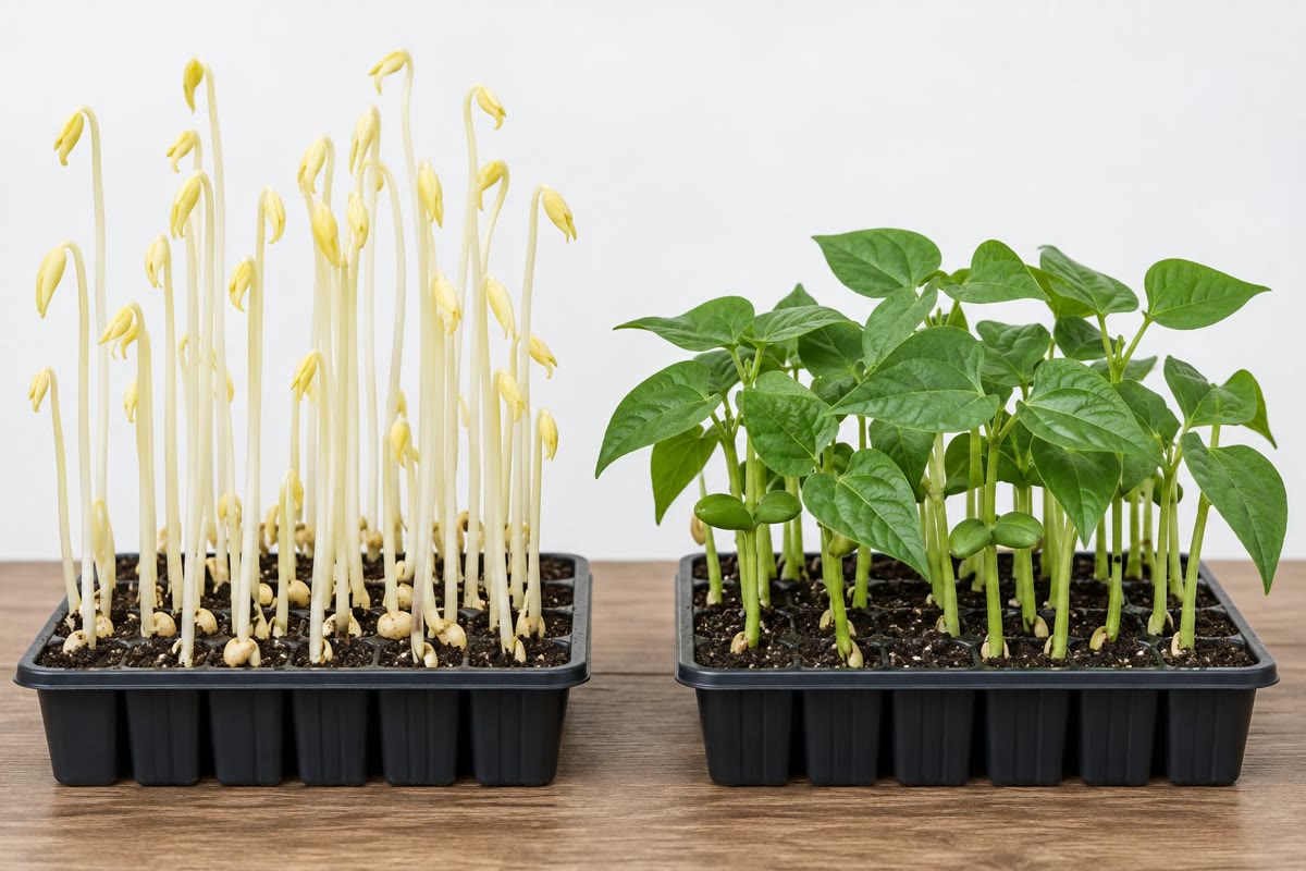
Plants sense light with three families of proteins, none of them chlorophyll. Phytochromes are dimers with a bilin pigment switched by red light (660nm) from the inactive Pr form to the active Pfr, and back by far-red (730nm); Pfr moves into the nucleus and marks a family of transcription factors, the PIFs, for degradation, releasing the genes of light-grown development; in darkness Pfr slowly reverts. Cryptochromes (blue, 450nm) are flavoproteins related to DNA-repair enzymes, controlling de-etiolation, the clock and flowering; phototropins (blue) are membrane kinases that direct bending toward light, the opening of stomata and the movement of chloroplasts. A seedling in the dark is etiolated — long hypocotyl, closed cotyledons, no chlorophyll, all resources spent on reaching the surface; light switches it to photomorphogenesis, and the switch is thrown by Pfr and cryptochrome inactivating a single repressor complex (COP1) that in darkness destroys the transcription factors of the light programme. Light is thus read twice: as energy, by the chloroplast, and as signal, by receptors sensitive to photon fluxes a million times smaller.
Examples
Example 22.5 (Gravitropism and phototropism)
Turn a seedling on its side. In the root cap, dense starch-filled plastids settle onto the new lower side of the columella cells within minutes; the PIN3 carriers of those cells relocate to the lower face; auxin flows preferentially down the lower flank of the root, where, above the optimum for root cells, it inhibits elongation, and the root curves downward. In the shoot the same lateral flow to the lower side promotes elongation (shoot cells’ optimum is higher), and the shoot curves upward. Phototropism: phototropin on the lit side of a shoot alters PIN placement so that auxin accumulates on the shaded side, which grows faster, bending the shoot toward the light. Both movements are slow — hours — because they are growth, not motion, and both are irreversible in the tissue that has grown. Darwin (1880) showed the tip of a grass seedling perceives the light and the region below bends; Went (1928) collected the influence in an agar block placed on a cut tip and showed a block placed asymmetrically on a decapitated shoot made it bend: the influence was a diffusible substance, which was then named auxin.
Example 22.9 (The plant clock and the length of the day)
Plants keep a circadian clock of the same design as the animal one and of unrelated parts: morning factors (CCA1, LHY) repress an evening gene (TOC1) whose product represses them, with further loops that make a robust 24-hour cycle even in constant light, anticipating dawn by opening stomata and switching on photosynthesis genes an hour before the sun. The clock’s most consequential output is the measurement of day length. In Arabidopsis, a long-day plant, the clock makes the CONSTANS protein accumulate in the late afternoon; in a long day that afternoon is still lit, phytochrome and cryptochrome stabilise the protein, and it switches on FT in the leaf; in a short day the protein is made in darkness and destroyed. FT protein, the florigen that grafting experiments had chased since Chailakhyan (1936), travels in the phloem to the shoot apex and, with a partner there, converts the apex from making leaves to making flowers. Short-day plants (rice, soybean) use the same clock with the sign reversed. A plant thus reads the calendar by comparing an internal rhythm with the external light — the “external coincidence” that Bünning proposed in 1936 and that molecular genetics made literal seventy years later.
 Coach
Coach