High School Biology · Grades 10–12
21The Eye and Its Photoreceptors
On a moonless night, away from any lamp, you can see the path but not its colours; a friend’s red jacket is a grey shape. Look straight at a faint star and it vanishes; look slightly to one side and it reappears. Both oddities come from the retina, the thin sheet at the back of the eye where light becomes a nerve signal: two kinds of receptor cell, unequally distributed, one of them blind to colour and the other blind in the dark. This chapter takes the eye apart from the cornea to the optic nerve, and reads, in the genes of the retina’s pigments, a piece of our evolutionary history.
21.1 The eye as an optical instrument
Definition 21.1 (The parts of the eye)
The eye is a globe about across. Light enters through the transparent cornea, passes the pupil — the opening in the coloured iris, which widens in the dark and narrows in bright light — and the lens, crosses the clear jelly of the vitreous body, and reaches the retina, the light-sensitive layer lining the back of the globe. Cornea and lens together form, on the retina, a small inverted image of the scene; the lens, whose curvature muscles can change, adjusts the focus from distant to near objects (accommodation). The optic nerve leaves the back of the eye carrying the retina’s signals to the brain.
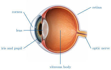
Example 21.2 (Focus, and its faults)
A relaxed eye focuses distant objects on the retina; to read at the lens must bulge. An eye slightly too long focuses distant objects in front of the retina — short sight, corrected by a diverging spectacle lens; an eye too short, or a lens that has stiffened with age, cannot bring near objects to focus — long sight, corrected by a converging lens. In each case the retina is intact: the fault is optical, and glasses put the image back where the receptors are.
21.2 The retina
Definition 21.3 (Photoreceptors)
The retina contains two kinds of photoreceptor cells, named for their shape. The rods, about 120 million, respond to very dim light but all with the same pigment: they give vision in shades of grey at night. The cones, about 6 million, need brighter light and come in three types, each with a pigment most sensitive to a different part of the spectrum: they give daylight vision and colour. Each photoreceptor contains a photopigment — a protein, an opsin, holding a small light-absorbing molecule derived from vitamin A — whose change of shape on absorbing a photon starts the cell’s electrical response.
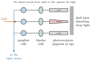
Proposition 21.4 (Organisation of the retina)
The photoreceptors lie at the back of the retina, against a dark layer; the light must cross the other layers to reach them. Their signals pass to bipolar cells, then to ganglion cells, whose long fibres — about a million — gather into the optic nerve. The distribution is uneven: at the fovea, the small central pit the eye points at, there are only cones, packed tightly and each connected to its own ganglion cell, giving the sharpest vision; away from the centre, rods dominate and many of them share one ganglion cell, giving sensitivity at the price of detail. Where the optic nerve leaves, there are no receptors at all: the blind spot.
Proof. Admitted at this level. ∎
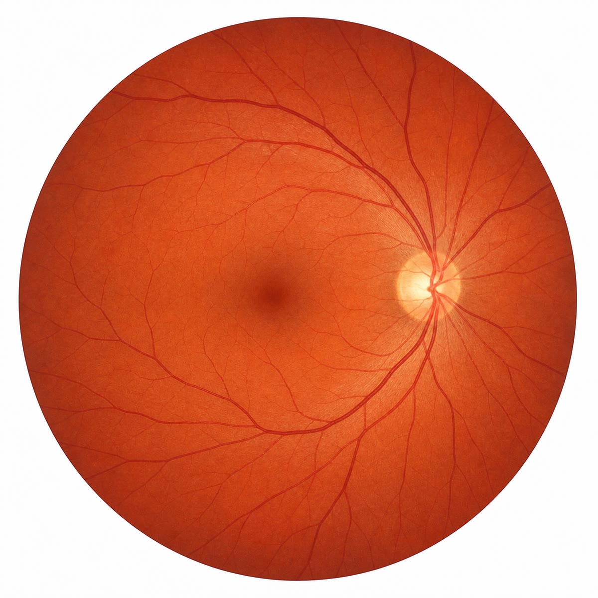
Example 21.5 (The two oddities explained)
The faint star vanishes when looked at directly because the fovea has only cones, which need more light than the star provides; looked at sideways, its light falls on rods, which detect it. The jacket is grey at night because the rods that see it have a single pigment: they can report how much light, not which wavelength. Colour is a comparison between the three cone types, and the cones are silent in the dark.
21.3 Colour: three pigments
Proposition 21.6 (Colour vision)
Each cone type contains one of three opsins, whose absorption peaks near (S cones, blue), (M cones, green) and (L cones, red). A given light excites the three types in proportions that depend on its wavelength; the brain reads the colour from those proportions. A person lacking one cone type confuses the colours that type distinguished: without working L or M cones, red and green are hard to tell apart — red–green colour blindness, which affects about 8% of men and 0.5% of women.
Proof. Admitted at this level. ∎
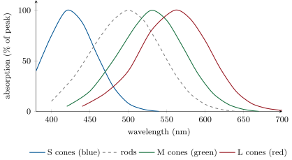
Example 21.7 (Reading the curves)
Light of : S cones at 8%, M at 75%, L at 40% — seen as blue-green. Light of : S at 0, M at 10%, L at 42% — red. A person without L cones receives, for both, only an S and an M value, and the second light gives S 0, M 10: barely distinguishable from a dim green. The colours that the missing type would have told apart collapse into one.
21.4 The opsin genes: a family with a history
Proposition 21.8 (Genes of the opsins)
Each opsin is encoded by its own gene. The S-opsin gene lies on an ordinary chromosome; the M- and L-opsin genes lie side by side on the X chromosome. The L and M proteins differ at only 15 of their 364 amino acids (96% identical), the S protein at more than half of its positions from either. Being on the X, a non-working M or L allele is expressed in every male who carries it and only in females carrying two — the pattern of Chapter 16, and the reason red–green colour blindness is sixteen times commoner in men.
Proof. Admitted at this level. ∎
Proposition 21.9 (A family of genes from duplications)
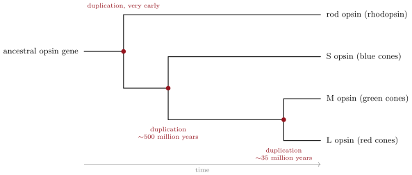
The three opsin genes, and the rod pigment’s, are versions of one ancestral gene copied by duplication and then diverged by mutation. Their degrees of similarity date the copies: the S and L/M lines separated very early in vertebrate history, some 500 million years ago; the L and M genes arose from one duplication about 30 to 40 million years ago in the ancestor of the Old World primates, whose descendants — monkeys, apes, humans — have three cone types while most other mammals have two.
Evidence. Sequence comparison: the L and M genes are 96% identical and adjacent on the X — the signature of a recent tandem duplication; each is about 43% identical to the S gene, and all three about 40% identical to the rod opsin gene, the more distant copies. Dogs, cattle and most mammals have only one X-linked opsin gene, in the position where primates have two; New World monkeys have one, with several alleles, and only females carrying two different alleles see three colours. The gene count matches the cone count in each lineage, and the tree of the genes matches the tree of the species. ∎
Example 21.10 (What one duplication changed)
A monkey with two cone types sees the forest in two dimensions of colour, and ripe fruit is hard to pick out of foliage. After the L/M duplication, one copy could mutate towards longer wavelengths while the other kept the original — a third cone, and ripe red against green. The mutation that later breaks one copy in 8% of men simply returns the eye to the ancestral two-cone state.
Method 21.11 (Reasoning about a colour-vision case)
- Which cone type is missing or altered? Read it from the colours confused (red–green: L or M; blue–yellow, rare: S).
- Which gene, and on which chromosome? L and M on the X, S elsewhere.
- Apply the inheritance: X-linked recessive for red–green — sons of carrier mothers affected one time in two, daughters of affected fathers all carriers.
- Relate to the family of genes: a lost copy, or an L and M copy made too alike by an exchange between the adjacent genes.
Remark 21.12 (From the eye to the brain)
The retina does not see; it converts and begins to sort. The million fibres of the optic nerve carry a coded description — contrasts, edges, ratios of cone responses — to the brain, where the next chapter picks it up. What we call seeing happens there, built on the signals of the cells described here and on nothing else: a person whose retinas work but whose visual brain is damaged is blind, and one whose visual brain works but whose photoreceptors have died is blind too.
21.5 Exercises
Exercise 21.1 ★
List, in order, the structures light crosses from the outside to the photoreceptors.
Solution
Solution of Exercise 21.1.
Cornea, pupil (through the iris), lens, vitreous body, then the transparent layers of the retina (ganglion and bipolar cells) to the photoreceptors at the back.
Exercise 21.2 ★
Compare rods and cones: number, sensitivity, pigments, kind of vision.
Exercise 21.3 ★
What are the fovea and the blind spot?
Solution
Solution of Exercise 21.3.
The fovea is the central pit of the retina, made only of tightly packed cones, where vision is sharpest. The blind spot is where the optic nerve leaves, with no photoreceptors.
Exercise 21.4 ★
From the absorption figure, which pigments absorb light of , and how strongly?
Exercise 21.5 ★
On which chromosome are the L- and M-opsin genes, and what follows for the inheritance of red–green colour blindness?
Solution
Solution of Exercise 21.5.
On the X chromosome; the condition is X-linked recessive: expressed in every male carrying the allele, in females only with two copies, so it is far commoner in men.
Exercise 21.6 ★★
Explain why we cannot see colours at night, and why a faint star is better seen sideways.
Exercise 21.7 ★★
Two lights excite the cones as follows: light 1, S 0, M 60, L 100; light 2, S 0, M 30, L 50. Are they the same colour? The same brightness? Explain.
Solution
Solution of Exercise 21.7.
Same colour: the ratios S:M:L are 0:0.6:1 in both. Different brightness: light 2 excites every cone half as much. Colour is the ratio; brightness is the total.
Exercise 21.8 ★★
A colour-blind man marries a woman with no colour-blind relatives. Give the colour vision of their sons, their daughters, and their daughters’ sons.
Solution
Solution of Exercise 21.8.
Sons receive their X from the mother: all normal. Daughters receive the father’s X: all carriers, normal vision. Each carrier daughter’s sons are colour-blind with probability one half.
Exercise 21.9 ★★
Why is red–green colour blindness sixteen times commoner in men than in women? Compute the expected frequency in women from 8% in men.
Solution
Solution of Exercise 21.9.
A man needs one mutant X, a woman two. If the allele’s frequency is 0.08, a woman carries two with probability , about 0.6% — sixteen times less than 8%.
Exercise 21.10 ★★
The L and M genes are 96% identical, each 43% identical to S. Explain how these two numbers date the duplications relative to each other.
Solution
Solution of Exercise 21.10.
Differences accumulate with time since a duplication: 4% between L and M means a recent copy; 57% between either and S means a much older one. The L/M duplication is the latest event, the S split far earlier.
Exercise 21.11 ★★
Most mammals have two cone types, Old World primates three. Explain the difference with a duplication, and say why it may have been kept.
Solution
Solution of Exercise 21.11.
The ancestor of Old World primates duplicated its single X-linked opsin gene; one copy shifted its peak to longer wavelengths, giving a third cone type and a new dimension of colour. It was kept, probably because it helped find ripe fruit and young leaves against foliage.
Exercise 21.12 ★★★
A person lacks the S cones. Which colours does he confuse? Explain why this condition is rare and equally frequent in both sexes.
Solution
Solution of Exercise 21.12.
Blues and yellows (and blue-green from green). The S gene is on an ordinary chromosome, so both alleles must be mutant in either sex — rare and sex-independent.
Exercise 21.13 ★★★
Cones at the fovea are apart and the eye’s focal length is about . Compute the smallest angle two points can subtend and still fall on separate cones, and the size of the smallest detail resolvable at . (Use the small-angle approximation: angle in radians size distance.)
Solution
Solution of Exercise 21.13.
, about half a minute of arc. At : .
Exercise 21.14 ★★★
In New World monkeys the single X-linked opsin gene has several alleles of different peak wavelengths. Explain why only some females of those species see three colours, and no male.
Solution
Solution of Exercise 21.14.
A male has one X, hence one allele, hence one M/L-type cone: two cone types with S. A female with two different alleles expresses one in some cones and the other in others: three types, hence three-colour vision; a female with two identical alleles has two types like a male.
Exercise 21.15 ★★★
A disease destroys the photoreceptors but spares the rest of the retina. Explain why an implant that stimulates the ganglion cells electrically can restore some vision, and what limits its quality.
Solution
Solution of Exercise 21.15.
The ganglion cells and the optic nerve are intact, so electrical pulses delivered to them reach the visual brain as signals. Quality is limited by the number of electrodes (hundreds, against a million fibres and millions of receptors) and by the loss of the retina’s own sorting of the image.
21.6 Problem: Three Pigments and One Duplication
Problem 21.1
Weekend problem — colour vision taken apart: the cone responses to a spectrum, a family with colour blindness, the opsin genes compared, and the sharpness a fovea can reach
Use the absorption figure. A test presents lights of , , and .
Part I — The cones respond.
- For each light, read the approximate response of the S, M and L cones.
- Which light excites all three types? Which excites only one?
- A person lacking L cones receives only the S and M values. Which two of the four lights become hard to distinguish for him? Justify with the numbers.
- A person lacking M cones: which pair does she confuse?
- Explain why neither person is "blind to red" but both are poor at telling red from green.
Part II — A family. Nora sees colours normally; her father is red–green colour-blind. She marries Sam, who sees normally.
- Give Nora’s genotype for the X-linked opsin gene, and justify.
- Give the genotypes and colour vision of their possible sons and daughters, with probabilities.
- Their daughter Ines, whose colour vision is normal, marries a colour-blind man. Compute the probability that Ines is a carrier, then the probability that a daughter of theirs is colour-blind.
- Explain why a colour-blind daughter always has a colour-blind father.
- Nora’s father’s mother saw colours normally. Was she necessarily a carrier? Explain.
Part III — The genes. Identities between opsin protein sequences: L–M 96%, L–S 43%, M–S 43%, S–rhodopsin 41%.
- Draw, or describe, the tree of the four genes implied by these numbers.
- If differences accumulate at a steady rate and the L/M duplication is 35 million years old, estimate the age of the S versus L/M split.
- The L and M genes lie side by side on the X. Explain how an unequal exchange between them during meiosis can produce an X carrying only one of the two, and what colour vision results.
- Such exchanges are the commonest cause of red–green colour blindness. Why are two adjacent, nearly identical genes especially prone to them?
- The L/M duplication is found in all Old World primates and in no other mammal. What does this say about when and in whom it occurred?
Part IV — The sharpness of the fovea. Foveal cones are apart; the eye’s focal length is . Use angle (radians) size distance.
- Compute the angle subtended by two neighbouring cones, in radians and in minutes of arc ( rad).
- What is the smallest letter detail resolvable at ? Compare with the strokes of the letters on an eye-test chart, about at that distance for "normal" vision.
- Away from the fovea, 100 rods share one ganglion cell. Estimate the resolvable angle there, and explain what is gained in exchange.
- An eagle’s foveal cones are twice as densely packed and its eye slightly larger. By what factor is its resolution finer?
- State the result: the three cone types and the light each is blind to, the single genetic event that gave primates the third, and the angle the fovea resolves.
Solution
Solution of Problem 21.1.
1. : S 40, M 35, L 18. : S 0, M 100, L 80. : S 0, M 40, L 90. : S 0, M 2, L 12.
2. excites all three; essentially only L.
3. With S and M only: gives (0, 40) and gives (0, 2); gives (0, 100). The (orange) and (green) lights differ only in M intensity, like a dim and a bright green, and are the hard pair; looks like a very dim light.
4. With S and L only: gives (0, 80) and gives (0, 90) — nearly identical: green and orange are confused.
5. Both still detect red light, with the cone type they keep; what is lost is the comparison between M and L responses, which is what separates red from green.
6. : her father gave her his only X, which carries the mutant allele; her normal vision shows the other X is normal.
7. Sons: normal or colour-blind, one half each. Daughters: or , both normal vision, one half each (the second carriers).
8. Ines is a carrier with probability . A daughter is colour-blind if she receives from both parents: from the father certainly, from Ines with probability : .
9. A colour-blind daughter has two mutant X chromosomes, one from each parent; a father passes his only X to every daughter, so his X is mutant and he is colour-blind.
10. Yes: her son received his X from her, and it carries the mutant allele; with normal vision, she was .
11. Rhodopsin branches off first; then S separates from the L/M line; L and M split last: (rhodopsin, (S, (L, M))).
12. 4% difference for 35 million years; 57% would take about million years — consistent with the early vertebrate origin (the relation is not exactly proportional at large differences, so this is an order of magnitude).
13. The two nearly identical adjacent genes can misalign during the pairing of the two X chromosomes; an exchange then gives one X with three copies and one with a single copy. A male receiving the single-copy X has only one M/L-type cone: red–green colour blindness.
14. Pairing relies on sequence similarity: two adjacent 96% identical genes can pair out of register, gene 1 with gene 2, and exchange unequally.
15. It occurred once, in the common ancestor of the Old World primates, after their separation from other mammals and New World monkeys, some 35 million years ago, and was inherited by all their descendants.
16. rad, about .
17. : finer than the strokes, which is why normal vision reads that line comfortably (in practice optical blur brings the limit close to ).
18. Ten times coarser at least (100 rods share one signal, about 10 by 10): several minutes of arc; in exchange, a hundred receptors pool their light and detect far dimmer sources.
19. Twice the density is closer spacing, and a larger eye adds a longer focal length: about twice as fine.
20. S, M and L cones, each nearly blind to the far end of the spectrum away from its peak; one tandem duplication of the X-linked opsin gene, 35 million years ago, gave primates the third; the fovea resolves about half a minute of arc.