 Coach
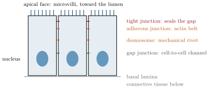
CoachAn epithelium is a sheet of closely packed cells covering a surface or lining a cavity, resting on a thin layer of extracellular matrix, the basal lamina. Its cells are polarised: the apical face looks at the outside or the lumen, the basal face at the lamina, and the two carry different proteins. They are joined by cell junctions: tight junctions near the apex, which seal the space between cells so that nothing crosses the sheet except through the cells; adherens junctions and desmosomes, which fasten the cells mechanically; gap junctions, channels that let ions and small molecules pass from cell to cell. Epithelia are classified by the number of layers (simple, stratified) and the shape of the cells (squamous, cuboidal, columnar), and include the glands, which are epithelial cells specialised in secretion. They cover, protect, absorb and secrete.
Examples
Example 4.12 (Where the tissues come from)
Epithelia derive from all three germ layers (skin from ectoderm, gut lining from endoderm, the lining of vessels and of the coelom from mesoderm); connective tissue, muscle and blood from mesoderm; nervous tissue from ectoderm. The frog’s skin is an ectodermal epithelium on mesodermal connective tissue; its gut, an endodermal epithelium wrapped in mesodermal muscle; its brain, ectoderm folded inward.
Example 4.15 (The villus as an exchange surface)
A villus of the human small intestine is a finger high and across, covered by a single layer of columnar cells whose apical faces each carry some microvilli. Villi multiply the surface of the tube about sevenfold and microvilli twentyfold again: the of a plain tube becomes tens of square metres of absorptive epithelium, one cell thick, each cell within a few micrometres of a capillary and a lymphatic in the connective core of its villus (Chapter 22). The epithelium is renewed from the glands at its base every four days: the sheet, its junctions and its polarity are rebuilt continuously.