University Biology — Year 1 · Bachelor Year 1
4Animal Body Plans and Tissues
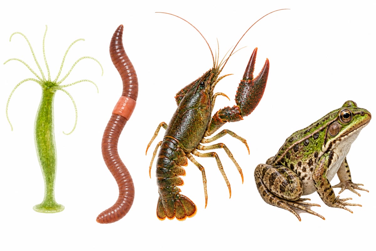
Four animals on a laboratory bench: a hydra a centimetre long, an earthworm, a crayfish, a frog. Under the differences of size and habit they are built on a handful of shared decisions — whether the body has a front and a back, how many layers of cells it grew from, whether there is a cavity around the gut, whether the body is made of repeated segments, where the skeleton is — and every one of them is built from the same four kinds of tissue. This chapter sets out the body plans of animals and the tissues that build them, and ends where the previous chapters left off: at the wall of a mammalian gut, seen as tissues assembled into an organ.
4.1 Body plans
Definition 4.1 (Symmetry and cephalisation)
An animal has radial symmetry when its body is organised around a central axis with no left and right, like a hydra or a jellyfish: it meets its environment equally from all sides, and is usually fixed or drifting. It has bilateral symmetry when one plane divides it into mirror halves, defining a front (anterior) and rear (posterior), a back (dorsal) and a belly (ventral): the plan of an animal that moves head first, with sense organs and nervous centres concentrated at the front (cephalisation).
Definition 4.2 (Germ layers, body cavities)
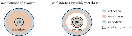
The embryo of an animal forms two or three layers of cells, the germ layers, from which all tissues derive: the outer ectoderm (skin, nervous system), the inner endoderm (lining of the gut and its outgrowths) and, in all but the sponges and cnidarians, a middle mesoderm (muscle, skeleton, connective tissue, blood, kidneys, gonads). Cnidarians are diploblastic, the rest triploblastic. Among triploblastic animals, the mesoderm may fill the space between gut and body wall (acoelomate: flatworms), or leave a cavity between them: a coelom when the cavity is entirely lined by mesoderm (annelids, molluscs, arthropods, echinoderms, vertebrates). The coelom suspends the gut in its own compartment, lets it move independently of the body wall, and gives the soft-bodied a hydrostatic skeleton.
Definition 4.3 (Protostomes and deuterostomes)
Among coelomate animals, the fate of the first opening of the embryonic gut divides two great lineages: in protostomes (annelids, molluscs, arthropods) it becomes the mouth, the coelom forms by splitting of the mesoderm, and the early cleavages of the egg are spiral and their fate fixed early; in deuterostomes (echinoderms, vertebrates) it becomes the anus, the coelom forms by outpocketing of the gut, and the early cells remain flexible in fate.
Definition 4.4 (Segmentation, skeletons)
A body is segmented when it is built of repeated units along its axis (the rings of an earthworm, the segments of a crayfish, the vertebrae and muscle blocks of a fish), each with its own set of muscles, nerves and often appendages. Segments may be alike or specialised into regions (head, thorax, abdomen). The skeleton, against which the muscles pull, is hydrostatic (a fluid under pressure in a coelom, as in the earthworm), an exoskeleton (a rigid cuticle outside the body, jointed, moulted to grow, as in arthropods), or an endoskeleton (cartilage or bone inside the body, growing with it, as in vertebrates).
Proposition 4.5 (Four plans compared)
| hydra | earthworm | crayfish | frog | |
|---|---|---|---|---|
| symmetry | radial | bilateral | bilateral | bilateral |
| germ layers | 2 | 3 | 3 | 3 |
| coelom | none | yes | reduced | yes |
| lineage | — | protostome | protostome | deuterostome |
| segments | no | alike | specialised | vertebrae, muscle blocks |
| skeleton | hydrostatic | hydrostatic | exoskeleton | endoskeleton |
| gut | one opening | mouth to anus | mouth to anus | mouth to anus |
| circulation | none | closed | open | closed, heart |
| nervous system | net | ventral cord | ventral cord | dorsal cord |
The plans are not steps on a ladder: each is a complete and successful way of being an animal, and the crayfish and the frog are each other’s contemporaries, not ancestors.
Example 4.6 (Reading an animal from its plan)
An animal with a head end, a through gut, a body of similar rings and no hard parts: a coelomate protostome moving by a hydrostatic skeleton, each segment’s coelomic fluid pressurised by circular and longitudinal muscles in turn — an earthworm burrowing. An animal with a jointed cuticle and specialised segments: an arthropod, whose growth must pass through moults. Bilateral, segmented muscle blocks around a dorsal axis of vertebrae: a vertebrate, whose endoskeleton grows continuously.
4.2 The four tissue types
Definition 4.7 (Tissue)
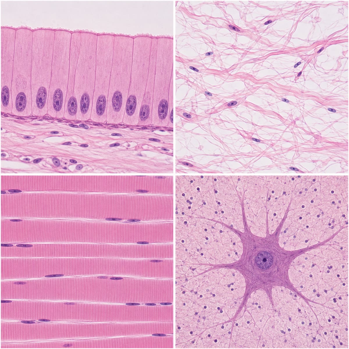
A tissue is a group of cells of similar structure and origin, together with the extracellular material they produce, organised to perform a common function. Animal tissues fall into four types: epithelial, connective, muscle, nervous. Every organ is a combination of at least two of them.
Definition 4.8 (Epithelial tissue)
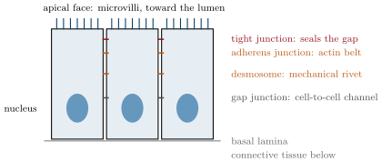
An epithelium is a sheet of closely packed cells covering a surface or lining a cavity, resting on a thin layer of extracellular matrix, the basal lamina. Its cells are polarised: the apical face looks at the outside or the lumen, the basal face at the lamina, and the two carry different proteins. They are joined by cell junctions: tight junctions near the apex, which seal the space between cells so that nothing crosses the sheet except through the cells; adherens junctions and desmosomes, which fasten the cells mechanically; gap junctions, channels that let ions and small molecules pass from cell to cell. Epithelia are classified by the number of layers (simple, stratified) and the shape of the cells (squamous, cuboidal, columnar), and include the glands, which are epithelial cells specialised in secretion. They cover, protect, absorb and secrete.
Definition 4.9 (Connective tissue)
Connective tissue is made of scattered cells in an abundant extracellular matrix that they secrete: protein fibres — collagen (tensile strength; a quarter of all the protein in a mammal), elastin (recoil) — embedded in a hydrated ground substance of polysaccharides. The proportions define its kinds: loose connective tissue under epithelia and around organs (fibroblasts, few fibres, much ground substance); dense connective tissue of tendons and ligaments (parallel collagen); adipose tissue (fat-storing cells); cartilage (a firm gel of collagen and polysaccharides, no vessels); bone (collagen mineralised with calcium phosphate, living cells in cavities, vessels); and blood, whose matrix is the plasma. It binds, supports, stores, transports and defends.
Definition 4.10 (Muscle tissue)
Muscle tissue is made of elongated cells packed with contractile protein filaments (actin and myosin). Three kinds: skeletal muscle, of long multinucleate fibres showing cross-striations, attached to the skeleton and under voluntary control; cardiac muscle, of branched striated cells joined end to end by junctions that pass the beat from cell to cell, contracting on its own rhythm; smooth muscle, of spindle-shaped unstriated cells in the walls of hollow organs (gut, vessels, uterus), contracting slowly and involuntarily. The mechanism of contraction is treated in the Year 2 volume.
Definition 4.11 (Nervous tissue)
Nervous tissue is made of neurons, cells with a body and long extensions (dendrites receiving signals, an axon carrying them away) that conduct electrical signals and pass them to other cells at synapses, and of glial cells, several times more numerous, which support, insulate and feed the neurons. It senses, integrates and commands.
Example 4.12 (Where the tissues come from)
Epithelia derive from all three germ layers (skin from ectoderm, gut lining from endoderm, the lining of vessels and of the coelom from mesoderm); connective tissue, muscle and blood from mesoderm; nervous tissue from ectoderm. The frog’s skin is an ectodermal epithelium on mesodermal connective tissue; its gut, an endodermal epithelium wrapped in mesodermal muscle; its brain, ectoderm folded inward.
4.3 From tissues to an organ: the wall of the gut
Proposition 4.13 (The four layers of the gut wall)
From the lumen outward, the wall of the vertebrate digestive tube is built of four concentric layers, each a combination of tissues: the mucosa (an epithelium — absorptive and secretory — on a loose connective tissue carrying vessels and lymphatics, with a thin sheet of smooth muscle beneath); the submucosa (dense connective tissue with larger vessels and a nerve network); the muscularis (two layers of smooth muscle, inner circular and outer longitudinal, with a nerve network between them, producing the mixing and propulsive movements); and the serosa (a thin connective layer covered by the epithelium of the coelom). The same four layers run from oesophagus to rectum; what changes is the mucosa — folded into villi in the small intestine, glandular in the stomach.
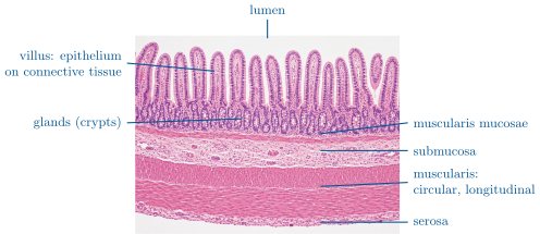
Method 4.14 (Reading a histological section)
- Find the free surface or the lumen: the epithelium lining it is the first layer. Count its rows of nuclei (simple or stratified) and note the shape of the cells.
- Below the epithelium look for the pale, fibrous zone with scattered nuclei and vessels: connective tissue.
- Muscle appears as dense bundles of elongated cells: striated with peripheral nuclei (skeletal), striated and branched (cardiac), unstriated with a central nucleus (smooth). Note the direction of the fibres relative to the section.
- Nervous tissue in an organ wall appears as clusters of large pale neuron bodies between the muscle layers.
- Name the organ from the sequence of layers and the specialisations of the mucosa (villi, glands, keratin).
Example 4.15 (The villus as an exchange surface)
A villus of the human small intestine is a finger high and across, covered by a single layer of columnar cells whose apical faces each carry some microvilli. Villi multiply the surface of the tube about sevenfold and microvilli twentyfold again: the of a plain tube becomes tens of square metres of absorptive epithelium, one cell thick, each cell within a few micrometres of a capillary and a lymphatic in the connective core of its villus (Chapter 22). The epithelium is renewed from the glands at its base every four days: the sheet, its junctions and its polarity are rebuilt continuously.
Remark 4.16 (The tissues of the two model organisms)
The plant’s three tissue systems of Chapter 3 have no exact counterparts here. Its epidermis is an epithelium in function but its cells are held by walls, not junctions; its ground tissue is at once storage, support and photosynthetic; it has neither muscle nor nerve. The animal’s four tissues are the tool-kit of a body that moves and senses; the plant’s three, of a body that grows.
4.4 Exercises
Exercise 4.1 ★
Define radial and bilateral symmetry and give an animal of each. Which goes with cephalisation, and why?
Solution
Solution of Exercise 4.1.
Radial: organised around an axis, no left and right (hydra, jellyfish). Bilateral: one mirror plane, front and rear, back and belly (earthworm, frog). Cephalisation goes with bilateral symmetry: an animal that moves head first meets the world at its front, where sense organs and nervous centres concentrate.
Exercise 4.2 ★
Name the three germ layers and one tissue derived from each.
Solution
Solution of Exercise 4.2.
Ectoderm: skin epidermis, nervous tissue. Mesoderm: muscle, bone, blood, connective tissue. Endoderm: the lining of the gut.
Exercise 4.3 ★
List the four tissue types with their defining feature and one function each.
Solution
Solution of Exercise 4.3.
Epithelial: packed polarised cells in a sheet on a basal lamina; covering, absorption, secretion. Connective: scattered cells in an abundant matrix; binding, support, transport. Muscle: cells packed with contractile filaments; movement. Nervous: neurons and glia; sensing, integration, command.
Exercise 4.4 ★
From the epithelium figure, name the four junctions from apex to base and the job of each.
Solution
Solution of Exercise 4.4.
Tight junction (seals the intercellular gap), adherens junction (fastens, linked to an actin belt), desmosome (mechanical rivet linked to intermediate filaments), gap junction (channel between the two cytoplasms).
Exercise 4.5 ★★
An animal has three germ layers, a body cavity lined by mesoderm, a through gut, similar segments and no hard parts. Give its plan features one by one and name a group that fits.
Solution
Solution of Exercise 4.5.
Triploblastic, coelomate, with a through gut, segmented, soft-bodied with a hydrostatic skeleton: an annelid (earthworm).
Exercise 4.6 ★★
Explain how an earthworm moves using its coelom as a skeleton, with the two muscle layers of each segment.
Solution
Solution of Exercise 4.6.
The coelomic fluid of each segment is incompressible. Contracting the circular muscles makes the segment thin and long (it pushes forward); contracting the longitudinal muscles makes it short and fat (it anchors against the burrow wall with its bristles). Waves of the two contractions passing backward along the body pull the worm forward.
Exercise 4.7 ★★
Compare the exoskeleton of the crayfish and the endoskeleton of the frog: growth, protection, attachment of muscles, and the size limit each imposes.
Solution
Solution of Exercise 4.7.
Exoskeleton: rigid, protective, muscles attached inside; cannot grow, so the animal must moult and is soft and vulnerable meanwhile; on land its mass grows as the cube of size while its strength as the square, which limits arthropods to small sizes. Endoskeleton: grows with the animal, no moulting, less surface protection, muscles attached outside; permits large sizes.
Exercise 4.8 ★★
Why does an epithelium need tight junctions to be an exchange surface under the organism’s control? What would happen to intestinal absorption without them?
Solution
Solution of Exercise 4.8.
Tight junctions force every substance to pass through the cells, whose membranes carry selective transporters; the epithelium can then choose what crosses and in which direction. Without them the lumen contents would leak between the cells into the internal environment (bacteria, toxins, undigested molecules), and absorbed nutrients would leak back.
Exercise 4.9 ★★
A section shows, from a lumen outward: a simple columnar epithelium with villi, loose connective tissue, a thin muscle sheet, dense connective tissue, two thick muscle layers, a thin outer layer. Name the organ and each layer.
Solution
Solution of Exercise 4.9.
Small intestine. Mucosa (epithelium with villi, its loose connective tissue, the thin muscularis mucosae), submucosa (dense connective tissue), muscularis (inner circular, outer longitudinal smooth muscle), serosa.
Exercise 4.10 ★★★
Cartilage has no blood vessels and bone has many. Relate this to how each is nourished, how fast each heals, and where each is used in the body.
Solution
Solution of Exercise 4.10.
Cartilage cells are fed by diffusion through the gel from surrounding vessels, so the tissue is thin or slowly metabolising, heals badly and is used where a smooth, compressible, avascular surface is wanted: joint surfaces, the ear, the nose, the growing ends of bones. Bone’s cells sit within a mineral that nothing diffuses through and must be fed by vessels in canals; bone heals well and remodels throughout life, and forms the load-bearing skeleton.
Exercise 4.11 ★★★
A drug blocks the formation of tight junctions in the intestinal epithelium. Predict its effects on absorption, on the composition of the internal environment, and on infection, using the properties of epithelia.
Solution
Solution of Exercise 4.11.
Absorption becomes unselective: nutrients cross between cells down their gradients and the transporters’ directional work is lost, while absorbed molecules leak back. The internal environment receives whatever is in the lumen — ions, water, undigested peptides, bacterial products — and loses water and ions into the gut: diarrhoea, oedema or dehydration, disturbed plasma composition. Bacteria and toxins crossing the barrier cause inflammation and infection.
Exercise 4.12 ★★★
“A body plan is a set of constraints, not a design.” Discuss in a paragraph with the moulting of arthropods, the size of insects, the segments of vertebrates, and the meaning of the table in this chapter.
Solution
Solution of Exercise 4.12.
A plan is inherited, not chosen for the animal’s needs, and it fixes what later evolution can do: an exoskeleton demands moulting and keeps insects small; a vertebrate must build its body from segments even when it has no use for them; a radial animal cannot easily evolve a head. The table lists the alternatives that lineages were handed, each complete and successful within its constraints — not a ladder, since the frog’s plan is no improvement on the crayfish’s, only a different set of possibilities.
4.5 Problem: The Wall of the Small Intestine
Problem 4.1
Weekend problem — a histological series of the human small intestine read from the tube down to the microvillus: layers, villi, cells, junctions and renewal, ending on the absorptive surface of a single villus
The human small intestine is a tube long and in internal diameter. Circular folds of its wall triple the surface of the plain tube. Its mucosa bears villi per square millimetre of folded surface, each a cylinder high and in diameter. The villi are covered by columnar cells whose apical face is a square of side ; each apical face carries microvilli, cylinders long and in diameter. The epithelium is renewed every days.
Part I — The layers. A transverse section is examined under the microscope.
- List the four layers of the wall from the lumen outward and the tissue types in each.
- The section shows villi. Which region of the digestive tube is it? What would a section of the stomach show instead?
- Between the two muscle layers of the muscularis lie clusters of large pale cells. What are they, and what do they control?
- Explain why the two muscle layers run in perpendicular directions.
Part II — Surfaces.
- Compute the internal surface of the plain tube.
- Compute the surface after the circular folds.
- Compute the lateral surface of one villus (cylinder, ignoring the tip).
- Compute the villus surface per square millimetre of folded mucosa and the factor by which the villi multiply the surface.
- Compute the surface after the villi.
- Compute the lateral surface of one microvillus and of the on one cell.
- Compute the factor by which the microvilli multiply the apical surface of a cell.
- Compute the total absorptive surface of the small intestine.
Part III — Cells.
- Compute the number of epithelial cells covering one villus.
- Compute the number of villi in the small intestine and the number of epithelial cells covering them.
- The epithelium is renewed every days. How many cells are shed into the lumen per day, and per second?
- Where are the replacement cells produced, and what does this say about the position of the dividing cells relative to the absorbing ones?
- An epithelial cell is tall. Compute the volume of epithelium in the small intestine and its mass (density ).
Part IV — Junctions, matrix and the villus.
- Each cell is sealed to its neighbours by a tight junction running around its apex. Compute the length of tight junction per cell and the total length in the small intestine.
- Explain why a substance in the lumen can reach the blood only by crossing two cell membranes.
- The core of the villus is loose connective tissue with a capillary network and a central lymphatic vessel. Name the two kinds of extracellular fibre it contains and the cell that secretes them.
- Name the germ layer of origin of the epithelium and of the core.
- The villus also contains a strand of smooth muscle that makes it shorten rhythmically. Propose its function.
- A disease flattens the villi (the surface returns to that of the folded tube). By what factor does the absorptive surface fall, and what are the consequences?
- Compute the absorptive surface of one villus with its microvilli, in square millimetres.
- State the result: the absorptive surface of a single villus, and the number of villi in the small intestine.
Solution
Solution of Problem 4.1.
1. Mucosa: epithelium, loose connective tissue, smooth muscle sheet. Submucosa: dense connective tissue (vessels, nerves). Muscularis: smooth muscle in two layers, nervous tissue between. Serosa: connective tissue with an epithelium. 2. The small intestine; the stomach mucosa has no villi but deep tubular glands. 3. Neuron bodies of the gut’s own nerve network; they control the contractions (mixing and propulsion) and the secretions. 4. The circular layer narrows the tube (segmentation, squeezing); the longitudinal layer shortens it. Their alternation propels the contents along. 5. . 6. . 7. . 8. per mm: factor . 9. . 10. ; per cell. 11. Apical face : factor . 12. . 13. cells. 14. villi; cells. 15. per day, per second. 16. In the glands (crypts) at the base of the villi; the dividing cells are protected at the bottom and their products migrate up the villus, absorbing as they go, and are shed from the tip. 17. , about . 18. Perimeter , i.e. per cell (each edge shared); . 19. The tight junctions close the paracellular path; the substance must enter a cell through its apical membrane and leave through its basal membrane. 20. Collagen and elastin, secreted by fibroblasts. 21. Epithelium: endoderm; core (connective tissue, vessels, muscle): mesoderm. 22. Pumping: shortening the villus squeezes the lymphatic and the capillaries and renews the fluid in the core, keeping the gradient across the epithelium steep. 23. By the villus factor, (the microvilli of the flat epithelium remain): malabsorption of fats, sugars and vitamins, weight loss, diarrhoea. 24. . 25. About of absorptive membrane on a villus high, and some forty million villi in the small intestine.