 Coach
CoachThree kinds of vessel make each loop (Definition 27.3, now named):
- arteries: thick-walled, elastic, carrying blood away from the heart under pressure — the pulse (Proposition 27.6) is their stretch at each surge;
- capillaries: the hair-fine vessels threading every organ — walls one cell thin, where all the exchanges happen: alveoli, villi, muscles, skin;
- veins: wide, thinner-walled, returning blood to the heart at low pressure, with one-way valves of their own — the bluish lines of Remark 27.4.
Examples
Example 52.5 (One drop, one full tour)
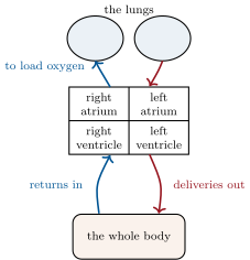
Example 27.2’s delivery round, with the map’s names: right ventricle — lung capillaries (oxygen aboard at the alveoli, bright red) — left atrium, left ventricle — artery — a thigh muscle’s capillaries (oxygen and glucose off, carbon dioxide on, darker) — vein, valves easing the uphill trip — right atrium. Two circuits, under a minute, no wrong turns available: the valves see to that.
Example 52.7 (Measurements at the roadside)
Three everyday readings of the network: the pulse — artery stretch, counting the surges (Method 27.8); blood pressure, the cuff’s two numbers — the artery pressure at surge and between surges; and the stethoscope’s lub-dub — the valves’ doors heard closing. Three windows, no incision.