Primary & Middle School Biology · Grades 1–9
52The Heart and Circulation
Chapter 27 introduced the pump and its delivery rounds; since then the blood has acquired responsibilities — nutrients at the villi (Proposition 50.6), oxygen at the alveoli (Proposition 51.4), bills at every working muscle (Proposition 49.1). Time to map the road network properly: the heart’s four rooms, the two circuits, and the three kinds of vessel.
52.1 The heart’s four rooms
Definition 52.1 (Atria and ventricles)
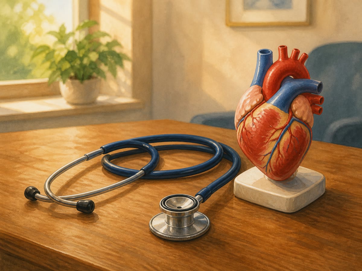
The heart’s two sides (Definition 27.5) each hold two rooms: an upstairs atrium that receives incoming blood, and a downstairs ventricle — the thick-walled pumping room — that drives it out. Between and beyond them, valves — one-way doors — snap shut behind each surge; their paired snaps are exactly the “lub-dub” a stethoscope (or an ear on a chest) hears.
Proposition 52.2 (One beat, in order)
Each beat runs the same sequence on both sides at once: the atria contract, topping up the ventricles; the ventricles contract, driving blood out — right side toward the lungs, left side toward the body — while the entry valves slam (lub); the ventricles relax, the exit valves slam (dub), and the rooms refill. Then again, times a day (Remark 27.9).
Proof. Admitted at this level. ∎
52.2 Two circuits, three vessels
Definition 52.3 (The double circulation)
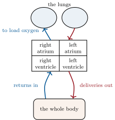
The blood’s road network is two loops through one pump (Definition 27.5’s two sides, now as circuits):
- the lung circuit: right ventricle lungs (loading oxygen, unloading carbon dioxide) left atrium;
- the body circuit: left ventricle the whole body (delivering, collecting) right atrium.
Every drop alternates circuits: lungs, body, lungs, body — which is why the left ventricle, pushing round the far larger body loop, is the heart’s thickest-walled room.
Definition 52.4 (Arteries, capillaries, veins)
Three kinds of vessel make each loop (Definition 27.3, now named):
- arteries: thick-walled, elastic, carrying blood away from the heart under pressure — the pulse (Proposition 27.6) is their stretch at each surge;
- capillaries: the hair-fine vessels threading every organ — walls one cell thin, where all the exchanges happen: alveoli, villi, muscles, skin;
- veins: wide, thinner-walled, returning blood to the heart at low pressure, with one-way valves of their own — the bluish lines of Remark 27.4.
Example 52.5 (One drop, one full tour)
Example 27.2’s delivery round, with the map’s names: right ventricle — lung capillaries (oxygen aboard at the alveoli, bright red) — left atrium, left ventricle — artery — a thigh muscle’s capillaries (oxygen and glucose off, carbon dioxide on, darker) — vein, valves easing the uphill trip — right atrium. Two circuits, under a minute, no wrong turns available: the valves see to that.
Remark 52.6 (Why capillaries are the point)
Arteries and veins are only highways; every actual delivery — every exchange this year has studied — happens across capillary walls. The specification of Proposition 47.1 lives here too: one-cell walls (thin), threading every tissue by the tens of thousands of kilometres (Definition 27.3 — large), bathed in the body’s moisture. The heart exists to keep blood moving through capillaries; all the rest is plumbing to and from.
52.3 Reading and keeping the network
Example 52.7 (Measurements at the roadside)
Three everyday readings of the network: the pulse — artery stretch, counting the surges (Method 27.8); blood pressure, the cuff’s two numbers — the artery pressure at surge and between surges; and the stethoscope’s lub-dub — the valves’ doors heard closing. Three windows, no incision.
Proposition 52.8 (Keeping the network)
The network’s long-term enemies and friends are known:
- exercise strengthens the pump (Proposition 49.7) and keeps the highways supple;
- balanced eating (Method 18.8) protects the artery walls, which too-rich blood slowly furs up and stiffens;
- smoke attacks vessels as well as lungs (Proposition 51.6) — narrowing, stiffening, seat-stealing;
- the heart’s own supply — it feeds by its own small arteries, not from the blood in its rooms — depends on all three.
The habits of Proposition 36.1 are, concretely, vessel maintenance.
Proof. Admitted at this level. ∎
Method 52.9 (Mapping any organ into the network)
For any organ this book meets:
- place it on the body circuit: artery in, vein out, capillaries within;
- list what crosses its capillary walls, in and out (villi: nutrients in; muscle: fuel and oxygen in, carbon dioxide out; skin: heat out);
- note its special connection, if any (intestine’s blood calls at the liver first; the lungs sit on their own circuit);
- check the flows against the organ’s job — the network diagram should retell the organ’s chapter.
52.4 Exercises
Exercise 52.1 ★
Name the heart’s four rooms and each one’s role.
Exercise 52.2 ★
What are the heart’s valves for, and what is the lub-dub?
Solution
Solution of Exercise 52.2.
They are one-way doors that stop blood surging backward. The lub-dub is their paired closings: entry valves slamming as the ventricles drive (lub), exit valves slamming as they relax (dub).
Exercise 52.3 ★
Trace the two circuits, naming start, destination and return for each.
Solution
Solution of Exercise 52.3.
Lung circuit: right ventricle to the lungs’ capillaries (load oxygen, unload carbon dioxide), returning to the left atrium. Body circuit: left ventricle to the whole body’s capillaries (deliver, collect), returning to the right atrium.
Exercise 52.4 ★
Give the three vessel kinds with one structural feature and one job each.
Exercise 52.5 ★
Why is the left ventricle the thickest-walled room?
Solution
Solution of Exercise 52.5.
It drives the body circuit — the far larger loop, out to toes and brain and back — so it must push hardest, and its wall is built to match.
Exercise 52.6 ★
Where — in which vessel kind — do all the body’s exchanges happen? Name three organs’ capillary exchanges.
Solution
Solution of Exercise 52.6.
In the capillaries. Lungs’ capillaries: oxygen in, carbon dioxide out; villi’s: nutrients in; muscles’: fuel and oxygen off, carbon dioxide on (skin’s: heat out).
Exercise 52.7 ★★
Run Example 52.5 for a drop serving the brain instead of the thigh. What does the brain’s capillary exchange take and leave? (Proposition 33.9 lists its appetites.)
Solution
Solution of Exercise 52.7.
Right ventricle — lung capillaries (oxygen aboard) — left atrium — left ventricle — neck arteries — the brain’s capillaries — neck veins — right atrium. The brain’s exchange takes a large, steady share of oxygen and glucose and leaves carbon dioxide — the demanding commander’s standing order.
Exercise 52.8 ★★
The pulse is felt at arteries, never at veins. Explain with the two vessels’ builds and pressures.
Solution
Solution of Exercise 52.8.
The pulse is the artery wall’s elastic stretch at each ventricular surge — high pressure meeting a stretchy thick wall. By the veins the surge has been spent crossing the capillaries: low, smooth pressure in a soft wide tube — nothing left to throb.
Exercise 52.9 ★★
Veins have one-way valves; arteries none. What problem do the vein valves solve — and why do soldiers standing stock-still sometimes faint?
Solution
Solution of Exercise 52.9.
The uphill return: from the legs, venous blood must climb against gravity at low pressure, and the valves hold each gain. The still-standing soldier’s calf muscles never squeeze the veins, the climb stalls, blood pools in the legs and the brain’s supply dips — the faint. Marching feet are vein pumps.
Exercise 52.10 ★★
Apply Method 52.9 to the small intestine, special connection included.
Solution
Solution of Exercise 52.10.
Artery in from the body circuit; within, the villi’s capillaries take up the meal’s nutrients (Proposition 50.6); the special connection: its vein runs first to the liver for sorting and banking, and only then does the blood rejoin the general return to the right atrium. The diagram retells the digestion chapter.
Exercise 52.11 ★★
Why does the heart need its own small feeding arteries, with its rooms full of blood? And why does their furring matter so much?
Solution
Solution of Exercise 52.11.
The rooms’ blood rushes through; the heart’s own thick walls, working without rest, need their own capillary delivery like any muscle — so small feeding arteries branch over the heart itself. Furred, they throttle the pump’s own fuel line: the one organ that can never pause is the one their blockage starves.
Exercise 52.12 ★★★
“The heart is plumbing in the service of capillaries.” Defend the claim using Remark 52.6 and two exchanges from earlier chapters.
Solution
Solution of Exercise 52.12.
Every exchange the body lives by — oxygen at the alveoli, nutrients at the villi, fuel and wastes at every muscle — happens only across capillary walls; arteries and veins merely bring blood to those walls and take it away, and the heart merely keeps it moving. Pump and highways exist for the sake of the hair-fine lanes: stop the capillary traffic anywhere and that organ dies, however sound the plumbing.
52.5 Problem: The Cardiologist’s Morning
Problem 52.1
Weekend problem — four consultations, one map of the network
Shadow a cardiologist for a morning of four (invented) consultations, map in hand.
Part I — The healthy baseline.
- Patient 1, a fit teenager, gets the three roadside readings of Example 52.7. Name each, and what part of the machinery each reads.
- Her resting pulse is . The cardiologist smiles. Why (Exercise 27.11)?
- Her lub-dub is crisp and double. What exactly snapped, twice, in every beat?
- Sketch her blood’s path from right atrium round both circuits and back, in eight named stops.
Part II — Three troubles.
- Patient 2 has a leaky valve between left atrium and ventricle: some blood surges backward at each beat, with a whooshing murmur. Predict the consequences for the body circuit’s deliveries and the patient’s stamina.
- Patient 3, a heavy smoker with a rich diet, has furred, stiffened arteries. Which readings of consultation 1 will have worsened, and why does the left ventricle work overtime?
- Patient 3’s furring includes the heart’s own feeding arteries. Explain the cardiologist’s special concern in one sentence.
- Patient 4 has swollen ankles after long standing days: the veins’ uphill return is struggling. Which structures are failing, and why does walking — calf muscles squeezing the veins — relieve it?
Part III — The wall chart.
- The clinic’s wall chart shows the double loop. Explain to a waiting patient why blood must visit the lungs between every two body tours.
- Add the vessel legend: artery, capillary, vein — one line each on build, pressure and job.
- The chart’s footer reads: “all treatment serves the capillaries.” Justify it with Remark 52.6.
- Close the morning with the prevention prescription the cardiologist hands every patient — three habits, each tied to its mechanism in Proposition 52.8.
Solution
Solution of Problem 52.1.
1. The pulse: the arteries’ surge count — the pump’s rate read at a wrist. Blood pressure: the artery pressure at surge and between — the highways’ load. The stethoscope’s lub-dub: the valves heard closing — the doors’ soundness.
2. A low resting pulse in a fit patient means a strong ventricle moving more blood per beat: the trained heart idles calm, with reserve in hand.
3. The valve doors: entry valves at the ventricles’ drive (lub), exit valves at their relaxation (dub) — four doors closing in two pairs, cleanly.
4. Right atrium, right ventricle, lung capillaries, left atrium, left ventricle, body arteries, body capillaries (say, a muscle), veins — and home to the right atrium.
5. Each beat loses part of its surge backward through the leak, so the body circuit’s deliveries per beat shrink; the heart compensates with more beats and harder work, and stamina drops — the murmur is delivery lost as backwash.
6. Blood pressure up (stiff, narrowed highways resist the surge) and the pulse’s artery stretch changed; the left ventricle, pushing every round against the stiffened circuit, works overtime and thickens.
7. Furring in the heart’s own feeding arteries throttles the fuel line of the muscle that can never rest — the classic path to a heart attack.
8. The veins’ one-way valves and the missing muscle-pump of movement: standing still, the calves never squeeze, the valves’ gains stall, and the ankles pool. Walking squeezes the veins rhythmically — the relief is mechanical.
9. Because the body’s tour spends the blood’s oxygen: only the lung circuit can reload it. Skipping the lungs would send spent blood on deliveries with an empty van — the double loop is refuel-then-deliver, enforced by the heart’s two sides.
10. Artery: thick, elastic, high pressure — outbound highways. Capillary: one cell thin, threading everything — the delivery lanes where all exchanges happen. Vein: wide, valved, low pressure — the return.
11. Every consultation’s trouble mattered exactly where it touched capillary service: the leak starved deliveries, the furring throttled supply lines, the pooling stalled returns. Readings, valves and highways are means; capillary traffic — the exchanges — is the end.
12. Move daily — the pump strengthens and the highways stay supple; eat balanced — the artery walls stay clear of furring; no smoke — vessels neither narrow nor stiffen, and the blood’s seats stay free for oxygen.