 Coach
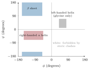
CoachThe secondary structure of a protein is the local, regular folding of its backbone, held by hydrogen bonds between backbone C=O and N–H groups. Two patterns dominate. The helix: a right-handed coil of 3.6 residues per turn, rising per residue ( per turn), each C=O bonded to the N–H four residues ahead, with the side chains pointing outward. The sheet: chains stretched almost fully ( per residue) and laid side by side, parallel or antiparallel, bonded between neighbouring strands, with side chains alternating above and below the sheet. Between them, turns and loops reverse the chain’s direction. Proline, whose ring locks , breaks helices; glycine, with no side chain, allows turns that no other residue can make.
Examples
Example 12.6 (Helix and sheet by the numbers)
A membrane-spanning helix must cross of hydrophobic core: residues, and indeed the transmembrane segments of Chapter 7 are runs of about twenty hydrophobic residues. A strand of the same length spans : silk fibroin is stacked antiparallel sheets of glycine and alanine, and a silk thread is stronger than steel of the same weight because the covalent backbones lie along the fibre.