University Biology — Year 1 · Bachelor Year 1
7Membranes and Membrane Transport
Drop a red blood cell into pure water and it swells and bursts within a second; drop it into strong salt and it shrivels. Put it back into plasma and it keeps its biconcave shape for four months, pumping sodium out and potassium in every second of that time. The membrane that does this is five nanometres thick — a film of lipid two molecules deep, threaded with proteins — and it is the boundary across which every exchange of Chapter 1 finally happens. This chapter describes the membrane’s structure, the physics of what crosses it unaided, the proteins that carry, pump and channel the rest, the electrical potential this traffic produces, and the vesicles that move what no protein can.
7.1 The fluid mosaic
Definition 7.1 (The plasma membrane)
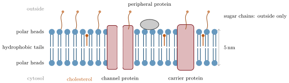
The plasma membrane — and every membrane of the cell — is a lipid bilayer about thick: two sheets of phospholipids (Chapter 9) with their hydrophobic tails facing each other and their polar heads facing the two aqueous sides, with cholesterol between the tails, and membrane proteins embedded in it or attached to it. In the fluid mosaic model (Singer and Nicolson, 1972) the bilayer is a two-dimensional fluid in which lipids and proteins diffuse laterally, and the proteins are a mosaic of independent units, not a continuous coat. The two faces differ: sugars attached to lipids and proteins face outward only (the glycocalyx), and some phospholipids are confined to one leaflet.
Proposition 7.2 (Membranes are bilayers and fluids)
The plasma membrane is exactly two lipid molecules thick, and its components move within its plane.
Evidence. Gorter and Grendel (1925) extracted the lipids of a known number of red blood cells and spread them as a monolayer on water: the area was twice the cells’ total surface, so the membrane is a bilayer. Electron microscopy shows every membrane as two dark lines (the stained heads) around a pale core (the tails), in all. Frye and Edidin (1970) fused a mouse cell with a human cell whose surface proteins had been labelled with dyes of two colours: after forty minutes at the two colours were completely intermixed over the hybrid cell, and not at , where the lipid is nearly solid. Bleaching a spot of a fluorescent membrane protein with a laser and watching the fluorescence return as unbleached molecules diffuse in (FRAP) measures the lateral diffusion: in a few seconds for lipids, slower for proteins, and zero for proteins anchored to the cytoskeleton. ∎
Definition 7.3 (Membrane proteins)
Integral membrane proteins span the bilayer with one or more hydrophobic helices and can only be extracted with detergents; peripheral proteins are bound to one face. By function: transporters (channels, carriers, pumps), receptors that bind a signal outside and act inside, enzymes, anchors linking the cytoskeleton to the extracellular matrix, and recognition proteins bearing the sugars by which cells identify one another. Proteins are half the mass of a typical membrane and nearly all of its function.
7.2 Crossing the membrane without help
Proposition 7.4 (Permeability of the bilayer)
A pure lipid bilayer is freely permeable to small non-polar molecules (, , , steroids), fairly permeable to small uncharged polar molecules (water, urea, ethanol), poorly permeable to larger polar molecules (glucose, amino acids), and practically impermeable to ions (, , , ) and to macromolecules. The permeability spans twelve orders of magnitude, from for water to for : a hydrophobic core thick lets through what dissolves in oil and stops what carries a charge.
Theorem 7.5 (Fick’s law of diffusion)
The net flux of a solute across a membrane of area (moles per second) is proportional to the concentration difference across it:
where the permeability coefficient (in ) lumps together the solute’s diffusion coefficient in the membrane, its solubility in the lipid and the membrane’s thickness (). Diffusion needs no energy and always runs down the gradient; it is passive transport.
Proof. Inside the membrane the solute diffuses down a linear concentration profile from to ( the partition coefficient between lipid and water); Fick’s first law in the bulk, , gives . ∎
Definition 7.6 (Osmosis, water potential)
Osmosis is the net diffusion of water across a membrane that lets water through but not the solutes, from the solution where water is more concentrated (fewer solutes) to the one where it is less. It is described by the water potential , the chemical potential of water expressed as a pressure, zero for pure water at atmospheric pressure:
where , the solute potential, falls with the total solute concentration (in osmoles per litre, the van ’t Hoff law) and , the pressure potential, is the hydrostatic pressure above atmospheric. Water moves from higher to lower . At , : a solution has . A solution is isotonic to a cell when no net water moves, hypotonic when water enters, hypertonic when it leaves.
Example 7.7 (A red cell in three solutions)
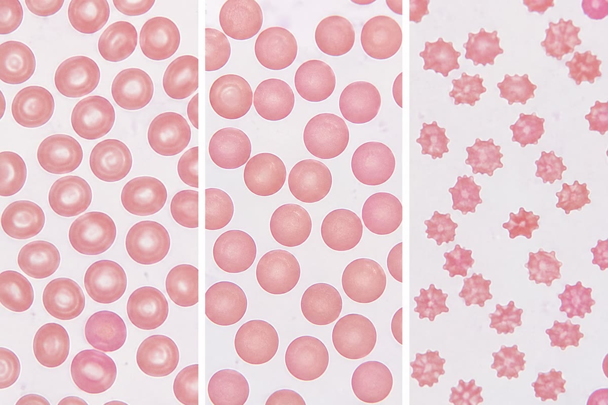
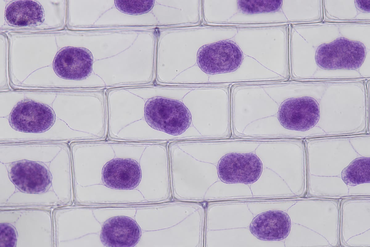
Plasma is : on both sides, no net flow. In pure water () the cell, at inside, takes up water until its membrane, which can stretch only a few percent, ruptures. In salt it loses water until its inside is as concentrated, at half its volume. A plant cell in pure water does not burst: as water enters, the wall is stretched and rises until , the water potential inside is zero, and the flow stops with the cell turgid at .
Method 7.8 (Water-potential bookkeeping)
- Convert every solute concentration to osmoles (a salt that dissociates into two ions counts twice) and compute .
- Add the pressure term: for a solution in an open vessel or an animal cell, positive for a turgid plant cell, negative for water under tension in a xylem vessel.
- Water flows toward the lower . Equilibrium is reached when the two are equal: by dilution of the cell’s contents (animal cell), or by a rise of pressure (plant cell).
- Read the sign of the result as the direction of flow, and its size as the driving force; the flow rate also depends on the membrane’s water permeability, raised a hundredfold by aquaporin channels.
7.3 Transport proteins
Definition 7.9 (Channels, carriers, pumps)
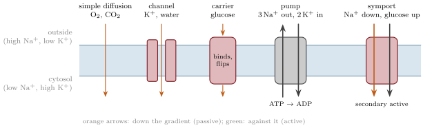
Channels are integral proteins with a water-filled pore through which a specific ion or small molecule diffuses down its gradient at up to per second; many are gated, opening in response to a voltage, a ligand or a mechanical force. Aquaporins are water channels. Carriers bind their solute on one face, change conformation, and release it on the other, a thousand times a second at most; the glucose transporter of the red cell is one. Channels and carriers working down a gradient perform facilitated diffusion, passive but selective and saturable. Pumps are carriers that couple the transport of a solute against its gradient to the hydrolysis of ATP: primary active transport. The sodium–potassium pump of animal cells exports three and imports two per ATP; the proton pump of plant, fungal and bacterial membranes exports .
Proposition 7.10 (Secondary active transport)
A gradient built by a pump is a store of energy that other carriers spend: a symporter moves a solute uphill by coupling it to an ion moving downhill in the same direction (the sodium–glucose transporter of the intestine and kidney; the proton–sucrose transporter of the phloem), an antiporter couples it to an ion moving the opposite way (the sodium–calcium exchanger). In animal cells the currency is the gradient; in plants, fungi and bacteria the gradient.
Example 7.11 (Kinetics tell the mechanism)
The flux of glucose into a red cell rises with the outside concentration, then levels off at a maximum, like an enzyme (Chapter 13): a carrier, saturable, with a of about ; a similar sugar, mannose, competes for it. The flux of urea rises in proportion to its concentration without limit: simple diffusion. The flux of glucose into an intestinal cell stops when sodium is removed from the lumen, or when the pump is poisoned with ouabain: secondary active transport.
7.4 The membrane potential
Definition 7.12 (Membrane potential)
Every living cell holds an electrical potential difference across its plasma membrane, the membrane potential , negative inside: in a neuron, in a muscle fibre, or more in a plant cell. It arises because the membrane is selectively permeable to ions whose concentrations differ across it.
Theorem 7.13 (The Nernst equation)
An ion of charge at concentrations and is at equilibrium across the membrane — no net flux, though the membrane is permeable to it — when the potential equals its equilibrium potential
For at outside and inside, ; for at outside and inside, .
Proof. Moving one mole of the ion from outside to inside changes the free energy by the sum of a concentration term and an electrical term:
At equilibrium , which gives . Converting to base-ten logarithms and inserting , , gives the numerical form. ∎
Proposition 7.14 (Origin of the resting potential)
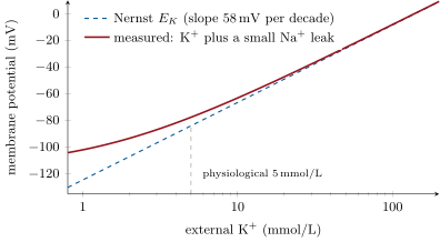
The resting membrane is far more permeable to (through open potassium channels) than to ; leaks out down its concentration gradient, leaving the inside negative, until the electrical pull back nearly balances the leak. The resting potential therefore lies close to , pulled a little toward by the small sodium leak (the Goldman equation weights each ion’s Nernst term by its permeability). The pump sustains the gradients that the leaks would otherwise dissipate; it also contributes a few millivolts directly, since it moves three charges out for two in.
Evidence. In a squid axon the resting potential follows when the outside potassium is varied over a wide range (a straight line of slope per decade at high concentrations), and departs from it at low concentrations exactly as a small sodium permeability predicts. Blocking the pump with ouabain leaves the potential almost unchanged for minutes, then lets it decay over hours as the gradients run down: the potential is a diffusion potential, the pump its long-term support. ∎
Example 7.15 (Where the pump’s energy goes)
Pumping one out against to and costs ; three of them, , plus two in at each: per cycle against the of one ATP under cellular conditions. The pump runs near its thermodynamic limit, and it consumes a third of a resting animal’s ATP, two thirds in the brain.
7.5 Bulk transport
Proposition 7.16 (Vesicular transport)
Macromolecules and particles cross the plasma membrane without passing through it: by exocytosis, a vesicle fuses with the membrane and empties outward; by endocytosis, the membrane engulfs material into a vesicle (Chapter 6). Both require ATP, both move membrane as well as contents, and both preserve the sidedness of the membrane. The three routes across a membrane — through the lipid, through a protein, inside a vesicle — are selective in three ways: by solubility, by molecular recognition, by the choice of what is engulfed.
Example 7.17 (Cholesterol delivery)
Cholesterol travels in the blood inside protein-coated particles too large for any carrier. A cell needing cholesterol displays receptors for the particle; particle and receptor gather in a coated pit, which pinches off, and the vesicle delivers the particle to a lysosome where the cholesterol is freed. A person whose receptors are defective cannot clear the particles: blood cholesterol doubles and arteries clog in early adulthood. One membrane protein, one disease.
7.6 Exercises
Exercise 7.1 ★
Describe the fluid mosaic model in four sentences: the lipids, the proteins, the fluidity, the asymmetry.
Solution
Solution of Exercise 7.1.
A bilayer of phospholipids, tails inward, with cholesterol, forms a sheet. Proteins are embedded across it or attached to one face, as separate units. Lipids and most proteins diffuse laterally in the plane, which is a two-dimensional fluid. The two leaflets differ in lipid composition, and the sugar chains are on the outer face only.
Exercise 7.2 ★
Rank , glucose, , water and ethanol by their permeability through a pure lipid bilayer, and explain the order.
Solution
Solution of Exercise 7.2.
(small, non-polar) ethanol (small, weakly polar) water (small, polar) glucose (large, polar) (charged, hydrated). Permeability falls with polarity, size and above all charge, because the solute must dissolve in the hydrophobic core.
Exercise 7.3 ★
Compute the solute potential of a sucrose solution and of a NaCl solution at .
Solution
Solution of Exercise 7.3.
Sucrose: . NaCl dissociates into two ions, : .
Exercise 7.4 ★
Compute the Nernst potential of at outside and inside, at . Is chloride at equilibrium in a cell at ?
Solution
Solution of Exercise 7.4.
. At chloride is within of equilibrium: it is distributed passively.
Exercise 7.5 ★★
Gorter and Grendel used red cells of surface each and obtained a lipid monolayer of . Compute the ratio of monolayer area to cell surface and conclude.
Exercise 7.6 ★★
A plant cell has a solute potential of and a pressure potential of . It is placed in a solution of solute potential in an open dish. Which way does water move, and what is the cell’s pressure potential at equilibrium (assume its solute potential does not change)?
Exercise 7.7 ★★
Glucose uptake by a cell is measured at increasing external concentrations: give . Show that the uptake saturates, estimate the maximum rate and the concentration giving half of it, and name the mechanism.
Solution
Solution of Exercise 7.7.
Doubling the concentration from 10 to 20 raises the rate by only : saturation. Plotting against (or noting that at and about at infinite ) gives and half-maximum at about : facilitated diffusion by a carrier.
Exercise 7.8 ★★
Explain why the resting potential of a cell is close to and not to , and what would happen to it if sodium channels suddenly opened.
Solution
Solution of Exercise 7.8.
At rest the membrane’s permeability is dominated by open potassium channels; potassium leaks out until the inside is negative enough to hold it, i.e. near . If sodium channels opened, the permeability would be dominated by sodium and the potential would swing toward , about — the action potential.
Exercise 7.9 ★★
The sodium–glucose symporter carries two per glucose. With at outside and inside and , compute the free energy available from the two sodium ions and the maximum glucose concentration ratio (inside/outside) the symporter can build at .
Solution
Solution of Exercise 7.9.
Per mole in: ; two: . Glucose uphill costs ; equality gives , a ratio of about .
Exercise 7.10 ★★★
A cell is cooled to . Predict, with reasons, the effects on: membrane fluidity, simple diffusion of , the pump, the ion gradients over hours, the membrane potential, and the volume of the cell. (Consider that the cell’s proteins exert an osmotic pull that the sodium gradient normally balances.)
Solution
Solution of Exercise 7.10.
Fluidity falls (tails pack, the bilayer approaches a gel); oxygen diffusion slows but continues; the pump, an enzyme, nearly stops; the leaks continue, so over hours sodium enters and potassium leaves and the gradients decay; the potential falls toward zero as the gradients fade; with sodium entering and the proteins’ osmotic pull no longer balanced, water enters and the cell swells — cold-stored cells and organs swell for exactly this reason.
Exercise 7.11 ★★★
Frye and Edidin’s fused cells showed complete mixing of surface proteins at in , none at , and none at when ATP synthesis was blocked — the last result was later shown to be wrong. Say what each result would imply about the mechanism of mixing, and why the correct version (mixing does not need ATP) matters for the fluid mosaic model.
Solution
Solution of Exercise 7.11.
Mixing at but not at implies a temperature-dependent process — diffusion in a fluid that becomes solid in the cold. No mixing without ATP would imply an active, energy-driven redistribution (motors, vesicle cycling) rather than diffusion. The corrected result, that mixing needs no ATP, is what the fluid mosaic model requires: proteins move by thermal diffusion in a two-dimensional fluid, without the cell doing work.
Exercise 7.12 ★★★
“The membrane potential is a by-product of the ion gradients, not something the cell builds directly.” Discuss in a paragraph, with the Nernst equation, the pump, and the number of ions actually needed to charge the membrane (a capacitance of ).
Solution
Solution of Exercise 7.12.
The potential is the value at which the electrical force balances the diffusion of the most permeant ion, given by Nernst; the cell sets the gradients (by the pump) and the permeabilities (by its channels), and the potential follows. A membrane of at carries , about ions per square centimetre — for a cell some ions, against potassium ions inside: a negligible fraction of the ions, moving a negligible distance, charges the capacitor. The gradients are the store; the potential is the reading on it, and the pump’s direct contribution (three charges out for two in) is a few millivolts.
7.7 Problem: The Enterocyte
Problem 7.1
Weekend problem — the cell that pulls glucose out of the gut: pump, symporter, carrier and the Nernst equation, ending on the highest glucose ratio the cell can build
An intestinal epithelial cell (enterocyte) is tall and wide; its apical membrane faces the lumen, its basolateral membrane the blood. Its cytosol holds of and of ; the lumen and the blood hold of and of . The membrane potential is . Take , , , , and in the cell.
Part I — The gradients.
- Compute the Nernst potentials of and .
- Which ion is nearer equilibrium at ? What does this say about the resting permeabilities?
- Compute the free energy change for one mole of entering the cell, and for one mole of leaving.
- Compute the free energy cost of one pump cycle ( out, in) and compare with the energy of one ATP. What is the efficiency?
- The pump is on the basolateral membrane only. Explain why this placement is necessary for the cell to move glucose from lumen to blood.
Part II — Glucose uphill. The apical membrane carries a symporter taking in one glucose with two ; the basolateral membrane carries a glucose carrier (facilitated diffusion).
- Compute the free energy released by two entering the cell.
- Write the free energy needed to move one glucose from the lumen (concentration ) to the cytosol ().
- At the symporter’s limit the two are equal. Compute the maximum ratio .
- If the lumen falls to of glucose late in absorption, what cytosolic concentration can the symporter still maintain?
- Blood glucose is . Explain how glucose leaves the cell into the blood without any further energy.
- Why is the symporter placed on the apical and the carrier on the basolateral membrane, and not the reverse?
- Oral rehydration solutions for cholera contain glucose and salt. Explain, from the symporter, why the glucose makes the salt — and the water — be absorbed.
Part III — Counting the traffic. The cell absorbs of glucose per second.
- Compute the entering per second through the symporters, and the number of pump cycles per second needed to export it.
- Compute the ATP spent per second on this export, and the fraction of the glucose absorbed that would have to be respired to pay for it (about 30 ATP per glucose).
- Each symporter cycles times per second. How many symporters does the apical membrane need? The apical membrane is with microvilli multiplying it by : compute the symporter density per square micrometre.
- The that enters brings water by osmosis. Two and one glucose, with the accompanying , are four osmoles; the cell keeps its osmolarity at . Compute the volume of water that must follow the solutes per second, and the time it would take to double the cell’s volume if the water did not leave through the basolateral membrane.
- Compute the number of water molecules absorbed per glucose molecule (, density ).
Part IV — Charging the membrane. The membrane is a capacitor of .
- Compute the area of the cell’s membrane (take a box by by without microvilli) and its capacitance.
- Compute the charge needed to hold and the number of monovalent ions it represents.
- Compute the number of excess ions per square micrometre of membrane that this represents.
- Compute the number of ions in the cell, and the fraction that must leave to charge the membrane. Comment.
- The pump moves a net charge out on each cycle. Using question 13, compute the current it carries and the change in potential it would produce in one second if nothing else moved. Why does the potential not in fact run away?
- A drug blocks the pump. Predict, in order, the changes in the sodium gradient, the glucose absorption, the membrane potential and the cell volume.
- A drug blocks the potassium channels instead. Predict the change in the resting potential.
- State the result: the maximum glucose concentration ratio the enterocyte can build, and the two membrane proteins that make it possible.
Solution
Solution of Problem 7.1.
1. ; . 2. ( away) is nearer than ( away): the membrane is much more permeable to . 3. in: . out: . 4. Cost per cycle against per ATP: efficiency . 5. The pump keeps cytosolic sodium low; the symporter on the apical face uses the inward sodium gradient to pull glucose in from the lumen; the pump must be on the other face so that the sodium it exports goes to the blood, not back into the lumen, which would leave the lumen’s sodium to recycle and the net movement zero. 6. of glucose. 7. (glucose is uncharged). 8. ; . 9. Up to in principle: the ratio is never reached because glucose leaves through the basolateral carrier; in practice the cytosol stays near . 10. The cytosol (above ) is more concentrated than the blood; the carrier lets glucose diffuse down that gradient into the blood, passively. 11. Reversed, the symporter would pump glucose from the blood into the cell and the carrier would let it leak into the lumen: the gut would secrete glucose. Polarity of the transporters is the direction of absorption. 12. The symporter takes up sodium only with glucose; glucose in the lumen therefore drives sodium absorption through it even when the cholera toxin blocks other routes; chloride follows sodium electrically and water follows the salt osmotically. 13. of , i.e. ions per second; at three per cycle, cycles per second. 14. ATP per second ; at 30 ATP per glucose, of glucose, of the glucose absorbed. 15. glucose per second at 50 per symporter: symporters; membrane : per square micrometre, i.e. one per square — a membrane packed with transporters. 16. Osmoles entering: ; at , water . Cell volume : doubled in . The water leaves as fast as it enters, across the basolateral membrane, and this is how the gut absorbs water. 17. of water is ; per glucose (): about 750 water molecules. 18. ; . 19. ; ions. 20. ions per square micrometre, one excess charge per square of membrane. 21. in the cell: ions; the charge needs of them: the concentrations are unchanged by charging the membrane. 22. Net charge per cycle out: current ; in one second the membrane would gain , i.e. . It does not, because every charge pumped out is immediately balanced by ions flowing back through channels (mainly leaking in less, or re-entering): the membrane’s conductance shorts the pump’s current, and the pump adds only millivolts. 23. Cytosolic sodium rises over minutes; the symporter loses its driving force and glucose absorption stops; the potential decays toward zero as the gradients run down; sodium and water enter and the cell swells. 24. With the potassium leak blocked, the sodium leak dominates: the potential moves toward , becoming much less negative (depolarisation). 25. A ratio of about (cytosol over lumen), set by two sodium ions per glucose at ; made possible by the sodium–potassium pump (basolateral) and the sodium–glucose symporter (apical).