University Biology — Year 1 · Bachelor Year 1
12Amino Acids and Proteins
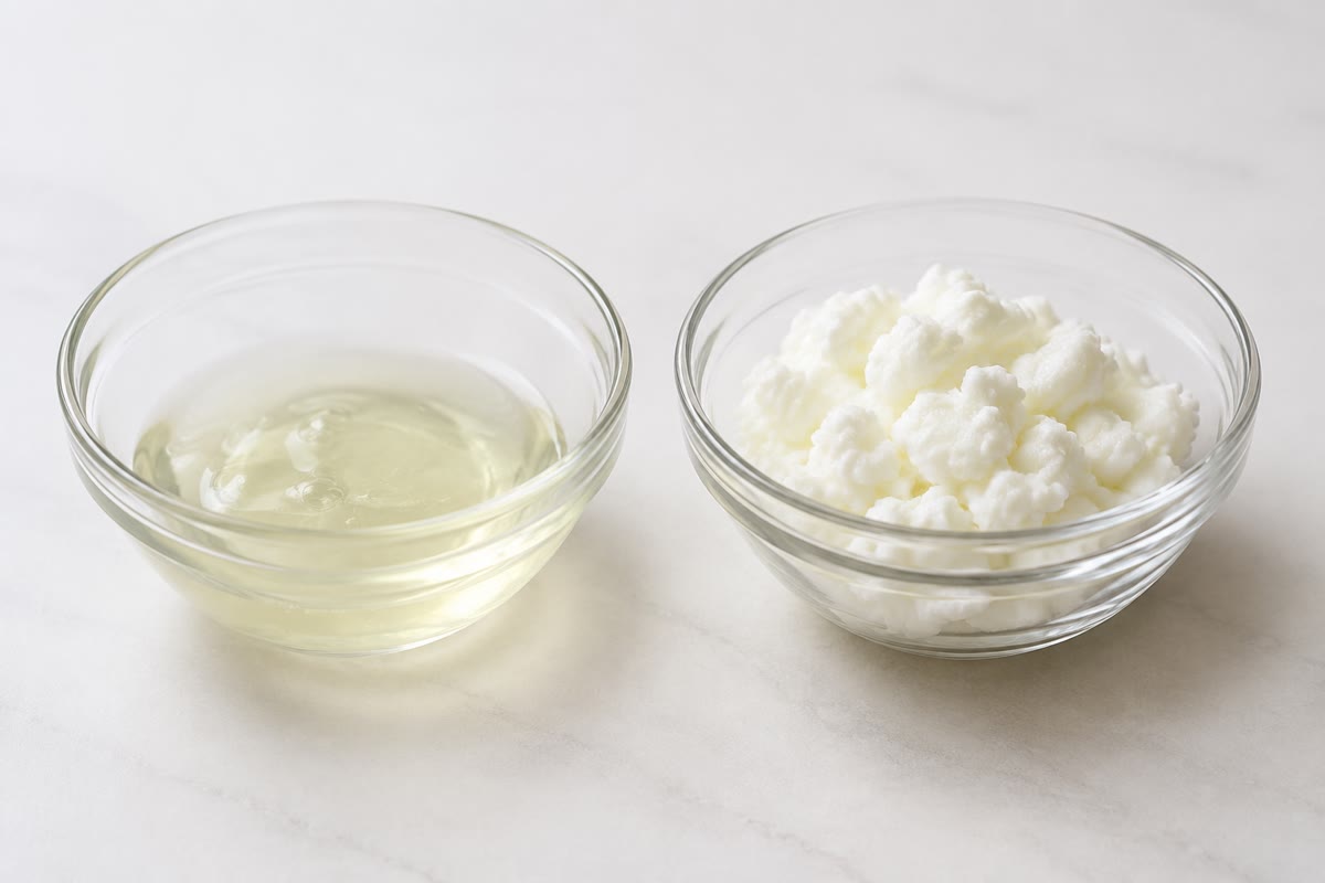
Boil an egg and the clear white turns opaque and firm; nothing has been added or removed, but every protein molecule in it has unfolded and tangled with its neighbours. Cool it and it stays cooked. Yet a small enzyme unfolded gently in a test tube, then left in plain buffer, folds back within minutes into its exact working shape, with no help from anything: the shape was written in the chain. Proteins are the working molecules of the cell — its enzymes, pumps, motors, scaffolds, antibodies, hormones — and every one of them is a chain of amino acids folded into a shape that its sequence dictates. This chapter describes the twenty amino acids, the bond that joins them, the four levels of protein structure, the folding that turns a chain into a machine, the cooperative behaviour of proteins with several subunits, and the methods by which proteins are separated and seen.
12.1 Amino acids
Definition 12.1 (Amino acid)
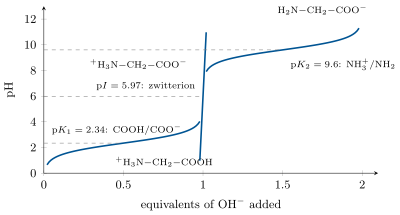
An amino acid has a central carbon bearing an amino group (), a carboxyl group (), a hydrogen and a side chain that distinguishes the twenty standard amino acids of proteins. At the pH of the cell the amino group is protonated and the carboxyl ionised: the molecule is a zwitterion, , with no net charge. The carbon is chiral in all but glycine, and living things use the L enantiomer only. Side chains group by their chemistry:
| class | amino acids (three- and one-letter codes) | side chain |
|---|---|---|
| non-polar, aliphatic | Gly G, Ala A, Val V, Leu L, Ile I, Met M, Pro P | hydrocarbon; Pro a ring |
| aromatic | Phe F, Tyr Y, Trp W | rings; Tyr and Trp absorb UV |
| polar, uncharged | Ser S, Thr T, Cys C, Asn N, Gln Q | OH, SH, amide |
| positively charged | Lys K, Arg R, His H | bases; His p 6.0 |
| negatively charged | Asp D, Glu E | carboxylates, p 4 |
Nine of them (His, Ile, Leu, Lys, Met, Phe, Thr, Trp, Val) are essential for humans: we cannot make them and must eat them.
Proposition 12.2 (Ionisation)
Each ionisable group of an amino acid has its own p: about 2 for the -carboxyl, 9.5 for the -amino, and for the side chains 3.9 (Asp), 4.1 (Glu), 6.0 (His), 8.3 (Cys), 10.1 (Tyr), 10.5 (Lys), 12.5 (Arg). The isoelectric point p is the pH at which the net charge is zero — the mean of the two p that bracket the neutral form; a protein’s p is set by its content of charged side chains, and at that pH it is least soluble and does not move in an electric field. Histidine, with a p close to 7, is the residue that changes charge within the physiological range, and it sits in the active sites of many enzymes.
12.2 The peptide bond
Definition 12.3 (Peptide bond, polypeptide)
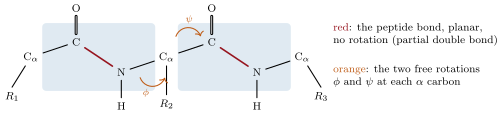
The peptide bond joins the carboxyl of one amino acid to the amino group of the next by condensation (an amide bond, ). A chain of amino acids so joined is a polypeptide: a backbone of repeating units from which the side chains project, with an N-terminus (free amino group) and a C-terminus (free carboxyl). Sequences are written from N to C, the order of synthesis (Chapter 19). A protein is one or more polypeptides folded into a definite shape; typical chains have residues of mean mass , so a protein of residues is .
Proposition 12.4 (Geometry of the peptide bond)
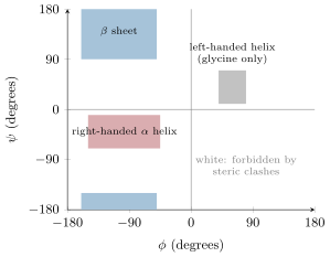
The C–N bond of a peptide has partial double-bond character (the amide resonance): it is short, cannot rotate, and holds the six atoms in one plane, almost always with the two carbons trans. The backbone is therefore a chain of rigid planes hinged at the carbons, each hinge having two rotations, (about N–) and (about –C), and steric clashes between side chains and backbone forbid most combinations of and . The conformation of a protein is essentially the list of its and angles.
12.3 Secondary structure
Definition 12.5 (Secondary structure)
The secondary structure of a protein is the local, regular folding of its backbone, held by hydrogen bonds between backbone C=O and N–H groups. Two patterns dominate. The helix: a right-handed coil of 3.6 residues per turn, rising per residue ( per turn), each C=O bonded to the N–H four residues ahead, with the side chains pointing outward. The sheet: chains stretched almost fully ( per residue) and laid side by side, parallel or antiparallel, bonded between neighbouring strands, with side chains alternating above and below the sheet. Between them, turns and loops reverse the chain’s direction. Proline, whose ring locks , breaks helices; glycine, with no side chain, allows turns that no other residue can make.
Example 12.6 (Helix and sheet by the numbers)
A membrane-spanning helix must cross of hydrophobic core: residues, and indeed the transmembrane segments of Chapter 7 are runs of about twenty hydrophobic residues. A strand of the same length spans : silk fibroin is stacked antiparallel sheets of glycine and alanine, and a silk thread is stronger than steel of the same weight because the covalent backbones lie along the fibre.
12.4 Tertiary and quaternary structure
Definition 12.7 (Tertiary and quaternary structure)
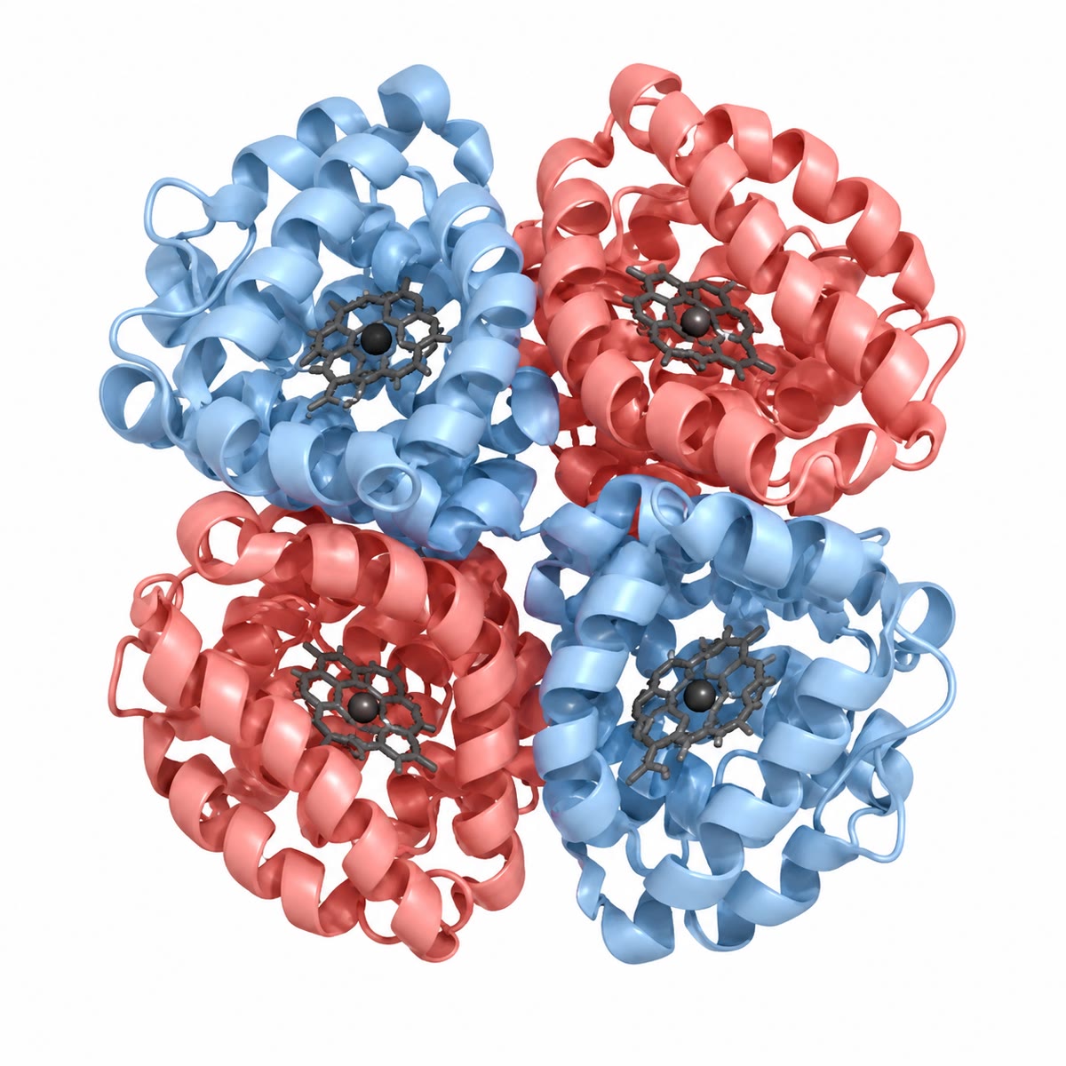
The tertiary structure is the complete three-dimensional arrangement of one polypeptide: its helices, sheets and loops packed together, often in several compact domains that fold independently. It is held by non-covalent forces — the hydrophobic effect burying non-polar side chains in a core away from water, hydrogen bonds, salt bridges between charged side chains, van der Waals packing — and sometimes by disulfide bridges, covalent S–S bonds between two cysteines, in proteins secreted to the oxidising outside. The quaternary structure is the assembly of several polypeptides (subunits) into one protein: haemoglobin is two and two chains. Globular proteins are compact and water-soluble (enzymes, carriers); fibrous proteins are extended and insoluble (collagen’s triple helix, the coiled coils of keratin and myosin, silk).
Theorem 12.8 (The sequence determines the fold)
The three-dimensional structure of a protein is determined by its amino-acid sequence: the native fold is the conformation of lowest free energy that the chain can reach in its cellular environment, and the information needed to reach it is contained in the chain itself.
Evidence. Anfinsen (1961) unfolded ribonuclease A, a -residue enzyme with four disulfide bridges, in concentrated urea with a reducing agent that broke the bridges: the chain lost all activity. Removing the urea and letting the cysteines re-oxidise slowly in air, he recovered the enzyme fully active, with its four bridges re-formed between the same eight cysteines out of the possible pairings — the chain had found its own fold. Re-oxidised while still in urea, it formed random bridges and stayed inactive; a trace of the reducing agent then let it shuffle them until the native set, the most stable, was reached. Since then thousands of sequences have been folded from scratch in the test tube, and the fold of a new sequence can now be computed from it. ∎
Proposition 12.9 (Denaturation)
Heat, extremes of pH, urea, detergents or organic solvents unfold proteins by overwhelming the weak bonds that hold the fold: the chain loses its shape and its function, exposes its hydrophobic core, and aggregates with its neighbours. Small proteins refold when the denaturant is removed; large ones, and proteins in a crowded cell, often cannot, and the cell keeps chaperones — proteins that bind unfolded chains, shield their hydrophobic surfaces, and give them repeated chances to fold correctly — and destroys what stays misfolded. Fevers of a few degrees, and the at which brain proteins begin to unfold, mark the narrow margin of stability: a typical fold is only more stable than the unfolded chain, the worth of a few hydrogen bonds.
Example 12.10 (Collagen)
A quarter of a mammal’s protein is collagen: three chains, each a left-handed helix of a thousand residues with glycine at every third position (Gly–X–Y, Y often hydroxyproline), wound together into a right-handed triple helix long, then packed side by side into fibrils cross-linked by covalent bonds. Glycine’s absence of a side chain is what lets the three chains pack at the axis; a single substitution of glycine by any other residue kinks the helix and gives brittle bones. Vitamin C is needed to hydroxylate the prolines; without it the helix is unstable and the fibrils fail: scurvy.
12.5 Allostery: haemoglobin
Definition 12.11 (Allostery, cooperativity)
A protein is allosteric when the binding of a ligand at one site changes the protein’s conformation and thereby its affinity at another site. When the sites are alike and the change raises the affinity of the others, binding is cooperative: the saturation curve is sigmoid rather than hyperbolic, and the protein switches from nearly empty to nearly full over a narrow range of ligand concentration. The Hill equation describes it:
where is the fraction of sites occupied, the ligand concentration (or partial pressure), the value at half saturation, and the Hill coefficient — 1 for independent sites, up to the number of sites for perfect cooperativity.
Proposition 12.12 (Haemoglobin and myoglobin)
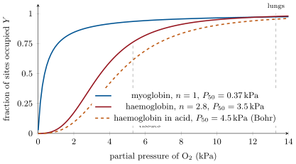
Myoglobin, one chain with one haem, binds with a hyperbola (, ): it is saturated at any pressure the tissues offer and serves as a store in muscle. Haemoglobin, four subunits, binds cooperatively (, ): the first bound shifts the tetramer from a low-affinity T state toward a high-affinity R state, so that it loads nearly fully at the lung’s and unloads a large fraction at the tissues’ . Its affinity is further lowered, and unloading favoured, by acid and (the Bohr effect) and by 2,3-bisphosphoglycerate (BPG), which binds the T state: in a working muscle, or at altitude, more oxygen is released for the same pressure. The fetus makes a haemoglobin that binds BPG weakly, so its blood draws oxygen from the mother’s across the placenta.
Method 12.13 (Using the Hill equation)
- Read from the curve at ; estimate from the slope of against (the Hill plot), which is a straight line of slope near .
- Compute at the loading pressure (lungs) and at the unloading pressure (tissues); the difference is the fraction of the carrying capacity delivered.
- Multiply by the capacity: blood holds of haemoglobin, each gram binding of , i.e. of per litre when saturated.
- To include the Bohr effect or BPG, use the shifted at the tissues only, where the acid is.
12.6 Seeing and separating proteins
Method 12.14 (Separating proteins)
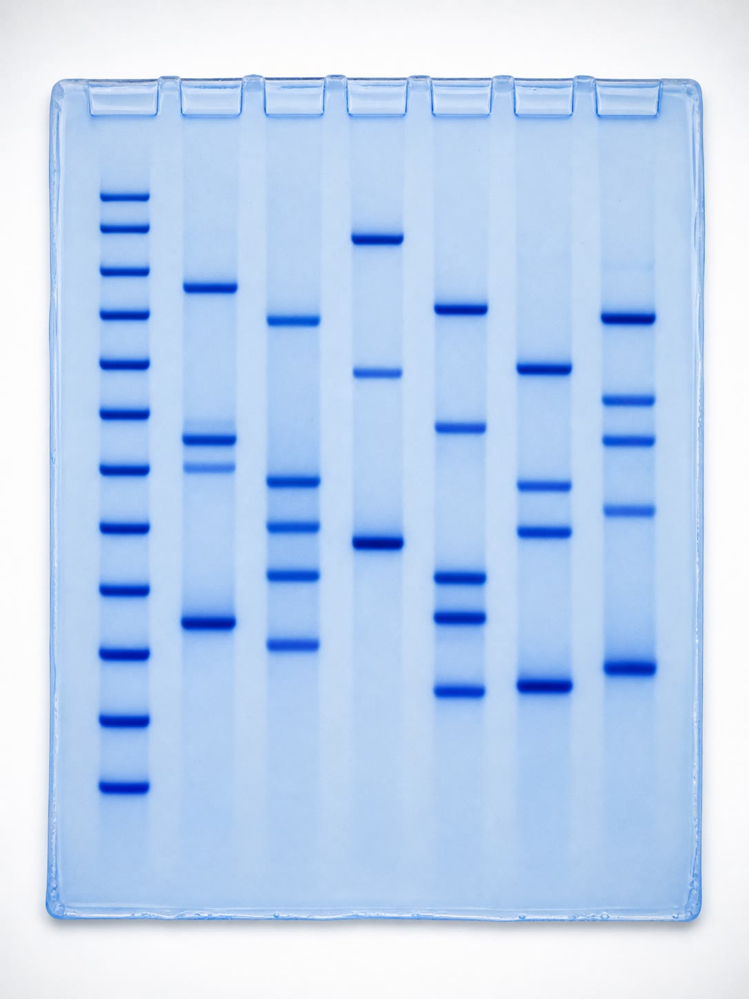
- Electrophoresis in SDS: the detergent coats every chain with negative charge in proportion to its length, so chains migrate through a polyacrylamide gel by size alone; a ladder of known masses calibrates the gel, and a stained band’s position gives the mass to a few percent.
- Chromatography: through a column of beads that retard proteins by size (gel filtration: large ones elute first), by charge (ion exchange), or by specific binding (affinity: an immobilised ligand holds one protein and lets the rest through). A purification is followed by the rise of specific activity (Chapter 5).
- Sequencing: the gene’s sequence, translated; or the protein itself, cut into peptides and read by mass spectrometry.
- Structure: X-ray diffraction of a crystal, or electron microscopy of frozen single particles, gives the position of every atom; the ribbon drawings of this chapter are their summaries.
Example 12.15 (Reading a gel)
A crude extract shows dozens of bands; after affinity chromatography one band remains at the position of the marker. Run without SDS and without reducing agent, the same protein migrates as a species: it is a dimer of two identical chains held by a disulfide bridge, which the reducing agent breaks and the detergent separates. Two gels, one line of reasoning, and the quaternary structure is known.
12.7 Exercises
Exercise 12.1 ★
Draw or describe the general structure of an amino acid at pH 7, and classify Leu, Ser, Lys, Glu and Phe by side chain.
Solution
Solution of Exercise 12.1.
: a zwitterion. Leu non-polar aliphatic; Ser polar uncharged; Lys positively charged; Glu negatively charged; Phe aromatic.
Exercise 12.2 ★
Define the four levels of protein structure and the bonds that hold each.
Solution
Solution of Exercise 12.2.
Primary: the sequence, peptide bonds. Secondary: local backbone folding (helix, sheet), backbone hydrogen bonds. Tertiary: the whole chain’s fold; hydrophobic effect, hydrogen bonds, salt bridges, van der Waals contacts, disulfide bridges. Quaternary: the assembly of subunits, by the same non-covalent forces.
Exercise 12.3 ★
A protein has residues. Estimate its mass. If it were a single helix, how long would it be? A single strand?
Solution
Solution of Exercise 12.3.
. Helix: . Strand: .
Exercise 12.4 ★
From the oxygen-binding figure, read the saturation of haemoglobin at and at , and the fraction delivered.
Solution
Solution of Exercise 12.4.
About and : a fifth of the load () is delivered.
Exercise 12.5 ★★
Compute the net charge of the peptide Lys–Gly–Asp–Ala at pH 7, at pH 2 and at pH 12, using the p values of the chapter, and estimate its isoelectric point.
Solution
Solution of Exercise 12.5.
Groups: N-terminal amino (9.5), Lys side chain (10.5), Asp side chain (3.9), C-terminal carboxyl (2). pH 7: . pH 2: (the terminal carboxyl half ionised). pH 12: (Lys mostly deprotonated). Net zero between the p of Asp (3.9) and of the amino group (9.5), where the charge is throughout: p (mean of 3.9 and 9.5).
Exercise 12.6 ★★
Explain why proline is rarely found inside helices and why glycine is found at tight turns, from the geometry of the backbone.
Solution
Solution of Exercise 12.6.
Proline’s side chain is bonded back to its own nitrogen: the nitrogen carries no hydrogen to donate the helix’s hydrogen bond, and is locked at about , which is tolerated only at the first turn of a helix. Glycine, with a hydrogen for side chain, suffers no steric clash and can adopt , values forbidden to every other residue, including those of tight turns.
Exercise 12.7 ★★
In Anfinsen’s experiment, what fraction of random re-oxidations would give the native set of four disulfides? (Count the ways of pairing eight cysteines.) What did the actual recovery of full activity prove?
Solution
Solution of Exercise 12.7.
Pairings of eight cysteines: ; random re-oxidation gives the native set about of the time. The recovery of nearly full activity showed that the chain, not chance, chose the pairing: the sequence encodes the fold and the disulfides merely lock it.
Exercise 12.8 ★★
Using the Hill equation with and , compute at , and . Repeat with . Which protein delivers more between and ?
Solution
Solution of Exercise 12.8.
. At 2: , ; at 3.5: ; at 8: , . With : : , , . Delivery between 8 and 2: cooperative , non-cooperative : the cooperative protein delivers twice as much.
Exercise 12.9 ★★
A protein runs as one band of on an SDS gel and elutes from a gel-filtration column at . Give its quaternary structure and say how you would check for disulfide bridges between the subunits.
Solution
Solution of Exercise 12.9.
A tetramer of four identical subunits. Run the SDS gel without reducing agent: if bands appear at 100, 150 or , disulfide bridges link the subunits; if the band alone remains, they are held non-covalently.
Exercise 12.10 ★★★
Sickle-cell haemoglobin has valine instead of glutamate at position 6 of the chain, on the surface. Explain, from the chemistry of the two side chains, why the mutant protein polymerises when deoxygenated, and why the disease shows in the tissues rather than the lungs.
Solution
Solution of Exercise 12.10.
Glutamate is charged and hydrated at the surface; valine is hydrophobic and creates a sticky patch that water excludes. In the deoxygenated T state a complementary hydrophobic pocket is exposed on a neighbouring molecule, so molecules stick end to end into fibres that deform the red cell. In the lungs the R state hides the pocket and the fibres dissolve; in the tissues, where haemoglobin is deoxygenated, they form — so the cells sickle and block the small vessels there.
Exercise 12.11 ★★★
A fold is stable by only . Compute the fraction of molecules unfolded at equilibrium at (), and at if has fallen to . Why is such marginal stability useful to a cell?
Solution
Solution of Exercise 12.11.
At , : : one molecule in five million unfolded. At with , : : unfolded, and rising steeply. Marginal stability lets proteins change shape when they work (allostery, catalysis), be unfolded for transport across membranes, and be degraded and replaced quickly when damaged or no longer needed.
Exercise 12.12 ★★★
“A protein is a sequence that folds into a shape, and the shape is the function.” Discuss in a paragraph with Anfinsen, chaperones, sickle cell and allostery: where the sentence holds and where the cell must intervene.
Solution
Solution of Exercise 12.12.
Anfinsen showed that the sequence suffices to specify the fold, and allostery shows that the shape — and its changes — is the function. But the cell intervenes at every step: chaperones rescue chains that would aggregate before folding, because the crowded cytosol is not a dilute test tube; a single substitution (sickle cell) gives a correctly folded protein whose surface is wrong, so “shape” must include the surface chemistry, not the backbone alone; and many proteins need modifications, partners or ligands added after folding before they work. The sentence states the principle; the cell supplies the conditions.
12.8 Problem: Haemoglobin at Work
Problem 12.1
Weekend problem — oxygen carried from lung to muscle, at rest, in a sprint, on a mountain and before birth, ending on the oxygen delivered per litre of blood
Blood holds of haemoglobin; binds of . Haemoglobin obeys the Hill equation with and at pH 7.4; myoglobin has and . Partial pressures of : in the lungs at sea level, in resting tissues. Take , , , , , , , .
Part I — At rest.
- Compute the oxygen capacity of a litre of blood.
- Compute for , and kPa (as ).
- Compute the saturation of haemoglobin in the lungs and in the tissues.
- Compute the fraction of the load delivered and the oxygen delivered per litre of blood, in millilitres.
- A resting human consumes of per minute. What cardiac output does this delivery require?
- Compute the saturation of myoglobin at the same two pressures and the oxygen it would deliver per litre if it replaced haemoglobin. Conclude.
- Explain in one sentence what cooperativity buys.
Part II — A sprint. In a sprinting muscle the pH falls to 7.2 and the local pressure of to ; the Bohr effect raises to there.
- Compute at with (no Bohr effect).
- Compute at with .
- Compute the oxygen delivered per litre in the two cases (loading in the lungs unchanged) and the gain from the Bohr effect.
- The muscle’s myoglobin is at . What is its saturation, and what happens to it if the pressure falls to during a contraction?
- Explain why the Bohr effect is a form of allostery and where the protons act.
Part III — A mountain. At the lungs’ pressure is . Within days the red cells raise their BPG, shifting to ; within weeks the marrow raises haemoglobin to .
- Compute the saturation in the lungs at with , and the delivery per litre (tissues at ). Compare with sea level.
- Compute the lung and tissue saturations with , and the delivery.
- Explain why lowering the affinity helps although it lowers the loading in the lungs.
- Compute the delivery per litre with and of haemoglobin.
- Would a still lower affinity () help at ? Compute the two saturations and decide.
Part IV — Before birth. Fetal haemoglobin has ; in the placenta the pressure of is on both sides.
- Compute the saturation of maternal haemoglobin at .
- Compute the saturation of fetal haemoglobin at .
- Explain how oxygen moves from mother to fetus although the pressure is the same on both sides.
- Fetal haemoglobin has chains instead of , which bind BPG weakly. Explain how this produces the lower .
- At birth the fetal blood must unload in tissues at . Compute the fetal delivery per litre from lungs at , and say why the switch to adult haemoglobin over the first months is useful.
- A mutation raises the adult to . Predict the person’s arterial saturation at sea level, and the consequence for delivery.
- Explain why a carrier with and no cooperativity () would be a poor oxygen carrier: compute its delivery.
- State the result: the oxygen delivered per litre of blood at rest and in a sprinting muscle, and the two properties of haemoglobin that make the difference.
Solution
Solution of Problem 12.1.
1. of per litre. 2. ; ; . 3. Lungs: . Tissues: . 4. : per litre. 5. — close to the resting cardiac output. 6. Myoglobin: lungs ; tissues ; delivers per litre, six times less: a hyperbolic carrier that loads well cannot unload at tissue pressures. 7. Cooperativity places the steep part of the curve between the two working pressures, so that a small drop in pressure releases a large fraction of the load. 8. ; . 9. ; . 10. Without Bohr: ; with: per litre; gain , . 11. saturated; at , : it releases a third of its store into the mitochondria during the contraction, and reloads between contractions. 12. Protons bind at sites (histidines, the N-termini) away from the haems, and by binding stabilise the T state, lowering the affinity at the haems: a ligand at one site changing the affinity at another — allostery. 13. ; lungs ; tissues ; delivery : about half of sea level. 14. : lungs ; tissues ; delivery . 15. The loading falls a little (0.875 to 0.828) but the unloading falls more (0.762 to 0.688), so the difference grows: at the lungs are still on the flat top of the curve while the tissues are on its steep part. 16. Capacity ; per litre — three quarters of sea level restored. 17. : lungs , tissues : delivery , better still on paper — but the arterial blood would be only saturated, and any further fall of lung pressure or any demand for a higher tissue pressure would leave the brain short; the real adjustment stops near . 18. . 19. ; . 20. At the same pressure the fetal protein binds more: as the mother’s blood unloads toward and the fetal blood loads toward , oxygen flows down the pressure gradient that the difference of affinities maintains across the placental membrane. 21. BPG binds the T state and lowers affinity; the chains bind it weakly, so fetal haemoglobin sits nearer its intrinsic high affinity: lower . 22. Lungs: ; tissues ; delivery , half the adult’s: good for taking oxygen from the mother, poor for delivering it once the lungs work, hence the switch. 23. : lungs , tissues : delivery , nearly double the normal — the person is mildly cyanotic at the lips but tolerates it well; the price is a smaller reserve at altitude. 24. : lungs , tissues : delivery — and in the sprinting muscle at , : delivery against . Without cooperativity the carrier loads badly and unloads badly. 25. of per litre at rest, per litre in a sprinting muscle; the difference comes from cooperativity (the sigmoid curve) and from the Bohr effect (the shift of in acid).