
সব বই

পেশাদার

1 Markets I: The Ecosystem and Exchange-Traded Marketsবাজার অনলাইন 2 Markets II: Rates, FX and Creditবাজার অনলাইন 3 Markets III: Commodities, Energy and Cryptoবাজার অনলাইন 4 Quantitative Methodsপদ্ধতি অনলাইন 5 Derivatives and Volatilityডেরিভেটিভস অনলাইন 6 Rates, Credit, XVA and Riskসুদহার, ঋণ ও ঝুঁকি অনলাইন 7 Research Craft: Predictors, Backtests, Measurement, Portfoliosগবেষণা অনলাইন 8 Strategies I: Equities and Futuresকৌশল অনলাইন 9 Strategies II: Volatility, Relative Value, Macro and the Bank Desksকৌশল অনলাইন 10 Microstructure and Executionএক্সিকিউশন অনলাইন 11 Market Making and High-Frequency Tradingমার্কেট মেকিং অনলাইন 12 Machine Learning for Marketsমেশিন লার্নিং অনলাইন 13 Low-Latency Softwareপ্রযুক্তি অনলাইন 14 Networks, Hardware and Trading Infrastructureপ্রযুক্তি অনলাইন 15 Research, Data and Risk Platformsপ্রযুক্তি অনলাইন 16 The Desk and the Firmপ্রতিষ্ঠান অনলাইন 17 The Industry: Firms, Roles and Careersক্যারিয়ার অনলাইন 18 Interviews I: Maths, Probability and Speedক্যারিয়ার অনলাইন 19 Interviews II: Markets, Trading Games and the Final Roundক্যারিয়ার অনলাইন 20 Interviews III: Codeক্যারিয়ার অনলাইন
অ্যাপ পরিচিতি Coach সাইন ইন পড়া শুরু করো

বইগুলো এখনো বাংলা ভাষায় পাওয়া যায় না — ইংরেজি সংস্করণ দেখানো হচ্ছে।

Biology · পরিভাষা

Secondary structure কী?

অন্য নাম: alpha helix · beta sheet

Definition 12.5 University Biology — Year 1 · অধ্যায় 12 — Amino Acids and Proteins

The secondary structure of a protein is the local, regular folding of its backbone, held by hydrogen bonds between backbone C=O and N–H groups. Two patterns dominate. The α\alpha helix: a right-handed coil of 3.6 residues per turn, rising 0.15 nm0.15\,\mathrm{nm} per residue (0.54 nm0.54\,\mathrm{nm} per turn), each C=O bonded to the N–H four residues ahead, with the side chains pointing outward. The β\beta sheet: chains stretched almost fully (0.35 nm0.35\,\mathrm{nm} per residue) and laid side by side, parallel or antiparallel, bonded between neighbouring strands, with side chains alternating above and below the sheet. Between them, turns and loops reverse the chain’s direction. Proline, whose ring locks ϕ\phi, breaks helices; glycine, with no side chain, allows turns that no other residue can make.
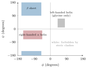
The Ramachandran plot: the combinations of  and  that a residue can adopt. Two large allowed regions correspond to the  sheet and the right-handed  helix; most of the plane is forbidden by collisions between atoms.
The Ramachandran plot: the combinations of ϕ\phi and ψ\psi that a residue can adopt. Two large allowed regions correspond to the β\beta sheet and the right-handed α\alpha helix; most of the plane is forbidden by collisions between atoms.
Linus Pauling (1901–1994), who deduced the  helix and the  sheet in 1951 from the planarity of the peptide bond and the geometry of hydrogen bonds, before either had been seen in a protein. Photograph: Nobel Foundation, 1962, public domain.
Linus Pauling (1901–1994), who deduced the α\alpha helix and the β\beta sheet in 1951 from the planarity of the peptide bond and the geometry of hydrogen bonds, before either had been seen in a protein. Photograph: Nobel Foundation, 1962, public domain.

উদাহরণ

Example 12.6 (Helix and sheet by the numbers)

A membrane-spanning α\alpha helix must cross 3 nm3\,\mathrm{nm} of hydrophobic core: 3/0.15=203/0.15 = 20 residues, and indeed the transmembrane segments of Chapter 7 are runs of about twenty hydrophobic residues. A β\beta strand of the same length spans 7 nm7\,\mathrm{nm}: silk fibroin is stacked antiparallel sheets of glycine and alanine, and a silk thread is stronger than steel of the same weight because the covalent backbones lie along the fibre.

প্রসঙ্গসহ পড়ো →