University Biology — Year 2 · Bachelor Year 2
11Organogenesis: Building the Tetrapod Limb
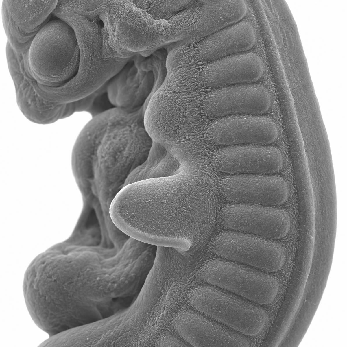
Three days after a hen’s egg is laid, a bump the size of a pinhead appears on each flank of the embryo. Four days later it is a wing, with a humerus, a radius and ulna, a wrist and three digits, in the right order, of the right lengths, with muscles attached to the right bones. Cut off the bump’s rim of thickened skin and the wing stops growing; graft a sliver of its rear margin onto its front and it grows six digits in mirror image. The limb bud is the classic case of organogenesis — the building of an organ from a field of cells by signals that say how far out, how far forward and how far up each cell is — and the experiments that decoded it are among the clearest in biology. This chapter follows the bud from its outgrowth to its skeleton.
11.1 The bud and its three axes
Definition 11.1 (The limb bud)
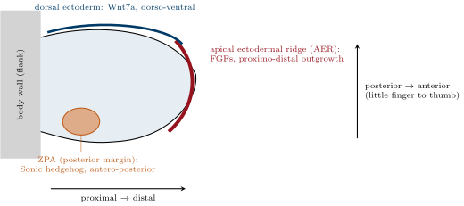
The limb bud is a swelling of lateral plate mesoderm covered by ectoderm, arising at a fixed position along the flank (the limb field) where the Hox code of the body axis permits it; its mesoderm will make bone, cartilage, tendon and dermis, and cells that migrate into it from the somites will make the muscles. The bud has three axes, each governed by a signalling centre. Proximo-distal (shoulder to fingertip): the apical ectodermal ridge (AER), a thickened strip of ectoderm along the tip, secretes fibroblast growth factors (FGFs) that keep the mesoderm beneath it dividing and undifferentiated. Antero-posterior (thumb to little finger): the zone of polarising activity (ZPA), a block of mesoderm at the posterior margin, secretes the morphogen Sonic hedgehog (Shh). Dorso-ventral (back of the hand to palm): the dorsal ectoderm secretes Wnt7a. Forelimb and hindlimb buds look alike and use the same signals; they differ in a transcription factor expressed before the bud appears (Tbx5 in the forelimb, Tbx4 in the hindlimb), which is why a wing is a wing.
11.2 Outgrowth: the proximo-distal axis
Proposition 11.2 (The apical ectodermal ridge)
The AER is required for outgrowth and its FGFs are sufficient: the mesoderm under it, the progress zone, divides every twelve hours or so, and cells that leave the zone as the tip advances stop dividing, condense and differentiate. The skeleton forms in proximo-distal order — stylopod (humerus or femur), zeugopod (radius and ulna, tibia and fibula), autopod (wrist and digits) — each segment specified in turn as the bud grows, and the segments correspond to nested domains of Hox gene expression (Hoxa/d 9–10 in the stylopod, 11 in the zeugopod, 13 in the autopod). A limb bud grows some tenfold in length in four days while the reach of the ridge’s signal stays the same few hundred micrometres, so that the ridge patterns the limb not all at once but as a moving front, the way a printer lays down a page line by line.
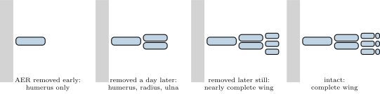
Evidence. Saunders (1948) removed the AER from chick wing buds at successive stages: removed early, the bud yielded only a stump of humerus; removed a day later, humerus and forearm but no hand; removed later still, a nearly complete wing — the later the operation, the more distal the level of truncation, so the ridge is needed for each segment in turn. Grafting a second ridge produced a second outgrowth; a bead soaked in FGF, placed on a bud whose ridge had been removed, restored a complete limb (Niswander, 1993), and a bead placed on the flank between the limb fields induced an extra limb. The mice lacking the ridge FGFs have no limbs. ∎
Theorem 11.3 (Growth by division)
A bud whose cells divide every hours multiplies its cell number by in a time ; four days of twelve-hour cycles give . If the bud’s volume grows a thousandfold in those four days, division supplies a factor of 256 and the remaining factor of four must come from cell enlargement and the matrix that cartilage cells secrete around themselves. Conversely, a segment that takes one cell cycle to specify — the roughly twelve hours by which the successive truncation levels of Saunders’ experiment are separated — corresponds to one doubling of the progress zone.
Proof. ; with and , . The ratio of the required factor to the division factor, , is what growth other than division must provide. ∎
11.3 Digits: the antero-posterior axis
Proposition 11.4 (The zone of polarising activity)
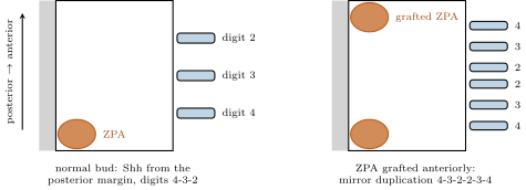
The ZPA specifies which digit forms where. Its cells secrete Sonic hedgehog, which spreads across the bud from the posterior margin, and mesoderm cells read the concentration — and the time they have been exposed to it — as a position: the highest dose gives the most posterior digit, lower doses more anterior ones, and below a threshold no digit at all. The gradient has the exponential form of Chapter 10, with a decay length of a few tens of micrometres, and its thresholds cut the bud into the primordia of the digits: in the chick wing, from posterior to anterior, digits 4, 3 and 2. A second source of the signal at the anterior margin produces a second, mirror-image set; a weaker source, or a shorter exposure, produces only the anterior digits of the extra set. The same gene, in the same role, patterns the digits of every tetrapod, and a mutation that adds an anterior source of Shh gives polydactyly in cats, chickens, mice and people.
Evidence. Saunders and Gasseling (1968) grafted a block of posterior-margin mesoderm from one chick wing bud to the anterior margin of another: the host grew a wing with six digits in the order 4-3-2-2-3-4, a mirror image about the middle. Tickle (1975) showed that the number of extra digits depended on the number of grafted cells — a dose. Riddle and Tabin (1993) found that the grafted tissue expressed the gene Sonic hedgehog, that the gene was expressed nowhere else in the bud, and that cells engineered to express it, grafted anteriorly, reproduced the mirror duplication; a bead of the protein did the same. Chick mutants with an anterior ectopic patch of Shh have extra anterior digits. ∎
Theorem 11.5 (Digit boundaries from the gradient)
With Shh at concentration at the posterior margin and a decay length , the boundary of the territory of a digit whose threshold is lies at from the margin. With and thresholds of , and of for digits 4, 3 and 2, the territories are –, – and – micrometres, and the tissue beyond forms no digit. Doubling the source moves every boundary outward by — enough, in a bud of a few hundred micrometres, to add a digit at the anterior edge; a second source at the anterior margin lays down the same territories in reverse.
Proof. From , at : , , . Replacing by adds to each . ∎
11.4 Coordination and sculpting
Proposition 11.6 (The axes talk to each other)
The centres maintain one another: FGF from the ridge keeps the ZPA expressing Shh, and Shh, through a relay in the mesoderm, keeps the ridge expressing FGF; remove either and the other fades within a day. Wnt7a from the dorsal ectoderm is needed for full Shh expression as well, and it drives the dorsal fate of the mesoderm (a mouse without it has two palms). This mutual dependence is a positive feedback loop that locks the three axes together: the limb grows only where it is being patterned, and is patterned only where it is growing, so that a bud never makes a hand without an arm or digits of the wrong sort. When outgrowth is complete the loop is broken — the mesoderm stops relaying, Shh stops, the ridge regresses — and the limb stops growing at its proper size.
Proposition 11.7 (From pattern to bone)
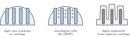
Once specified, mesoderm cells leaving the progress zone condense into rods and plates the shape of the future bones, differentiate into cartilage cells that secrete a matrix rich in collagen and proteoglycans, and lay down a cartilage model of each skeletal element; joints form where a band of cells is told not to become cartilage; blood vessels invade the models and bone replaces cartilage from the centre outward, leaving growth plates at the ends that keep the bone lengthening until adulthood (endochondral ossification). Muscle precursors from the somites migrate in, divide, fuse into fibres (Chapter 12) and attach through tendons made by the limb’s own mesoderm. Between the digit rays, the tissue is removed by programmed cell death: the cells of the interdigital webs, told by a bone morphogenetic protein (BMP) signal, activate their own destruction and are cleared in a day, which separates the fingers. A duck keeps its webs because a BMP inhibitor is expressed in its interdigital tissue; a chick given the same inhibitor on a bead grows webbed feet.
Evidence. Interdigital mesoderm cut from a chick leg bud and grafted elsewhere dies on schedule — the death is intrinsic, decided before the tissue is removed; the same tissue from a duck survives. Beads of BMP placed between the toes of a duck cause the web to die; beads of the inhibitor gremlin placed between the toes of a chick save it. A mouse lacking a BMP receptor in the limb keeps its webs. ∎
Example 11.8 (The same limb, differently grown)
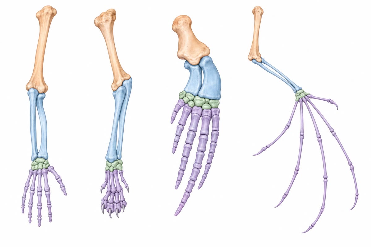
The bat’s wing is a hand whose digits 2 to 5 grew several times longer than a mouse’s — an enhancer of a limb gene, transplanted from bat to mouse, lengthens the mouse’s forelimb bones — and whose interdigital tissue survived, because the web expresses the BMP inhibitor the duck uses. The whale’s flipper is a hand with extra phalanges and no interdigital death. The horse’s leg is a single digit with the others reduced to splints. The snake has no limbs because its flank never expresses the signals that start a bud. And the fins of fish, which lack an autopod, are built by the same AER and ZPA but stop their Hox programme before the late phase that makes digits; the fossil Tiktaalik (375 million years old) shows the wrist appearing in a fin. Limb evolution is mostly the modulation of a conserved programme — how long each phase runs, how far each signal reaches, which cells die.
11.5 Exercises
Exercise 11.1 ★
Name the three axes of the limb bud and, for each, the signalling centre, its position and its signal.
Solution
Solution of Exercise 11.1.
Proximo-distal: the apical ectodermal ridge along the tip, FGFs. Antero-posterior: the zone of polarising activity at the posterior margin, Sonic hedgehog. Dorso-ventral: the dorsal ectoderm, Wnt7a.
Exercise 11.2 ★
Give the three segments of the limb skeleton and the bones of each in a human arm and leg. Which Hox genes mark them?
Solution
Solution of Exercise 11.2.
Stylopod: humerus, femur (Hoxa/d 9–10). Zeugopod: radius and ulna, tibia and fibula (Hox 11). Autopod: wrist and hand, ankle and foot (Hox 13).
Exercise 11.3 ★
List, in order, the steps from a patch of lateral plate mesoderm to a finger with its bone, joint and muscle.
Solution
Solution of Exercise 11.3.
Limb field specified (Tbx5); bud grows out under the ridge’s FGF; the ZPA and dorsal ectoderm pattern it; cells leaving the progress zone condense into rays; cartilage differentiates; joints form where cartilage is suppressed; blood vessels invade and bone replaces cartilage; interdigital cells die; muscle precursors from the somites migrate in, fuse and attach by tendons.
Exercise 11.4 ★
What does the limb look like if the AER is removed (a) as soon as the bud appears, (b) after the forearm has been specified, (c) not at all but replaced by a bead of FGF?
Solution
Solution of Exercise 11.4.
(a) A stump with little or no humerus; (b) humerus, radius and ulna but no hand; (c) a complete limb — FGF replaces the ridge.
Exercise 11.5 ★★
With and thresholds of , and of the source concentration for digits 4, 3 and 2, compute the three boundaries and the width of each digit territory.
Solution
Solution of Exercise 11.5.
, , : territories of 28, 36 and .
Exercise 11.6 ★★
Predict the digit pattern of a chick wing after (a) a full ZPA grafted to the anterior margin, (b) a graft of a quarter as many ZPA cells, (c) a bead of Shh at the anterior margin removed after a short exposure. Explain each from dose and time.
Solution
Solution of Exercise 11.6.
(a) 4-3-2-2-3-4, a full mirror set. (b) A weaker source gives a lower peak, so only the low-threshold digit is added: 2-2-3-4. (c) A short exposure likewise specifies only the most anterior extra digit: 2-2-3-4 — cells integrate concentration over time.
Exercise 11.7 ★★
A chick limb bud has cells when the ridge forms and its cells divide every . How many cells after four days? If the limb at that stage has cells, what must be true of the cell cycle or of cell death?
Solution
Solution of Exercise 11.7.
. Reaching needs doublings, so the cycle must average about , or the count must include the muscle precursors that migrate in from the somites; cell death would make the discrepancy worse, not better.
Exercise 11.8 ★★
Explain why a chick has free toes and a duck webbed feet, and what a bead of BMP inhibitor between the toes of a chick embryo does. What does the intrinsic timing of interdigital death (it happens on schedule in a graft) tell you about how the cells were instructed?
Solution
Solution of Exercise 11.8.
Chick interdigital cells receive a BMP signal and die; duck interdigital cells express a BMP inhibitor and survive, so the web persists. The inhibitor on a bead saves the chick web: a webbed foot. Death on schedule in a graft means the cells were already instructed before removal — the decision is made early and executed later, autonomously.
Exercise 11.9 ★★
A mouse lacks both Hoxa13 and Hoxd13; another lacks Hoxa11 and Hoxd11. Predict the skeleton of each forelimb and say what the result shows about the relation between Hox domains and segments.
Solution
Solution of Exercise 11.9.
Without Hoxa13 and Hoxd13: no autopod — the forelimb ends at the wrist. Without Hoxa11 and Hoxd11: a much shortened radius and ulna with a hand attached almost directly to the humerus. Each segment needs its own Hox pair; the domains are not merely markers but instructions.
Exercise 11.10 ★★★
Explain the positive feedback between the AER and the ZPA, why it makes the limb robust, and what would happen to a bud in which Shh could not maintain FGF. Contrast it with the positive feedback of labour in Chapter 8: what ends each loop?
Solution
Solution of Exercise 11.10.
FGF from the ridge keeps Shh on in the ZPA; Shh, through a mesodermal relay, keeps FGF on in the ridge. Each centre’s signal therefore certifies that the other is working, so a limb never grows without being patterned or is patterned without growing, and a transient loss of either is repaired by the other. If Shh could not maintain FGF, the ridge would regress within a day and outgrowth would stop at whatever segment had been reached: a truncated limb. Labour’s loop is ended by the event it drives — delivery removes the stimulus; the limb’s loop is ended by differentiation — the mesoderm stops relaying when the limb is complete.
Exercise 11.11 ★★★
A bat’s third digit is eight times longer than a mouse’s relative to body size. If digit cartilage lengthens exponentially at rate per day during a growth window, and the bat’s window is four days longer than the mouse’s, what is needed? Give two developmental changes (one in growth, one in cell death) that make a wing out of a hand.
Solution
Solution of Exercise 11.11.
: . Growth: a longer, faster proliferation of the digit cartilage (a limb enhancer that raises a growth gene’s expression in the digits). Death: survival of the interdigital tissue through expression of a BMP inhibitor, which keeps the membrane.
Exercise 11.12 ★★★
Sonic hedgehog patterns the neural tube from the floor plate (Chapter 10) and the digits from the ZPA. Discuss why evolution reuses a few signals for many jobs, how the same molecule can mean different things in different tissues, and what this implies for the effect of a mutation in such a gene.
Solution
Solution of Exercise 11.12.
Signals are reused because a working receptor–transduction module is cheap to redeploy: what changes between tissues is not the signal but the set of genes its receiving cells are competent to switch on, so the same concentration profile means motor neuron in one place and digit 4 in another. A mutation in such a gene therefore affects many organs at once — Shh mutations give holoprosencephaly and limb defects together — unless it lies in a tissue-specific enhancer, in which case it changes one organ only, which is how most evolutionary change in these genes occurs.
11.6 Problem: A Wing Built in a Week
Problem 11.1
Weekend problem — the chick wing followed from bud to skeleton: its growth by division, Saunders’ ridge removals timed, the Shh gradient computed and its duplications predicted, the webs sculpted and the wing compared with a bat’s, ending on the number of doublings, the digit boundaries and the width a bud needs for a mirror duplication
A chick wing bud appears at 3 days of incubation with mesoderm cells and a length of ; by 7 days the wing is long and its volume is times the bud’s. Progress zone cells divide every . Ridge removals: at 3.0 days the limb is a stump; at 3.5 days a humerus only; at 4.0 days humerus, radius and ulna; at 4.5 days a wing lacking only the digit tips. Shh: , half-life ; thresholds , , of for digits 4, 3, 2. Bud width (posterior to anterior) . Interdigital webs: three regions of , cells of , cleared in .
Part I — Growth.
- How many doublings occur in four days? How many cells does that give from ?
- Compare with the thousandfold volume increase: what factor must cell enlargement and matrix provide?
- By what factor did the length grow? If the bud grew as a cylinder of constant radius, what volume factor would that be, and what does the difference say about the shape?
- The ridge’s FGF reaches into the mesoderm. What fraction of the bud is under its influence at 3 days and at 7 days?
- Explain why a signal of fixed reach in a growing organ produces a proximo-distal sequence.
- A cell leaves the progress zone at 4 days. Which segment does it join?
Part II — Timing the segments.
- From the removal series, give the time by which each segment (stylopod, zeugopod, autopod) has been specified.
- How many cell cycles separate the specification of successive segments?
- Explain the result in terms of the progress zone and the Hox domains.
- An FGF bead is placed on a bud whose ridge was removed at 3.5 days. Predict the limb.
- A second ridge is grafted onto the dorsal surface of an intact bud at 3.5 days. Predict.
- Why does the bud stop growing at 7 days rather than continuing indefinitely?
Part III — The gradient.
- Compute and for Shh.
- Compute the three digit boundaries and territory widths.
- How wide is the anterior region that forms no digit in a bud?
- A mutation doubles Shh output. Recompute the boundaries and say whether an extra digit forms and which.
- A ZPA is grafted to the anterior margin. What minimum bud width allows a complete mirror set 4-3-2-2-3-4 without overlap of the two digit-2 territories? Is the chick bud wide enough?
- Predict the pattern if the bud were only wide.
- Why does a graft of a quarter of the ZPA give only 2-2-3-4?
Part IV — Sculpting and evolution.
- Compute the number of cells in the three webs and the rate of cell death during clearance, in cells per second.
- A duck’s interdigital cells express a BMP inhibitor. Predict the effect of a bead of BMP in a duck web and of the inhibitor in a chick web.
- A bat’s digit cartilage grows for four extra days at an exponential rate of . By what factor is it longer than the chick’s? Name a second change that makes the wing membrane.
- A fish fin bud has an AER and a ZPA but no digits. What is missing, and what does Tiktaalik show?
- If the web cells had instead been produced by division from founders at per cycle, how long would making them have taken? Compare with the day it takes to remove them.
- State the result: the doublings in four days, the three digit boundaries, and the minimum width for a mirror duplication.
Solution
Solution of Problem 11.1.
1. doublings: cells. 2. : cell enlargement and cartilage matrix. 3. Length ; a cylinder of constant radius would give in volume; the extra hundredfold means the radius also grew about tenfold — the bud widened into a paddle as it lengthened. 4. At 3 days ; at 7 days . 5. Only the tip is under the signal; as the tip advances, cells left behind leave the zone in the order they were made, so proximal structures are specified first and distal ones last. 6. The zeugopod (radius and ulna), specified between 3.5 and 4 days. 7. Stylopod by 3.5 days, zeugopod by 4.0 days, autopod by 4.5 days (digit tips later). 8. Half a day: one cell cycle. 9. Each cycle spent in the progress zone advances the Hox code (9–10, then 11, then 13); cells leaving after one, two or three cycles carry the code of the stylopod, zeugopod or autopod. 10. A complete wing: FGF is what the ridge provided. 11. A second outgrowth from the dorsal surface: duplicated distal structures, a supernumerary limb. 12. The mesoderm stops relaying Shh to the ridge, Shh expression ends, the ridge regresses, and cells differentiate instead of dividing: the loop that sustained growth is broken. 13. ; . 14. , , : territories of 26, 43 and . 15. . 16. Every boundary moves out by : 61, 104, 161. The three widths are 61, 43 and : only the digit-4 territory grows, the others are simply displaced. The pattern shifts anteriorly rather than gaining a digit, and the digit-free anterior region shrinks from 474 to . 17. Each set needs : at least ; the bud has room, with of digit-free tissue in the middle. 18. At the two gradients overlap and add: the middle cells see enough for digit 3 and the digit-2 territories vanish — 4-3-4 or 4-3-3-4, fewer digits than a full mirror set. 19. A quarter of the cells gives : no cell reaches the threshold of digit 4 or 3 ( at the margin itself), and the digit-2 threshold is passed out to : one extra digit 2. 20. : cells; over , cells per second. 21. BMP bead in the duck web: the web dies and the toes separate; inhibitor in the chick web: the web survives, a webbed foot. 22. . The second change is survival of the interdigital membrane by a BMP inhibitor. 23. The late phase of Hox 13 expression that specifies an autopod, and the persistence of the ridge: the fish’s fold makes fin rays instead. Tiktaalik’s fin contains wrist-like bones — a fin beginning to be a limb. 24. : doublings, about , three days to make what one day removes. 25. Eight doublings; digit boundaries at 26, 69 and ; a mirror duplication needs at least .