University Biology — Year 2 · Bachelor Year 2
19Chemical Messengers and Signal Transduction
A single molecule of adrenaline, arriving at the surface of a liver cell, never enters it. It binds a protein on the outside, changes the protein’s shape, and within a second the cell has made ten thousand molecules of a second messenger, activated a hundred thousand enzyme molecules, and begun releasing a million molecules of glucose — a chain of amplifications a million-fold long, started by a signal that stayed outside the door. This chapter is about the messengers cells send one another and how a cell converts a message at its surface into a change in its chemistry, its genes or its shape: the receptors, the second messengers, the cascades of kinases, and the two designs — the fast, wired signalling of nerves and the slow, broadcast signalling of hormones — whose contrast runs through the rest of this book.
19.1 Messengers
Definition 19.1 (Kinds of chemical signalling)
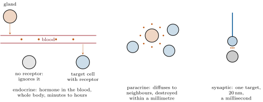
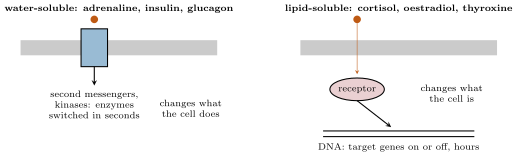
A cell signals to another with a secreted molecule, a messenger or ligand, that binds a receptor on or in the target. By range: endocrine signalling, in which a hormone is released into the blood by a gland and reaches every cell of the body, acting only on those with the receptor, over minutes to hours (insulin, cortisol, adrenaline, thyroxine); paracrine signalling, in which the messenger diffuses to neighbouring cells within a millimetre and is destroyed on the way (growth factors, histamine, prostaglandins, the morphogens of Chapter 10); autocrine, the cell signalling to itself; and synaptic, in which a neuron releases a neurotransmitter across a gap of onto one target cell, in a millisecond (Chapter 20). By chemistry: water-soluble messengers — amino-acid derivatives such as adrenaline, peptides and proteins such as insulin — cannot cross the membrane and act on surface receptors; lipid-soluble messengers — the steroids (cortisol, oestradiol, testosterone), thyroxine, retinoic acid, nitric oxide — cross it and act on receptors inside. The endocrine and nervous systems are the two ways a body of cells coordinates itself: the one broadcasts slowly to everyone, the other wires quickly to someone.
Theorem 19.2 (Receptor occupancy)
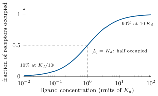
A receptor binds its ligand reversibly, , with a dissociation constant . At equilibrium the fraction of receptors occupied is
the same hyperbola as an enzyme’s saturation. is the concentration at which half the receptors are occupied and measures the affinity: the lower, the tighter. Hormone receptors have in the nanomolar to picomolar range, matched to the concentrations at which hormones circulate ( to mol/L): at a tenth are occupied, at nine tenths. A cell cannot respond to a hormone it does not have receptors for, and it tunes its sensitivity by changing the number of receptors, or — as in Chapter 9 — by withdrawing them when the hormone is constantly present.
Proof. With , , so , which is the formula. It is the Michaelis–Menten expression of the Year 1 volume without the catalytic step. ∎
19.2 Receptors at the surface
Proposition 19.3 (G-protein-coupled receptors and cyclic AMP)
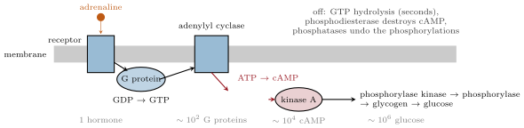
The largest family of receptors — some genes in a human, for hormones, neurotransmitters, odours, light — are proteins that cross the membrane seven times. A ligand binding outside changes their shape inside, where they activate a G protein: a three-subunit switch that swaps its bound GDP for GTP, splits, and in its GTP-bound form regulates an effector enzyme for a few seconds before hydrolysing the GTP and switching itself off. The classic effector is adenylyl cyclase, which makes cyclic AMP from ATP: a small, diffusible second messenger whose concentration rises within seconds from to several micromolar and which activates protein kinase A, an enzyme that phosphorylates — and so switches on or off — dozens of target proteins. The signal is ended by phosphodiesterase, which destroys cAMP, and by phosphatases, which remove the phosphates. Adrenaline on a liver cell acts this way: receptor G protein cyclase cAMP kinase A phosphorylase kinase glycogen phosphorylase glucose. Glucagon uses the same route; other G proteins inhibit the cyclase, or activate phospholipase C, which releases two other second messengers, inositol trisphosphate — which opens calcium stores — and diacylglycerol.
Evidence. Sutherland (1957) showed that adrenaline added to a liver homogenate released glucose only if membranes were present, that the membranes produced a heat-stable factor which alone activated phosphorylase in the membrane-free fraction, and that the factor was cyclic AMP — the first second messenger, and the first demonstration that a hormone acts at the cell surface through an intracellular intermediary. Rodbell and Gilman (1970s) found that the cyclase needed GTP and isolated the G protein that carries the signal between receptor and enzyme; cholera toxin, which locks the G protein in its on state, causes the gut to secrete litres of fluid a day — the disease is a signalling defect. ∎
Theorem 19.4 (Amplification in a cascade)
If each of stages of a cascade activates molecules of the next during the lifetime of its own activation, one molecule of messenger produces molecules of the final product. With four amplifying stages of a hundred each — a receptor activating a hundred G proteins, a cyclase making a hundred cAMP, a kinase phosphorylating a hundred enzymes, an enzyme releasing a hundred glucose molecules — ; in practice a million or so, in a few seconds, from a hormone at mol/L. Amplification is why hormones can be effective at concentrations a millionfold below those of the metabolites they control, and why the cascade must be switched off at every stage: an amplifier that cannot be turned off is a disease (cholera, and the many cancers in which a signalling protein is locked on).
Proof. Stage 1 produces active molecules; each of these produces at stage 2, giving ; by induction, after stages. The gain of each stage is the number of substrate turnovers an activated catalyst achieves before it is inactivated: its catalytic rate times its active lifetime. ∎
Proposition 19.5 (Receptor tyrosine kinases and calcium)
A second family of surface receptors — for insulin, for the growth factors that drive cell division, for the inductive signals of development — are receptor tyrosine kinases: single-pass proteins that, when their ligand brings two of them together, phosphorylate each other on tyrosines and so create docking sites for a set of adaptor proteins. From these run several cascades at once: a chain of three kinases (the MAP kinase cascade) that ends in the nucleus and switches on the genes of growth; a lipid kinase whose product recruits the kinase that mediates insulin’s metabolic effects — moving glucose transporters to the membrane, switching on glycogen synthesis; and others. A third universal second messenger is calcium: held in the cytosol at , ten thousand times below the outside, it floods in through channels opened by a signal or a voltage, or out of the endoplasmic reticulum in answer to inositol trisphosphate, rising to micromolar in milliseconds; bound to calmodulin it activates kinases and pumps, and bound to troponin it triggers contraction (Chapter 21). Pumps return it to the stores within a second. Whatever the receptor, the cell’s interior speaks a few languages — cAMP, calcium, phosphorylation — and the specificity lies in which proteins each cell type has for them to act on.

19.3 Receptors inside
Proposition 19.6 (Nuclear receptors)
Steroids, thyroxine, retinoic acid and vitamin D diffuse through the membrane and bind receptors in the cytosol or nucleus that are themselves transcription factors: the hormone–receptor complex binds specific DNA sequences and switches its target genes on or off. The response is slow — minutes to hours to make the mRNA and the protein — and lasting: cortisol induces the enzymes of gluconeogenesis for hours, oestradiol builds the uterine lining over days, testosterone makes muscle over weeks, thyroxine sets the metabolic rate of every cell. Some steroids also have fast actions through surface receptors, and some surface pathways also reach the nucleus (the MAP kinase cascade), so the distinction is not absolute; but the rule stands that a lipid-soluble messenger changes what a cell is by changing its genes, while a water-soluble one changes what it does by changing its enzymes.
19.4 A worked case: insulin, glucagon and the glucose set point
Proposition 19.7 (Two hormones and a set point)
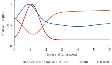
The blood’s glucose is held near () by two antagonistic hormones of the pancreatic islets. After a meal the rising glucose is sensed by the cells — which metabolise it, close a potassium channel as their ATP rises, depolarise, admit calcium and release insulin — and insulin, through its tyrosine-kinase receptor, makes muscle and fat take up glucose (by moving transporters to their membranes), the liver store it as glycogen and fat, and all tissues stop burning fat. Between meals the falling glucose lets the cells release glucagon, which, through cAMP in the liver, breaks glycogen down and makes new glucose from amino acids; adrenaline does the same faster in emergencies, and cortisol and growth hormone over hours. Each hormone is released in proportion to the deviation it corrects: a negative feedback with two effectors, one for each direction, and a set point defended to within a fifth. The brain, which burns of glucose a day and stores none, is the organ the loop protects; type 1 diabetes is the loss of the cells, and its untreated course — glucose at , fat burnt for want of insulin, acid in the blood — is what the loop looks like with one arm removed.
Example 19.8 (Speed, reach and cost of the two systems)
Adrenaline in the blood reaches every cell in about a minute (the circulation time) and acts for minutes; a nerve impulse reaches its target in milliseconds and acts for milliseconds. A hormone costs almost nothing per cell reached — a nanomole spread over the body — but cannot address one cell; a nerve addresses one cell exactly but must build and maintain a wire to it. The body uses both: the sympathetic nerves of Chapter 18 act on the heart and the arterioles in a second, and the adrenal medulla, itself a modified sympathetic ganglion, backs them up by pouring the same molecule into the blood for the whole body. And at the synapse the two designs meet, for a neurotransmitter is a messenger acting on a receptor by the same mechanisms — a channel that opens, a G protein that switches — only across twenty nanometres instead of a metre.
19.5 Exercises
Exercise 19.1 ★
Classify as endocrine, paracrine, autocrine or synaptic, and as water- or lipid-soluble: insulin, cortisol, acetylcholine at a synapse, histamine at a wound, oestradiol, adrenaline from the adrenal, nitric oxide from the endothelium.
Solution
Solution of Exercise 19.1.
Insulin: endocrine, water-soluble (peptide). Cortisol: endocrine, lipid-soluble. Acetylcholine at a synapse: synaptic, water-soluble. Histamine at a wound: paracrine, water-soluble. Oestradiol: endocrine, lipid-soluble. Adrenaline from the adrenal: endocrine, water-soluble. Nitric oxide: paracrine, lipid-soluble (a gas that crosses membranes).
Exercise 19.2 ★
List the steps from adrenaline at the liver cell membrane to glucose in the blood, and name the off-switch at each step.
Solution
Solution of Exercise 19.2.
Adrenaline binds the receptor (off: dissociation, receptor internalisation); receptor activates G protein, GDP for GTP (off: GTP hydrolysis in seconds); G protein activates adenylyl cyclase, which makes cAMP (off: phosphodiesterase); cAMP activates kinase A (off: cAMP loss); kinase A phosphorylates phosphorylase kinase, which phosphorylates glycogen phosphorylase (off: phosphatases); phosphorylase releases glucose-1-phosphate from glycogen, converted and exported as glucose.
Exercise 19.3 ★
Compare surface and nuclear receptors: what binds them, where they are, how fast they act, what they change.
Solution
Solution of Exercise 19.3.
Surface receptors bind water-soluble messengers at the membrane and act in seconds through second messengers and kinases, changing the activity of existing proteins. Nuclear receptors bind lipid-soluble messengers inside the cell and act in hours as transcription factors, changing which proteins are made.
Exercise 19.4 ★
What are the three universal second messengers named in this chapter, how is each raised, and how is each removed?
Solution
Solution of Exercise 19.4.
Cyclic AMP: made by adenylyl cyclase, destroyed by phosphodiesterase. Calcium: enters through channels or leaves the endoplasmic reticulum in answer to inositol trisphosphate, removed by pumps. Inositol trisphosphate (with diacylglycerol): made by phospholipase C from a membrane lipid, removed by phosphatases.
Exercise 19.5 ★★
A receptor has . Compute the occupancy at 0.2, 2, 20 and . A cell doubles its receptor number: what changes, the occupancy or the number of occupied receptors?
Solution
Solution of Exercise 19.5.
: , , , . Doubling the receptors leaves the fraction unchanged and doubles the number occupied — and so the response.
Exercise 19.6 ★★
A cascade has stages with gains of 50, 200, 100 and 300. Compute the overall amplification. If the hormone is at and of the receptors ( per cell) are occupied, how many product molecules does the cell make per second if each stage’s gain is per second?
Solution
Solution of Exercise 19.6.
. A hundred occupied receptors give product molecules a second — an upper bound, since the stages saturate.
Exercise 19.7 ★★
Cholera toxin locks the stimulatory G protein in its GTP-bound state in gut cells. Explain, step by step, why the patient loses litres of fluid a day, and why the effect persists for days after the bacteria are gone.
Solution
Solution of Exercise 19.7.
The locked G protein keeps the cyclase on, cAMP stays high, kinase A keeps the chloride channel of the gut cells phosphorylated and open, chloride is secreted, and sodium and water follow it into the lumen — litres a day. The lock is a covalent modification of the G protein, undone only when the cells themselves are replaced, days later.
Exercise 19.8 ★★
The cytosolic calcium rises from to in a cell of when a channel opens. How many calcium ions entered? If a single open channel passes ions a second, how long did one channel take, and why does a cell open many at once?
Solution
Solution of Exercise 19.8.
mol, ions (more in fact, since buffers bind most of what enters). One channel at a second would take a second; a thousand channels do it in a millisecond, which is the time scale a signal needs.
Exercise 19.9 ★★
Insulin and glucagon both act on the liver, one through a tyrosine kinase and one through cAMP. Explain how the liver cell integrates the two signals, what happens to glycogen when both are high, and why the ratio matters more than either level.
Solution
Solution of Exercise 19.9.
The two pathways converge on the same enzymes: glucagon’s kinase A phosphorylates glycogen synthase (off) and phosphorylase kinase (on); insulin’s cascade activates the phosphatase that removes the phosphates. The enzymes’ state is set by the balance of kinase and phosphatase activity, so what counts is the ratio of the two hormones; with both high the phosphatase usually wins and glycogen is stored.
Exercise 19.10 ★★★
Adrenaline raises cAMP in a liver cell within and glucose output within ; cortisol raises the liver’s gluconeogenic enzymes over . Using diffusion, transcription ( nucleotides a second on a -nucleotide gene), translation ( amino acids a second on a -residue protein) and the need to accumulate thousands of enzyme molecules, account for the two time scales.
Solution
Solution of Exercise 19.10.
Adrenaline: cAMP diffuses across a cell in under a second ( with ), and every enzyme of the cascade already exists — seconds. Cortisol: a transcript takes , a protein ; from a few dozen mRNAs each carrying a dozen ribosomes the cell makes a few hundred enzyme molecules a minute, so the thousands needed for a measurable effect take hours, after the minutes cortisol needs to enter and reach the DNA.
Exercise 19.11 ★★★
A mutation makes a growth-factor receptor tyrosine kinase dimerise, and so signal, without its ligand. Predict the behaviour of the cell, explain why this is a common step in cancer, and say what a drug that blocks the kinase’s ATP site would do.
Solution
Solution of Exercise 19.11.
The receptor signals continuously: the MAP kinase cascade drives the cell to divide without any growth factor, and it ignores the absence of the signal that normally restrains it. Since a single mutation removes a control on division, such receptors (and the kinases downstream) are among the commonest oncogenes. A drug that occupies the kinase’s ATP site stops the phosphorylations and silences the receptor — the principle of several cancer drugs.
Exercise 19.12 ★★★
“The endocrine system broadcasts; the nervous system wires.” Discuss the two designs in speed, reach, specificity and cost, and say where each is indispensable and where the body uses both for the same message.
Solution
Solution of Exercise 19.12.
Endocrine: reaches every cell in a minute, cheap per cell, cannot address one cell, acts for minutes to days — indispensable for whole-body states (fasting, growth, reproduction). Nervous: millisecond, one target, expensive wiring, acts for milliseconds — indispensable for movement and rapid reflexes. Both are used for the same message where speed and reach are both wanted: the sympathetic nerves and the adrenal medulla both deliver noradrenaline or adrenaline in a fright.
19.6 Problem: A Hormone from Blood to Gene
Problem 19.1
Weekend problem — adrenaline followed from the adrenal gland to a liver cell’s glucose, with its concentration, receptor occupancy, cascade gain and timing computed, then cortisol followed to a gene, and the glucose loop analysed, ending on the occupancy, the amplification and the time each hormone takes
Data: the adrenal medulla releases of adrenaline into of blood in a fright; adrenaline’s at the liver receptor is ; a liver cell () carries receptors. Cascade gains per second of activation: receptor G protein 20, G protein cAMP 100 (the cyclase’s turnover in the G protein’s 3-second lifetime), cAMP kinase A 1 (stoichiometric), kinase A phosphorylase kinase 50, phosphorylase 50, glucose 1000. The liver holds of glycogen. Cortisol: transcription at nucleotides a second on a gene of nucleotides, translation at residues a second on a -residue enzyme, ribosomes per mRNA, mRNAs made per hour, an effect needing enzyme molecules.
Part I — The hormone arrives.
- Compute the plasma concentration of adrenaline after the release, ignoring its removal.
- Compute the receptor occupancy and the number of occupied receptors per liver cell.
- Adrenaline is removed with a half-life of . What is its concentration and occupancy after ?
- How long does the adrenaline take to reach the liver from the adrenal, given a circulation time of about a minute? Why does the sympathetic nerve to the liver act faster?
- A cell with receptors and the same : what changes?
- Why is matched to the circulating concentration — what would a receptor with do with a nanomolar hormone?
Part II — The cascade.
- Compute the overall gain from one occupied receptor to glucose molecules per second.
- Compute the cAMP molecules the cell makes in the first second, and the concentration they reach in its volume.
- Compute the glucose output of one liver cell per second at the occupancy of question 2, and per minute.
- The liver has cells. Compute the output the gains predict, in grams per minute, and compare with the actually observed. What limits the real output?
- How long would the of glycogen last at this rate? Why does it not run out in a fright?
- Name the off-switch at each amplifying stage and say which acts fastest.
- A phosphodiesterase inhibitor (caffeine) is present. Predict the effect on the cascade and on the glucose output.
Part III — Cortisol to a gene.
- Compute the time to transcribe one mRNA.
- Compute the time to translate one enzyme molecule, and the number of enzyme molecules per mRNA per hour (each mRNA living an hour, ribosomes starting one after another).
- Compute the enzyme molecules made per hour from the mRNAs, and the time to reach .
- Add the time for cortisol to enter, bind, and reach the DNA (minutes) and explain the observed .
- Why would the body not use a cascade like adrenaline’s for cortisol’s job, and why not use cortisol’s mechanism for a fright?
- Cortisol also raises the number of adrenaline receptors in liver cells. Explain how a slow hormone can amplify a fast one, and what this does to the fast one’s occupancy curve.
Part IV — The loop.
- After a meal glucose rises from 5 to . Describe the cell’s response from glucose to insulin release, naming the coupling.
- Insulin acts through a tyrosine kinase, glucagon through cAMP. Explain why a single cell can obey both and how the liver’s glycogen enzymes are set by their ratio.
- A patient has no cells. Predict fasting and post-meal glucose, and explain why fat is burnt and acid appears.
- Injected insulin has no feedback. Explain the danger of an unusually long run with a usual dose.
- Compare the glucose loop with the baroreflex of Chapter 18: sensor, effectors, gain, speed.
- State the result: adrenaline’s occupancy after release, the cascade gain, the liver’s glucose output, and the time cortisol takes.
Solution
Solution of Problem 19.1.
1. . 2. : about 3300 occupied receptors. 3. Three half-lives: ; . 4. About half a minute to a minute, the time the blood takes to go round; the nerve delivers its transmitter directly to the liver within a second. 5. The same fraction, but occupied receptors: a tenfold larger response. 6. At a receptor with is a thousandth occupied: no response. Matching to the circulating range puts the steep part of the occupancy curve where the hormone actually varies. 7. glucose molecules a second per occupied receptor. 8. cAMP in the first second; in , mol, about . 9. a second, a minute per cell. 10. cells molecules a minute, 330 mol, a minute — ten thousand times the observed . The gains multiply only while every stage is unsaturated; in fact the phosphorylase is limited by its substrate and by the phosphatases, and the export of glucose by transporters. The cascade’s gain is spent on speed and sensitivity, not on output. 11. At , twenty minutes; the fright is over in minutes and the off-switches stop the cascade long before. 12. GTP hydrolysis by the G protein (seconds); phosphodiesterase on cAMP (seconds); phosphatases on the phosphorylated enzymes (seconds to minutes); receptor desensitisation (minutes). The fastest are the G protein’s own clock and the phosphodiesterase. 13. cAMP is not destroyed: it accumulates and persists, the kinase stays on, and the glucose output is larger and lasts longer — the alertness of coffee. 14. . 15. per enzyme per ribosome; 30 per ribosome per hour, 600 per mRNA per hour. 16. enzymes an hour; after about . 17. Ten to twenty minutes for cortisol to enter, bind and reach its genes, an hour before the first mRNAs are numerous and translated, then two hours of accumulation: about four hours. 18. A cascade changes the activity of enzymes that exist; cortisol’s job is to change which enzymes exist, for days, which only transcription can do. A fright cannot wait four hours for new enzymes. 19. More receptors means more occupied receptors at any adrenaline concentration: the occupancy fraction is unchanged but the response at each concentration is larger — the curve is scaled up, not shifted. A slow hormone sets the gain of a fast one. 20. Glucose enters the cell and is metabolised; ATP rises and closes an ATP-sensitive potassium channel; the membrane depolarises; voltage-gated calcium channels open; calcium triggers the exocytosis of insulin granules — metabolism coupled to secretion by an electrical step. 21. The two receptors start two pathways that converge on the same enzymes: glucagon’s kinase A phosphorylates them, insulin’s cascade activates the phosphatase that dephosphorylates them; the enzymes’ state, and so the direction of glycogen metabolism, follows the ratio of the two. 22. Fasting glucose high (the liver, unrestrained, keeps making it), post-meal glucose very high (no uptake by muscle and fat); without insulin the fat stores release fatty acids, which the liver converts to ketone acids — ketoacidosis. 23. Working muscle takes up glucose without insulin, so on a long run the glucose falls while the injected insulin, unregulated, goes on suppressing the liver: hypoglycaemia and collapse. 24. Glucose loop: sensor the and cells, two effector hormones for the two directions, a high gain holding the level within a fifth, minutes to act. Baroreflex: stretch receptors, the heart and vessels as effectors, gain about four, a second to act. 25. Occupancy after release; gain per second per occupied receptor; liver output in fact; cortisol takes about four hours.