University Biology — Year 2 · Bachelor Year 2
16Blood and the Circulatory System
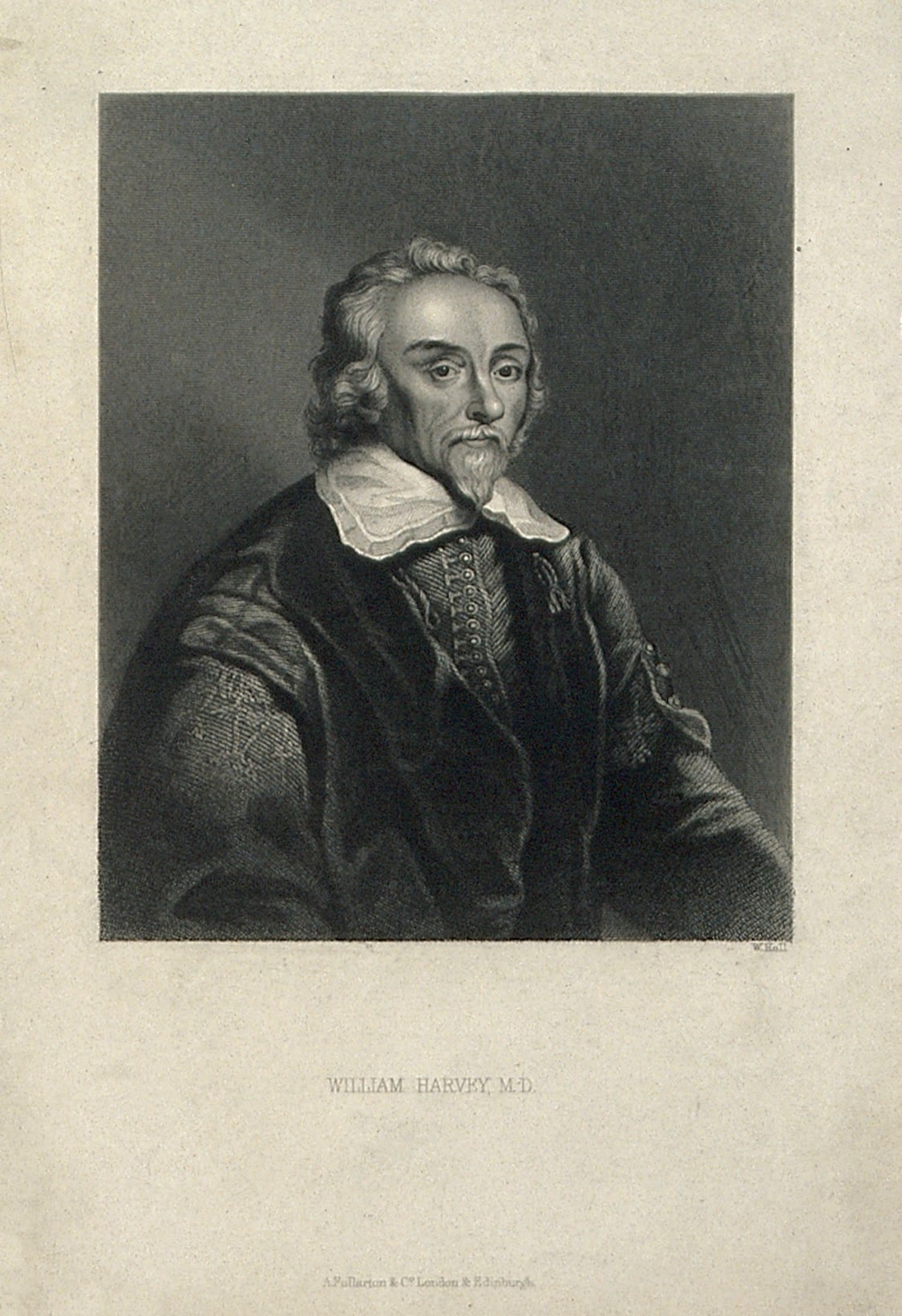
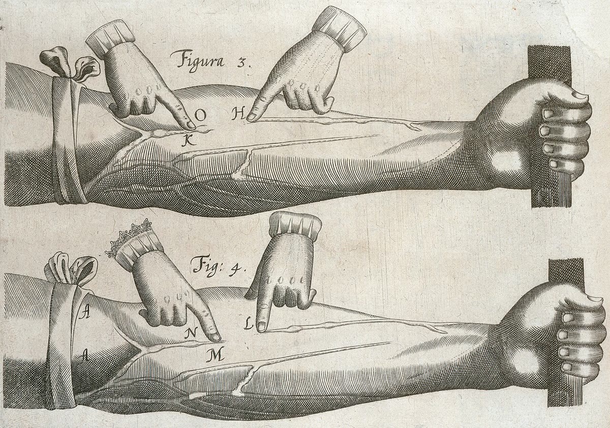
In 1628 William Harvey did a sum. The heart, he estimated, holds about two ounces of blood and beats seventy times a minute; even if only a fraction of that were expelled at each beat, the heart would pump in an hour several times the weight of the whole body. The blood could not be made and consumed at that rate, as Galen had taught for fourteen centuries; it must go round and round. Then he showed it: tie a cord round the arm, and the veins below the cord swell, not above; press the blood in a vein toward the hand, past a valve, and it will not go. The blood is pumped out through the arteries, returns through the veins, and the whole five litres pass through the heart every minute. This chapter is about that closed system — what the blood is, how it is routed, the physics of its flow through vessels from the aorta to a capillary, and the exchange, in the capillaries, that is the point of all of it.
16.1 Blood
Definition 16.1 (The composition of blood)
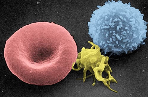
Blood is a tissue in suspension: about in an adult, of body mass. Spun down, it separates into plasma (: water, of proteins — albumin, globulins, fibrinogen — and the salts, sugars, lipids, gases and hormones it carries) and cells (, the haematocrit): red cells ( per litre, biconcave discs of without a nucleus, each packed with million molecules of haemoglobin, living 120 days and made in the marrow at two million a second), white cells ( per litre: neutrophils, lymphocytes, monocytes, eosinophils, basophils, the mobile arm of immunity), and platelets ( per litre, fragments of marrow cells that plug wounds and start clotting). The red cells carry oxygen — per litre of blood at full saturation, seventy times what water dissolves — and the plasma carries everything else, including the of that is not in the cells, the heat of the muscles to the skin, and the hormones of Chapter 19 to their targets. Blood is also a solution under oncotic pressure: its proteins, which cannot leave the capillaries, hold about of osmotic pull, and that pull is what keeps the plasma in the vessels.
Proposition 16.2 (Haemostasis)
A cut vessel is sealed in three steps within minutes. The vessel constricts; platelets stick to the exposed collagen, release signals that recruit and activate more platelets, and form a plug; and a cascade of plasma proteases, each activating the next (a dozen clotting factors, most made in the liver, several needing vitamin K), converts fibrinogen into insoluble fibrin, whose threads mesh the plug into a clot. A cascade amplifies: a trace of the trigger (tissue factor from the damaged wall) ends in grams of fibrin. It must also be contained: anticoagulants in the plasma (antithrombin, protein C) and on the intact endothelium confine the clot to the wound, and a second system, fibrinolysis, dissolves it as the vessel heals. Haemophilia is the lack of one factor; a thrombosis is a clot where none was wanted, and the leading cause of death in rich countries is a clot in a coronary or cerebral artery.
16.2 Circuits
Definition 16.3 (Open and closed, single and double)
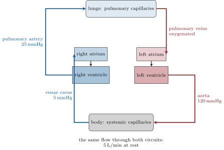
In an open circulation (insects, most molluscs) a heart pumps blood into a body cavity where it bathes the organs directly and returns at low pressure; flow is slow and poorly directed, and the insects have separated the gas problem from the blood by piping air straight to the tissues. In a closed circulation (annelids, cephalopods, vertebrates) the blood stays in vessels and is driven through capillaries at pressure, so that its distribution can be controlled organ by organ. Fish have a single circuit: heart gills body heart, so that blood reaches the tissues at the low pressure left after the gill capillaries. Mammals and birds have a double circuit: the right heart drives the pulmonary circulation (lungs, low pressure, systolic) and the left heart, after the blood returns, drives the systemic circulation (body, high pressure, ); the two pumps are in series and move the same flow, about a minute at rest. Amphibians and most reptiles have a heart with a single ventricle that partly mixes the two streams — the intermediate stage. The double circuit is what makes a warm-blooded metabolism possible: high pressure to every organ and a lung protected from it.
Evidence. Harvey (1628) argued from quantity — the output computed above — and from structure: the valves in the veins, which Fabricius had described, allow flow only toward the heart, as he showed by pressing on the veins of a ligatured arm; the valves of the heart allow flow only from atria to ventricles to arteries; and blood in the arteries spurts, in the veins seeps. He could not see the capillaries and inferred them; Malpighi saw them in a frog’s lung in 1661, four years after Harvey’s death. ∎
16.3 Vessels and the physics of flow
Definition 16.4 (The vascular tree)
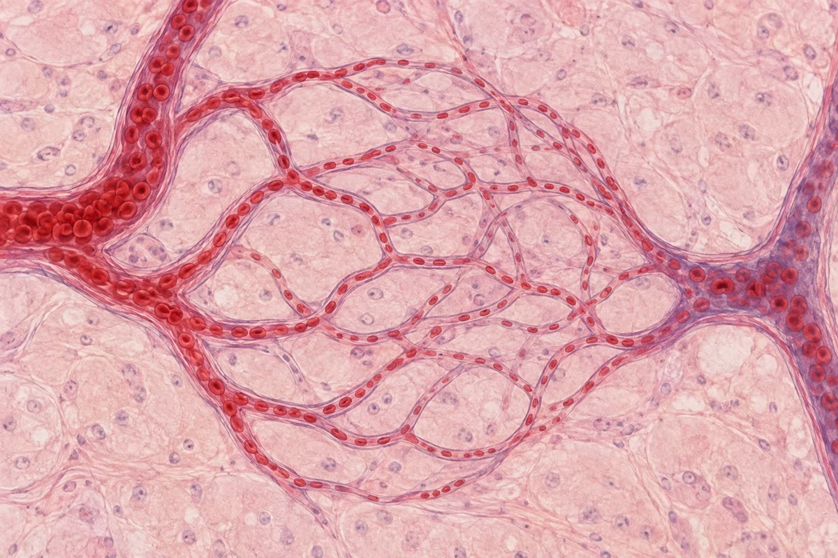
Blood leaves the heart through arteries: thick-walled tubes with elastic layers that stretch at each beat and recoil between beats, smoothing the pulses into a steadier flow (the aorta is across). They branch into arterioles, vessels of a tenth of a millimetre or less whose walls are mostly smooth muscle: by contracting or relaxing, an arteriole changes its radius and so, powerfully, its resistance — arterioles are the taps of the circulation and the site of most of the pressure drop. These open into capillaries: tubes of a single endothelial cell wrapped round a lumen of , about long, some forty billion of them with a total surface near , across whose walls all exchange takes place. Capillaries drain into venules and veins: thin-walled, distensible, holding two thirds of the blood at low pressure, with valves that keep the flow toward the heart while muscles squeeze them. Every vessel is lined with a single layer of endothelium, which is not a passive lining but a gland — it releases nitric oxide to relax the arteriole, and signals that recruit white cells — and a filter.
Theorem 16.5 (Poiseuille’s law and vascular resistance)
The steady flow of a fluid of viscosity through a tube of radius and length under a pressure difference is
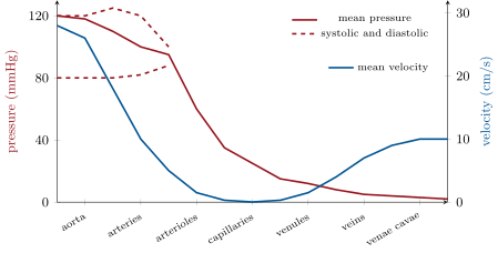
The resistance falls as the fourth power of the radius: an arteriole that narrows its radius by multiplies its resistance by , and one that halves it by 16. This is why a few millimetres of arteriolar muscle can redirect the blood of the whole body — to the gut after a meal, to the muscles in exercise, to the skin in heat — and why the pressure falls from in the small arteries to at the entrance of the capillaries, almost all of it across the arterioles, while the drop along the aorta is a fraction of a millimetre of mercury.
Proof. In steady laminar flow the fluid moves in concentric shells; the viscous force between shells balances the pressure. For the cylinder of radius the pressure force equals the viscous drag on its surface , so and, with , — a parabolic profile. Integrating the velocity over the cross-section, . (Blood is not quite a simple fluid and arteries are not rigid, but the fourth-power law holds well enough to run a body.) ∎
Theorem 16.6 (Continuity: where the blood slows down)
The same volume per second passes every level of the tree, so the mean velocity at a level is , where is the total cross-sectional area of all the vessels at that level. The aorta () carries at ; the capillaries, with a total cross-section near , carry it at , so that a red cell takes some three seconds to cross a capillary — time enough for its oxygen to diffuse out (Chapter 1: a micrometre in a millisecond). The veins, of smaller total section than the capillaries, speed the blood up again to in the venae cavae. The tree is built so that the blood is slow exactly where it must exchange and fast everywhere else.
Proof. Conservation of volume: what enters a level per second leaves it, so is the same at every level. ; ; . ∎
Theorem 16.7 (The elastic artery as a reservoir)
The heart ejects in bursts; the tissues receive a nearly steady flow. The large arteries do the smoothing: they stretch during ejection, storing part of the stroke volume, and recoil during diastole, driving it on. If the arteries have a compliance (volume stored per unit pressure) and the arterioles a resistance , then during diastole, with the aortic valve shut, the pressure decays as
so that the diastolic pressure after a diastole of duration is : with , and , a systolic falls to . A stiffer aorta (smaller , as in old age) lets the pressure fall further between beats and rise higher during ejection: the pulse pressure widens, and the heart works against a higher peak.
Proof. During diastole no blood enters the arteries and blood leaves them through the arterioles at ; the arterial volume falls at , and since , , whose solution is the exponential with time constant . With : . ∎
16.4 Exchange in the capillaries
Proposition 16.8 (Two kinds of exchange)
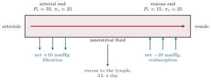
Across the capillary wall, solutes move by diffusion — gases and lipids through the cells, water and small solutes through the clefts between them, over the enormous area and the short distance that make the flux ample (Fick’s law, as for the placenta of Chapter 8) — and water moves by bulk flow, driven by the balance of two pressures. The hydrostatic pressure in the capillary, , pushes fluid out; the oncotic pressure of the plasma proteins, , pulls it in (Starling forces). At the arterial end exceeds and fluid filters out; at the venous end and fluid is reabsorbed; over the whole body about a day filter out and return, and the balance of , with the proteins that leaked, is collected by the lymphatic vessels and returned to the veins at the neck. Oedema — swelling by fluid in the tissues — follows whenever the balance tips: high venous pressure (heart failure), low plasma protein (starvation, liver or kidney disease), leaky capillaries (inflammation), or blocked lymphatics.
Theorem 16.9 (Oncotic pressure)
A solution of moles per litre of a solute that cannot cross a membrane exerts across it an osmotic pressure (van ’t Hoff). Plasma albumin, of a protein of molar mass , is and gives ; the other proteins and the extra ions that albumin’s negative charge retains bring the total to about . The sodium chloride of plasma, at , would exert — but it crosses the capillary wall freely and exerts none across it. What matters for the capillary is the protein: halve the albumin, as in kidney disease that loses it in the urine, and the reabsorbing pull halves, fluid accumulates in the tissues, and the patient swells.
Proof. ; ; , so . The van ’t Hoff law itself is the dilute-solution limit derived in the Year 1 volume. ∎
Example 16.10 (The circulation as a transport system)
Five litres a minute through of capillary wall is a delivery service without equal. It brings each cell oxygen within a few cell diameters (no cell of the body is more than from a capillary), removes its , lactate and urea, carries the glucose of the liver to the brain and the fatty acids of the fat stores to the muscles, distributes the hormones of Chapter 19 from one gland to every receptor in the body in a minute, moves the heat of the core to the skin and back, and ferries the white cells to a wound within hours. It is also the vulnerability: a clot, a haemorrhage or a failing pump stops all of it at once, and the brain, which stores no fuel, fails within seconds. The rest of this part of the book is about the pump (Chapter 17) and about how the pressure is regulated so that the service continues through standing up, running and bleeding (Chapter 18).
16.5 Exercises
Exercise 16.1 ★
Give the composition of blood by volume and the number, size and function of each cellular component.
Exercise 16.2 ★
Draw the circuit of a fish and of a mammal, mark the pressures, and say what the double circuit gains.
Solution
Solution of Exercise 16.2.
Fish: heart gills body heart, one circuit, the body receiving blood at the low pressure left after the gills. Mammal: right heart lungs () left heart body () right heart. The double circuit gives the body high pressure without exposing the lungs to it, and lets the two circuits be regulated separately.
Exercise 16.3 ★
For each vessel type — artery, arteriole, capillary, vein — give the wall structure and the function it serves.
Solution
Solution of Exercise 16.3.
Artery: thick wall with elastic layers and muscle — conducts blood at high pressure and smooths the pulse. Arteriole: wall mostly smooth muscle — sets resistance and distributes flow. Capillary: one endothelial cell thick — exchange. Vein: thin, distensible wall with valves — returns blood at low pressure and stores most of it.
Exercise 16.4 ★
Reconstruct Harvey’s argument from quantity, with modern numbers: per beat, beats a minute, of blood.
Exercise 16.5 ★★
Compute the pressure drop along the aorta (radius , length , ) carrying by Poiseuille’s law, in pascals and in mmHg. Comment.
Solution
Solution of Exercise 16.5.
, : the aorta costs nothing; the pressure is spent in the arterioles.
Exercise 16.6 ★★
An arteriole of radius constricts to , then dilates to . By what factor does its resistance change in each case, and its flow at constant pressure?
Solution
Solution of Exercise 16.6.
: resistance up 2.4-fold, flow down to . : resistance down to a third, flow up 3.2-fold.
Exercise 16.7 ★★
The aorta has a cross-section of and the capillaries a total of ; the flow is . Compute the mean velocity in each, and the time a red cell spends in a capillary. During exercise the output rises to and the muscle capillaries open: what happens to the transit time?
Solution
Solution of Exercise 16.7.
Aorta ; capillaries ; transit . In exercise the output rises fivefold and the open capillary area perhaps threefold, so the transit time falls to about — still enough for the oxygen to leave.
Exercise 16.8 ★★
With and at the two ends, , interstitial pressures negligible, compute the net filtration pressure at each end. What happens if venous pressure rises so that at the venous end is ?
Solution
Solution of Exercise 16.8.
Arterial end (filtration); venous end (reabsorption). With at the venous end the net is : fluid filters along the whole length and none is reabsorbed — oedema, the swollen ankles of heart failure.
Exercise 16.9 ★★
Compute the oncotic pressure of of albumin () at , and of . Why does the sodium of plasma, at , contribute nothing to the Starling balance?
Solution
Solution of Exercise 16.9.
; ; half the albumin, . Sodium crosses the capillary wall freely, so its concentration is the same on both sides and it exerts no osmotic pressure across it.
Exercise 16.10 ★★★
With and , compute the diastolic pressure after from a systolic of . Recompute for an aorta half as compliant. Explain why the pulse pressure of the elderly is wide, and what it costs the heart.
Solution
Solution of Exercise 16.10.
: . : , — and the same stroke volume raises the systolic pressure more in a stiff aorta. A wide pulse pressure means a higher peak against which the heart must eject, more work and more oxygen for the same output.
Exercise 16.11 ★★★
The systemic resistance is . Compute it at rest ( mmHg, ) and in exercise (, ). By what factor must the mean arteriolar radius have changed, if the arterioles carry the whole resistance?
Solution
Solution of Exercise 16.11.
Rest: ; exercise: , a fourfold fall. Since , has risen by : the arterioles have widened by on average.
Exercise 16.12 ★★★
“The circulation is built so that the blood is slow where it must exchange and fast everywhere else.” Discuss, with the continuity equation, Poiseuille’s law and the geometry of the tree, and say why an open circulation cannot do the same.
Solution
Solution of Exercise 16.12.
Continuity fixes the velocity at each level by the total cross-section, and the tree is built so that the section is a thousand times the aorta’s in the capillaries and small again in the veins; Poiseuille’s law puts the resistance in the arterioles, where muscle can change it, and leaves the wide vessels nearly free of loss. An open circulation has no capillaries to slow the blood at a defined place, no vessels in which to set a resistance, and no way to direct flow to one organ — it can only stir.
16.6 Problem: The Circulation in Numbers
Problem 16.1
Weekend problem — Harvey’s sum redone, the vessel tree’s pressures and speeds computed by Poiseuille and continuity, the capillaries’ daily filtration balanced, and the aorta’s smoothing modelled, ending on the cardiac output, the arteriolar pressure drop, the day’s filtrate and the diastolic pressure
Data: stroke volume , heart rate , blood volume , , . Aorta: radius , length . Arterioles: in parallel, each of radius and length . Capillaries: , radius , length , a quarter of them open at rest. Starling: from 35 to along a capillary, . Windkessel: , diastole .
Part I — Harvey’s sum.
- Compute the cardiac output in litres per minute and per day.
- How many times does the blood volume circulate in a day?
- Harvey’s estimate was 2 ounces () per beat, of which he supposed at least a quarter expelled, at 72 beats a minute. What mass of blood did that give per hour, and how did it compare with a man’s weight?
- Why was this an argument for circulation rather than for continuous production and consumption?
- Describe the ligature experiment and what the valves showed.
- What could Harvey not see, and who saw it?
Part II — The tree.
- Compute the mean velocity in the aorta.
- Compute the pressure drop along the aorta by Poiseuille’s law, in mmHg.
- Compute the resistance of one arteriole and of the in parallel.
- Compute the pressure drop across the arterioles at the resting output, in mmHg.
- Compute the total cross-section of the open capillaries and the mean velocity in them; then the transit time through one.
- The arterioles constrict so that their radius falls by . Recompute the drop. What must the heart do to keep the same output?
Part III — The capillaries.
- Compute the net filtration pressure at the arterial end, at the venous end, and at the midpoint (take to fall linearly).
- Split the capillary into the filtering half and the reabsorbing half and estimate the fluid filtered and reabsorbed per day over the whole body, taking the net pressures averaged over each half as and and each half moving a day per mmHg.
- Deduce the lymph flow.
- Plasma albumin falls to . Recompute (assume it scales with albumin) and the net pressures at the two ends. What happens?
- Venous pressure rises so that runs from 35 to . Recompute the average net pressure and the daily balance. Where does the fluid go?
- Compute the total capillary surface (cylinders, all of them) and the time a molecule takes to diffuse to a cell from a capillary ().
Part IV — The aorta as a reservoir.
- Compute the systemic resistance from a mean pressure of , venous and the resting output, in .
- Compute the time constant and the diastolic pressure after from a systolic of .
- Recompute for . What has happened to the pulse pressure?
- The heart rate rises to and diastole shortens to . Compute the diastolic pressure.
- Of the ejected, how much is stored in the arteries during systole if the pressure rises by ? Where does the rest go?
- Explain why the coronary arteries, which fill in diastole, depend on the aorta’s recoil.
- State the result: the cardiac output, the arteriolar pressure drop, the day’s net filtrate, and the diastolic pressure for and .
Solution
Solution of Problem 16.1.
1. ; a day. 2. times. 3. A quarter of at 72 beats a minute: an hour — about the weight of a man. 4. No food could supply, and no tissue consume, a man’s weight of blood every hour; the only escape is that the same blood returns to the heart. 5. A cord tied round the arm swells the veins below it, not above, so blood in the veins moves toward the heart; pressing the blood in a vein toward the hand, it stops at the valves and cannot be pushed back — the valves permit flow toward the heart only. 6. The capillaries joining arteries to veins; Malpighi saw them in the frog’s lung in 1661. 7. Area ; . 8. , . 9. ; in parallel, . 10. . 11. Open capillaries , each : ; ; transit . 12. Resistance : ; the heart must raise the arterial pressure by or the output falls by a third. 13. , and mmHg. 14. Filtration a day; reabsorption . 15. a day of lymph. 16. : net at the arterial end and at the venous end — filtration everywhere, no reabsorption: oedema. 17. Net and , average : filtration along the whole capillary, some a day; the lymphatics cannot carry it and the fluid accumulates in the tissues, feet first. 18. ; . 19. . 20. ; . 21. ; : the pulse pressure widens from 37 to . 22. : at a fast rate the pressure barely falls between beats. 23. stored; the other run off through the arterioles during systole itself. 24. The contracting ventricle squeezes its own vessels shut in systole, so the heart muscle is perfused in diastole, by the pressure that the recoiling aorta maintains; a stiff aorta that lets the diastolic pressure collapse starves the heart between beats. 25. Output ; arteriolar drop ; net filtrate a day to the lymph; diastolic for and for .