University Biology — Year 2 · Bachelor Year 2
20Neurons, Action Potentials and Synapses
Tap the tendon below the kneecap and the leg kicks, thirty milliseconds later. In that time a stretch has been sensed in the thigh, a signal has travelled a metre to the spinal cord, crossed one junction to a second cell, travelled a metre back and set a muscle contracting — the whole round trip faster than a blink. No hormone could do this; it is done by cells built to conduct, and by a signal that is not a substance moving but a wave of electricity regenerated along a membrane. This chapter is about that cell and that signal: the voltage a neuron keeps across its membrane, the impulse it fires and how the impulse travels, and the synapse where the electrical signal is turned into a chemical one and back again.
20.1 The neuron
Definition 20.1 (Neuron, axon, nerve)
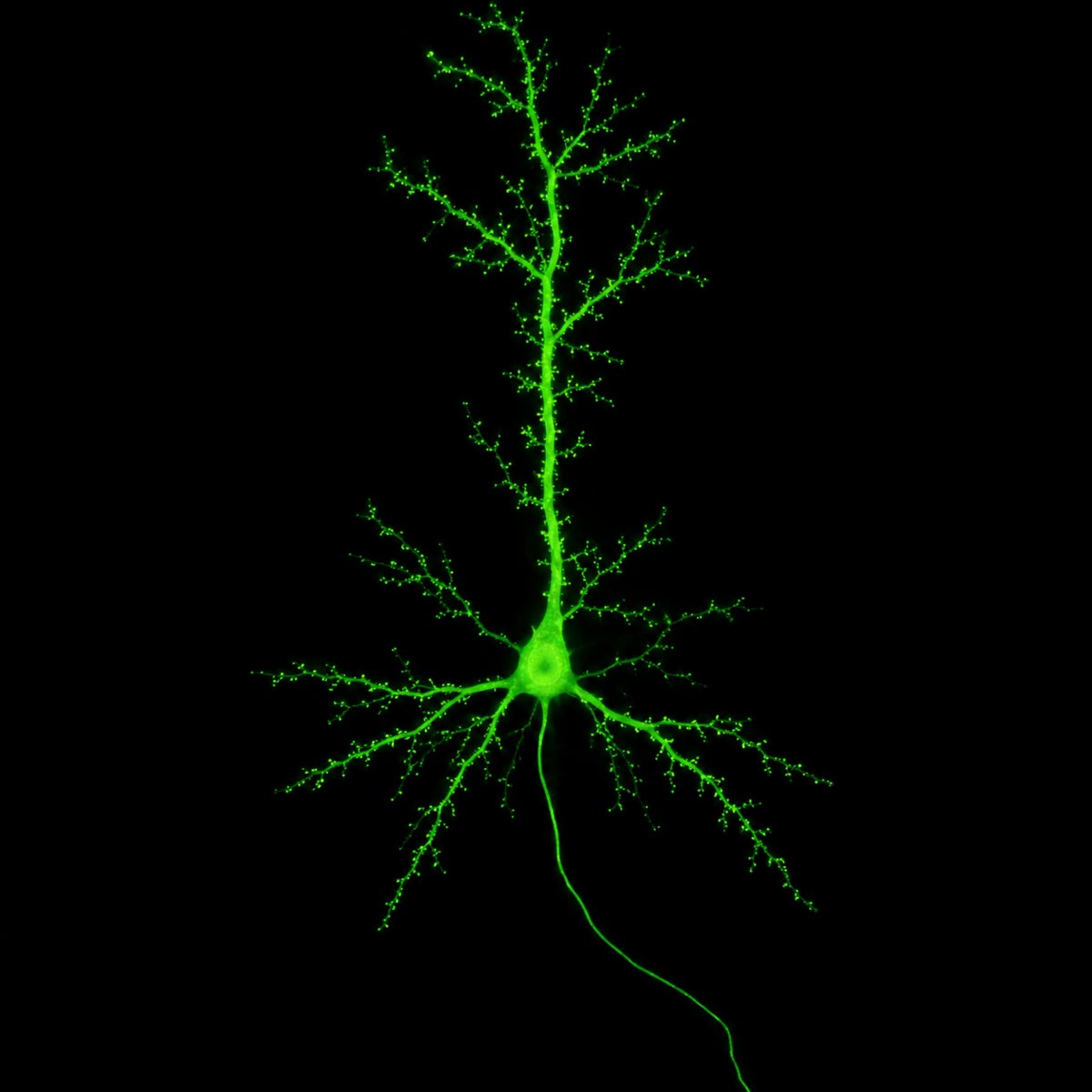
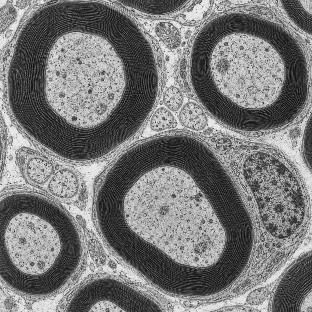
A neuron is a cell specialised for signalling: a cell body (soma) with the nucleus, branching dendrites that receive signals, and a single axon — a cable from a micrometre to twenty micrometres thick and up to a metre long — that carries its output to terminals on other cells. The human brain has some neurons and each makes about a thousand synapses. Neurons are outnumbered by glia: astrocytes that feed and buffer them, microglia that police them, and the cells that wrap axons in myelin — dozens of turns of their own membrane, nearly free of cytoplasm, forming an insulating sheath interrupted every millimetre or so at the nodes of Ranvier. A nerve is a bundle of thousands of axons, sensory and motor, running together in connective tissue. Neurons do not divide in the adult (with few exceptions), and an axon cut from its body dies; a neuron is a cell that must last a lifetime.
20.2 The resting potential
Theorem 20.2 (The Nernst potential)
A membrane permeable to one ion of charge separating concentrations and settles at the equilibrium potential at which the electrical force on the ion balances its diffusion:
A neuron holds of potassium inside against outside, and of sodium inside against outside, gradients built by the sodium–potassium pump at the cost of a third of the cell’s ATP. Hence and : if the membrane let only potassium through it would sit at , if only sodium at .
Proof. At equilibrium the electrochemical potential of the ion is the same on both sides: , so . At , and . (Derived in the Year 1 volume from the Boltzmann distribution; restated here.) ∎
Theorem 20.3 (The resting potential as a weighted mean)
If the membrane conducts several ions, each with conductance (the ease with which it passes, set by the number of open channels), the steady potential at which the currents cancel is
a mean of the equilibrium potentials weighted by the conductances. At rest a neuron’s membrane is about twenty times more permeable to potassium than to sodium, so ; with the chloride and the small leaks counted, the resting potential is . The membrane rests near because potassium channels are open; whatever opens sodium channels drags it toward — and that is the whole principle of nerve signalling.
Proof. Each ion carries a current (Ohm’s law, with the driving force measured from the ion’s own equilibrium). At a steady potential the total current is zero: , whence . (The full treatment, in which the permeabilities enter through the Goldman–Hodgkin–Katz equation, gives the same weighting to first order.) ∎
20.3 The action potential
Proposition 20.4 (The impulse)
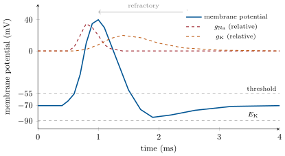
The axon membrane holds voltage-gated sodium channels, shut at rest and opened by depolarisation. If a stimulus raises the potential from past a threshold near , enough of them open to let in more sodium than the potassium leak can offset; the potential rises, more channels open, and within a fraction of a millisecond the membrane is swept toward — a positive feedback that is explosive and all-or-none. It peaks near because the sodium channels inactivate within a millisecond of opening and because slower voltage-gated potassium channels open and drive the potential back toward , overshooting to an undershoot before the potassium channels close. The action potential lasts about a millisecond; for another millisecond or two the inactivated sodium channels cannot reopen (the refractory period), which limits the firing rate to a few hundred per second and forces the impulse to travel one way. Because it is all-or-none, the impulse carries no information in its size: the neuron codes intensity in its frequency. Some sodium ions enter and as many potassium ions leave per impulse — a negligible change in concentration, restored by the pump at leisure.
Evidence. Hodgkin and Huxley (1952), using the squid’s giant axon (half a millimetre thick, so that a wire could be threaded inside it) and a feedback amplifier that held the membrane potential at any chosen value (the voltage clamp) while recording the current needed to hold it, separated the current into an early inward component that vanished when sodium was removed from the bath and a later outward component carried by potassium; measured how each conductance rose and fell with time at each voltage; and, fitting each with a few equations, computed the action potential’s shape, threshold, speed and refractory period from the two conductances alone. Every prediction held. The channels themselves were seen twenty-five years later, one at a time, with the patch clamp; tetrodotoxin (the pufferfish poison) blocks the sodium channel and abolishes the impulse, tetraethylammonium blocks the potassium channel and prolongs it. ∎
20.4 Conduction
Theorem 20.5 (The cable and its length constant)
An axon is a leaky cable: current injected at a point flows along the cytoplasm (resistance per unit length) and leaks out through the membrane (resistance times unit length). A steady depolarisation at decays along the axon as
where is the diameter and , the membrane’s and cytoplasm’s specific resistances. The length constant is the distance over which a passive signal falls to a third: about for a axon, growing as the square root of the diameter. An action potential propagates because the depolarisation at its front, spreading passively ahead by about , brings the next stretch of membrane to threshold, which regenerates it at full size; the further ahead it reaches, the faster the wave, so the conduction velocity also grows roughly as — from in a thin unmyelinated fibre to in the squid’s giant axon.
Proof. Let be the axial current and the membrane depolarisation. Ohm’s law along the axoplasm gives ; conservation of charge, with the leak through the membrane, gives . Combining, , whose decaying solution is with . For a cylinder, and , so . With , and : . ∎
Proposition 20.6 (Myelin and saltatory conduction)
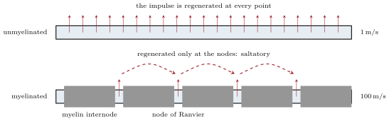
Myelin multiplies by the number of membrane turns and divides the membrane’s capacitance by the same factor, so that under the sheath almost no current leaks and little charge is needed to change the potential: the depolarisation spreads passively along an internode of a millimetre with little loss, and the action potential is regenerated only at the nodes, where the sodium channels are concentrated. The impulse thus jumps from node to node (saltatory conduction), at in a fibre — a speed an unmyelinated axon would need to be centimetres thick to reach — and at a fraction of the metabolic cost, since sodium enters only at the nodes. Myelin is the vertebrate invention that made a large, fast nervous system possible in a small body; in multiple sclerosis the sheath is destroyed patch by patch, the length constant collapses, and the impulses fail.
20.5 The synapse
Definition 20.7 (Chemical and electrical synapses)
At a synapse a neuron’s terminal meets a target — another neuron, a muscle fibre, a gland cell. In an electrical synapse gap junctions join the two cells and current passes directly — fast, bidirectional, without amplification, used where speed and synchrony matter (escape reflexes, the heart). In a chemical synapse the cells are separated by a cleft of and the signal is a molecule. An action potential arriving at the terminal opens voltage-gated calcium channels; calcium enters and, within a fraction of a millisecond, makes vesicles of neurotransmitter fuse with the membrane and empty into the cleft; the transmitter diffuses across in microseconds and binds receptors on the postsynaptic membrane — either ion channels that it opens (ionotropic, fast) or G-protein-coupled receptors (metabotropic, slower, Chapter 19); the resulting current shifts the postsynaptic potential by a few millivolts, up (an excitatory postsynaptic potential, from channels passing sodium) or down (an inhibitory one, from channels passing chloride or potassium); and the transmitter is removed within milliseconds by an enzyme or a transporter. The delay is half a millisecond; the price buys one-way transmission, amplification, inhibition, and modifiability — everything a nervous system needs beyond mere conduction.
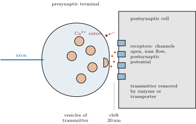
Theorem 20.8 (Quantal release)
Transmitter is released in packets — quanta, one vesicle each — and the number released by one impulse is a random variable. If the terminal holds release sites each releasing with probability , and is small, is approximately Poisson with mean :
The mean quantal content can therefore be read from the fraction of impulses that release nothing, and checked against the mean response divided by the size of one quantum. At a neuromuscular junction is a few hundred — transmission never fails; at a central synapse it is often near one, and the synapse transmits on a fraction of the impulses.
Proof. With independent sites each releasing with probability , is binomial; for large and small with fixed, the binomial tends to the Poisson law (the mathematics series, Year 2). gives from the failures. Katz’s check is that the same , put into the Poisson formula, predicts the observed fractions of responses of one, two and three quanta. ∎
Evidence. Fatt and Katz (1952) recorded from a frog muscle fibre at rest small spontaneous depolarisations of about , all the same size, at random intervals — the miniature end-plate potentials, each the effect of one vesicle. Lowering the calcium in the bath reduced the response to nerve stimulation until it fluctuated among 0, 1, 2 or 3 multiples of the miniature size; the frequencies of the multiples followed the Poisson law with the computed from the failures. Del Castillo and Katz thus showed that release is quantal, that calcium controls the probability of release and not the size of a quantum, and electron microscopy showed the vesicles that are the quanta. ∎
Proposition 20.9 (Integration: the neuron as a decision)
A central neuron receives thousands of synapses, excitatory and inhibitory, on its dendrites and soma. Each postsynaptic potential is small and decays passively, with the cable’s length constant in space and the membrane’s time constant (a few milliseconds) in time; they add — spatial summation of inputs arriving together, temporal summation of inputs arriving in quick succession — and the sum is read at the axon hillock, where the sodium channels are densest and the threshold lowest. If the sum crosses threshold the axon fires, at a rate that rises with the excess; inhibitory inputs subtract, and an inhibitory synapse close to the hillock can veto a distant excitatory one. The neuromuscular junction is the exception that shows the rule: one motor terminal, releasing hundreds of quanta of acetylcholine onto receptors that are also the channels, gives an end-plate potential of , far above threshold — every impulse in the motor nerve produces a muscle action potential (Chapter 21). Curare, which blocks the receptor, paralyses; nerve gases, which block the enzyme that removes acetylcholine, paralyse by the opposite excess.
Example 20.10 (Transmitters and drugs)
Acetylcholine at the neuromuscular junction and in the autonomic system; glutamate, the main excitatory transmitter of the brain, and GABA, the main inhibitory one; the monoamines noradrenaline, dopamine and serotonin, which modulate whole circuits from a few thousand cells; and dozens of peptides. Almost every drug that acts on the mind acts on a synapse: benzodiazepines strengthen GABA’s channel, antidepressants block serotonin’s transporter, cocaine blocks dopamine’s, opiates mimic a peptide, nicotine mimics acetylcholine on one receptor and atropine blocks it on another, botulinum toxin cuts the proteins that fuse the vesicle. The synapse is where the chemistry of Chapter 19 and the electricity of this chapter meet, and where a nervous system learns: synapses that are used strengthen, and that strengthening (the Year 3 volume) is memory.
20.6 Exercises
Exercise 20.1 ★
Name the parts of a neuron and the function of each, and say what myelin is made of and what it does.
Solution
Solution of Exercise 20.1.
Dendrites receive synapses; the soma holds the nucleus and integrates; the axon hillock decides; the axon conducts; the terminals release transmitter. Myelin is the wrapped membrane of a glial cell, nearly free of cytoplasm; it insulates the axon so that the impulse jumps between nodes and travels a hundred times faster at lower cost.
Exercise 20.2 ★
Describe the phases of an action potential and the channel events that cause each. Why is it all-or-none, and why does it not travel backwards?
Solution
Solution of Exercise 20.2.
Depolarisation to threshold; rising phase — voltage-gated sodium channels open, positive feedback; peak near — sodium channels inactivate; falling phase — voltage-gated potassium channels open; undershoot — potassium channels still open, near ; recovery as they close. All-or-none because the positive feedback either runs to completion or not at all; one-way because the membrane just traversed is refractory (sodium channels inactivated).
Exercise 20.3 ★
List the steps of transmission at a chemical synapse from the arrival of the impulse to the removal of the transmitter.
Solution
Solution of Exercise 20.3.
Impulse reaches the terminal; voltage-gated calcium channels open; calcium triggers vesicle fusion; transmitter diffuses across the cleft; binds postsynaptic receptors; channels open (or a G protein switches); postsynaptic potential; transmitter removed by enzyme or transporter; vesicle membrane retrieved.
Exercise 20.4 ★
Compare electrical and chemical synapses in speed, direction, amplification and the possibility of inhibition.
Solution
Solution of Exercise 20.4.
Electrical: no delay, usually both directions, no amplification, no inhibition (current only passes). Chemical: half a millisecond, one-way, amplification (one impulse releases hundreds of quanta), inhibition possible (channels for chloride or potassium), and modifiable.
Exercise 20.5 ★★
Compute the Nernst potentials at for potassium (140 in, 5 out), sodium (15 in, 145 out), chloride (10 in, 110 out) and calcium ( in, 2 out, in ). Which way does each ion flow when the membrane is at ?
Solution
Solution of Exercise 20.5.
From : potassium ; sodium ; chloride, with , ; calcium, with , . At : potassium leaves, sodium enters, chloride leaves slightly (the membrane is below its equilibrium), calcium enters strongly.
Exercise 20.6 ★★
With and , compute the membrane potential when (rest), (the peak of the impulse) and (the undershoot). Explain each state.
Solution
Solution of Exercise 20.6.
: , the resting state near . : , the peak, near . : , the undershoot with extra potassium channels open.
Exercise 20.7 ★★
Conduction velocity scales as in unmyelinated axons, with at . Compute the velocity of a squid axon. A myelinated fibre conducts at : how thick would an unmyelinated axon have to be to match it? How long does an impulse take from the spinal cord to the toe () in each fibre?
Exercise 20.8 ★★
In 200 stimulations of a synapse in low calcium, 74 produce no response. Compute the mean quantal content and the predicted fractions of responses of one, two and three quanta. The single quantum is : what mean response is expected?
Solution
Solution of Exercise 20.8.
; , , . Mean response .
Exercise 20.9 ★★
A neuron’s refractory period is . What is its maximal firing rate? A sensory neuron fires at 20 impulses a second for a light touch and 200 for a hard one: what is coded, and by what?
Solution
Solution of Exercise 20.9.
impulses a second. The intensity of the touch is coded by the frequency of impulses (and by how many neurons are recruited); the impulses themselves are identical.
Exercise 20.10 ★★★
Compute the length constant for , and diameters of 1, 10 and . Myelin multiplies by 200: recompute for . Explain why an internode of works and why a demyelinated stretch of blocks the impulse.
Solution
Solution of Exercise 20.10.
: , , . With myelin, : . An internode of loses only of the signal, so the next node is easily brought to threshold. Across a demyelinated the signal falls to — of a impulse, about the threshold: conduction fails or becomes unreliable.
Exercise 20.11 ★★★
Predict the effect on the action potential and on transmission at the neuromuscular junction of: tetrodotoxin; tetraethylammonium; a bath without calcium; curare; an acetylcholinesterase inhibitor; botulinum toxin.
Solution
Solution of Exercise 20.11.
Tetrodotoxin: no sodium current, no impulse, no transmission. Tetraethylammonium: no potassium current, prolonged impulse, more transmitter released per impulse. No calcium: impulses normal, no release, no transmission. Curare: release normal, receptors blocked, no end-plate potential — paralysis. Cholinesterase inhibitor: acetylcholine persists, receptors stay open, the muscle depolarises and then cannot repolarise — paralysis by excess. Botulinum toxin: vesicles cannot fuse, no release — paralysis.
Exercise 20.12 ★★★
“A chemical synapse costs half a millisecond and buys a nervous system.” Discuss what the delay pays for — one-way transmission, amplification, inhibition, plasticity — and where the body uses electrical synapses instead.
Solution
Solution of Exercise 20.12.
The delay buys: transmission in one direction only; amplification (a single impulse releases hundreds of quanta and can drive a larger cell); inhibition, which an electrical junction cannot do; and plasticity, since the number of quanta and receptors can change with use, which is how circuits learn. Electrical synapses are used where synchrony and speed matter more than computation: escape circuits, the heart’s muscle, some inhibitory networks.
20.7 Problem: The Knee Jerk Timed
Problem 20.1
Weekend problem — the stretch reflex followed from the tendon tap to the kick, with the neuron’s resting potential computed, the impulse’s conduction timed along the two limbs of the arc, the synapse’s quantal content measured, and the whole latency accounted for, ending on the resting potential, the conduction times, the quantal content and the reflex latency
Data at : potassium 140 in, 4 out; sodium 12 in, 145 out (); at rest. Sensory and motor axons: , myelinated, , each way. Synaptic delay ; muscle activation after its own action potential . Central synapse in low calcium: 300 trials, 111 failures. Quantum ; threshold above rest. , ; myelin multiplies by 250.
Part I — The resting neuron.
- Compute and .
- Compute the resting potential from the conductance ratio.
- The extracellular potassium rises to (as after intense exercise). Recompute and the resting potential, and say what this does to excitability.
- The pump stops. Explain what happens to the gradients and the potential over hours, and why the cell swells.
- How many sodium ions enter a axon per millimetre per impulse if the membrane’s capacitance is and the potential swings by ? (; one ion carries .)
- By what fraction does that change the internal sodium concentration (axon volume per millimetre)?
- The pump exports three sodium ions per ATP. How many ATP does it cost to undo one impulse per millimetre of axon?
Part II — Conduction.
- Compute the time for the sensory impulse to reach the spinal cord and for the motor impulse to reach the muscle.
- Compute the length constant of a bare axon and of the same axon under myelin.
- An internode is . What fraction of the nodal depolarisation reaches the next node passively, with and without myelin? Which case can bring the next node to threshold ( from a impulse)?
- Unmyelinated, the same axon would conduct at m/s. Compute the round trip and comment.
- A patch of loses its myelin. Compute the fraction of the signal crossing it and say whether the reflex survives.
- Why does the refractory period make the impulse travel in one direction only along the sensory axon?
Part III — The synapse.
- Compute the mean quantal content from the failures.
- Compute the predicted fractions of responses of 1, 2 and 3 quanta, and the expected numbers among the 300 trials.
- Compute the mean postsynaptic potential.
- In normal calcium the same synapse has . What is the probability of a failure, and the mean response? Does one such synapse fire the motor neuron?
- The motor neuron receives such sensory synapses active at once. Explain how they sum and whether threshold is reached.
- An inhibitory synapse on the same neuron opens chloride channels (, the resting potential). Explain how it can reduce the excitatory response without hyperpolarising the cell.
Part IV — The whole reflex.
- Add up: sensory conduction, synaptic delay, motor conduction, neuromuscular delay (), muscle activation. Compare with the measured .
- Where does the rest of the measured latency come from?
- The reflex has one synapse (monosynaptic). Explain why a withdrawal reflex, with two or three synapses in the cord, is slower but more flexible.
- The same tap in a patient with peripheral demyelination gives a reflex late. Estimate the conduction velocity in her nerves.
- A dose of tetrodotoxin blocks half the sodium channels. Predict the effect on the threshold, the impulse and the reflex.
- State the result: the resting potential, the sensory and motor conduction times, the quantal content , and the computed reflex latency.
Solution
Solution of Problem 20.1.
1. ; . 2. . 3. ; : closer to threshold, so more excitable at first — and, if it persists, less, as sodium channels inactivate at the depolarised level. 4. The gradients run down over hours as sodium leaks in and potassium out; the potential drifts toward zero; with the pump stopped the cell’s impermeant anions draw in chloride and water, and it swells. 5. Surface per millimetre ; ; : ions. 6. Volume per millimetre , holding mol, ions: a change of . 7. ATP per millimetre per impulse. 8. each way. 9. Bare: ; myelinated: . 10. Bare: , ; myelinated: , . Both exceed the threshold; but the bare axon must charge its whole membrane along the way, which is what makes it slow, whereas under myelin almost nothing is lost or charged. 11. : each way, nearly half a second for the round trip — too slow for a reflex that must catch a stumble. 12. : at the far node, below threshold — the impulse is blocked and the reflex is lost. 13. Behind the impulse the sodium channels are inactivated for a millisecond or two, so the membrane just traversed cannot be re-excited and the wave can only move forward. 14. . 15. , , : about 110, 55 and 18 of the 300 trials. 16. . 17. : never; mean response , above the threshold: one such synapse fires the motor neuron. 18. Twenty synapses active together add their currents (spatial summation), sublinearly as the potential approaches the reversal potential of the channels; even at each they would give , and at normal far more than threshold. 19. Opening chloride channels at adds conductance without moving the potential; the excitatory current now flows through a leakier membrane and produces a smaller depolarisation — shunting inhibition. 20. , against measured. 21. The receptor’s own activation in the muscle spindle, the propagation of the muscle fibre’s action potential along the fibre (metres per second over centimetres), and the mechanical latency before force appears. 22. Each synapse adds half a millisecond and an interneuron adds a cell; but interneurons allow the signal to be inverted (inhibiting the antagonist), combined with others, and modulated by the brain — a reflex that can be shaped. 23. of conduction for : about . 24. Half the inward current at every voltage: the threshold rises, the impulse is smaller and slower, the safety factor at each node falls, and the impulse may fail; the reflex weakens or disappears. 25. Resting potential ; each way; ; computed latency .