University Biology — Year 2 · Bachelor Year 2
1Diversity of Unicellular Organisms
A drop of pond water under the microscope holds a green cell rowing with two flagella, a slipper-shaped ciliate sweeping bacteria into its gullet, a glassy diatom sliding along the slide, and, too small to see well, a thousand bacteria. Each is one cell and each is a whole organism: it feeds, moves, senses, grows, reproduces and dies as a single cell. Most of the living world has always been like this. This chapter takes the unicellular organism as a design problem — what one cell can and cannot do — and surveys the answers that prokaryotes and eukaryotes have found, up to the point where cells begin to stay together.
1.1 One cell, all the functions
Definition 1.1 (Unicellular, colonial, multicellular)
An organism is unicellular when a single cell carries out every function of life — nutrition, exchange, movement, reproduction — and lives on its own. It is colonial when cells of one kind stay attached after dividing but each remains able to live alone if separated. It is multicellular when its cells are of several types, depend on one another, and only a few of them (the germ line) leave descendants. Unicellular organisms include all archaea, nearly all bacteria, and most lineages of eukaryotes; the informal name protist covers the unicellular eukaryotes, which are not one group but many.
Example 1.2 (Six inhabitants of a drop)
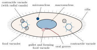
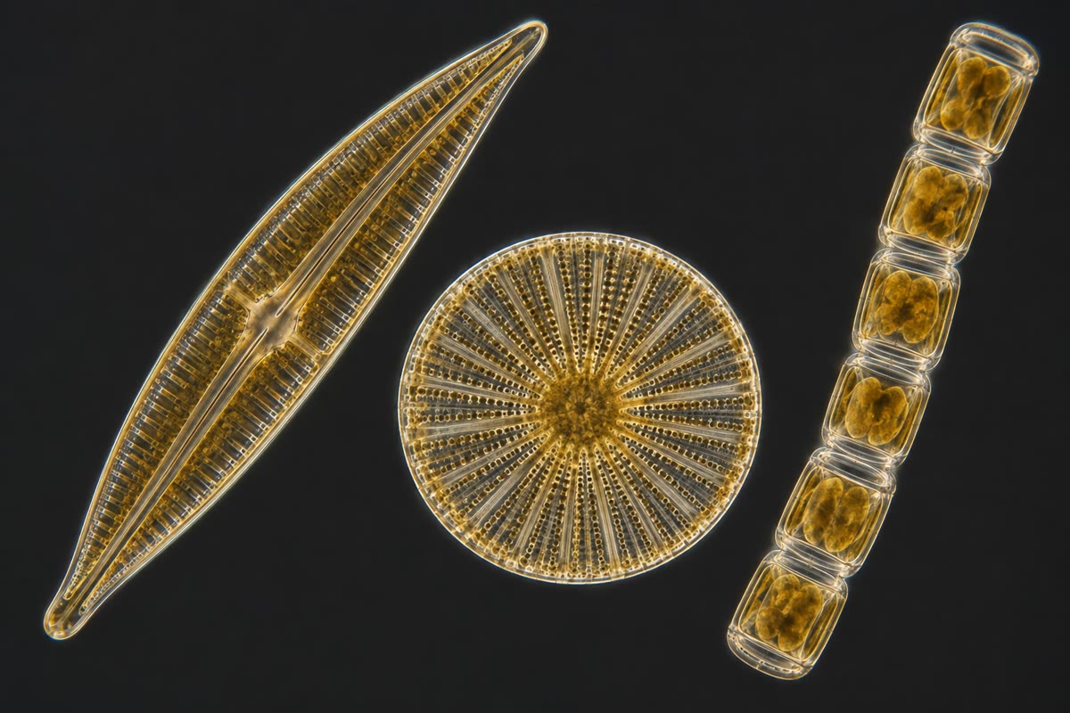
Escherichia coli, a rod long, swims with a tuft of flagella and divides every twenty minutes when fed. Chlamydomonas, a green alga across, rows with two flagella toward the light it detects with an eyespot. Paramecium, a ciliate of , is covered with thousands of cilia, feeds through a gullet and bails water out with two contractile vacuoles. A diatom lives in a two-part shell of silica, ornamented with pores. Amoeba flows by extending pseudopods and engulfs what it meets. A yeast cell buds a daughter from its side. Six answers, of widely different size and machinery, to one problem.
Proposition 1.3 (Surface, volume and the limit on size)
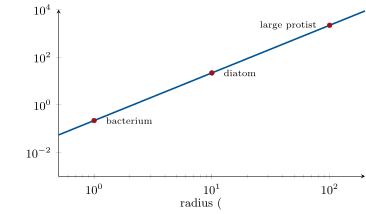
A cell exchanges with its surroundings through its surface and consumes in proportion to its volume. For a sphere of radius , the surface-to-volume ratio is
so that doubling the radius halves the surface available per unit of cytoplasm. A cell that relies on diffusion across its membrane to feed its interior therefore cannot grow indefinitely: at some radius the supply through the surface no longer meets the demand of the volume. The same law explains why small cells are metabolically the most active per gram, and why large cells are flattened, elongated, vacuolated or full of internal membranes.
Theorem 1.4 (Time to diffuse a distance)
A molecule with diffusion coefficient wanders in random steps; after a time its mean square displacement along one axis is . The time needed to cover a distance by diffusion alone is therefore
which grows with the square of the distance. With for a small molecule in water, crossing a bacterium () takes half a millisecond; crossing a cell would take eight minutes, and a metre, sixteen years.
Proof. Let the molecule take steps of length at random to the left or right every seconds. After steps its displacement is with independent, so and (the cross terms average to zero). With this is , and writing gives . Setting gives the time. ∎
Theorem 1.5 (Diffusive supply to a spherical cell)
A spherical cell of radius that absorbs every molecule of a solute reaching its surface, in a medium where the concentration far away is , receives at steady state a flux (molecules per second) of
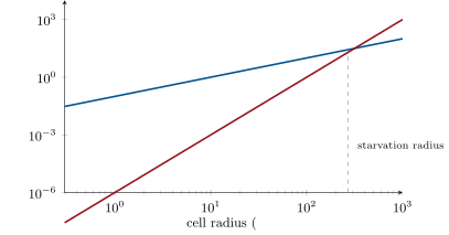
The supply grows only with , while the demand grows with : beyond a certain radius a cell fed by diffusion starves.
Proof. At steady state the number of molecules crossing every sphere of radius per second is the same, (Fick’s law, the flux directed inward). So is constant; integrating from , where , to infinity, where : , whence . ∎
Example 1.6 (How big can a diffusion-fed cell be?)
An aerobic cell consumes oxygen at about of cytoplasm (a brisk rate). Its demand is ; the supply from air-saturated water (, ) is . Equating, , so . Real active cells stay well below this because oxygen must also diffuse inside them; the sulfur bacterium Thiomargarita gets round the limit by being almost entirely a vacuole, its cytoplasm a thin shell a few micrometres deep.
1.2 Prokaryotic diversity
Definition 1.7 (The prokaryotic cell)
A prokaryote (the bacteria and the archaea, two domains as distinct from each other as either is from us) is a cell without a nucleus or membrane-bound organelles: a single circular chromosome in a nucleoid, often with small extra circles called plasmids, ribosomes free in a cytoplasm of , a plasma membrane that carries the respiratory or photosynthetic chains, and a rigid cell wall that resists the turgor of a cytoplasm several bars above its surroundings. Shapes are few and named: coccus (sphere), bacillus (rod), spirillum and spirochaete (helices), vibrio (comma); cells may stay in pairs, chains, tetrads or clusters after dividing.
Proposition 1.8 (Two envelopes: Gram-positive and Gram-negative)
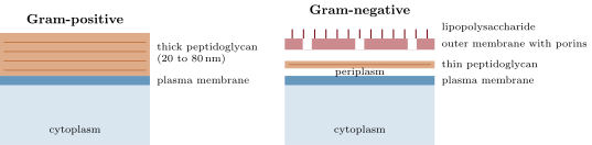
The bacterial wall is built of peptidoglycan, chains of two alternating sugars cross-linked by short peptides into one giant molecule around the cell. Gram-positive bacteria have a thick layer () outside a single membrane and retain the crystal-violet stain of the Gram procedure; Gram-negative bacteria have a thin layer (a few nanometres) sandwiched between the plasma membrane and an outer membrane whose outer leaflet is made of lipopolysaccharide, and they lose the stain. The outer membrane, pierced by porins, is a second barrier: it keeps out many antibiotics and detergents, which is why Gram-negative infections are harder to treat. Archaea have neither peptidoglycan nor the ester-linked lipids of every other cell: their membranes are built of ether-linked isoprenoid chains, sometimes spanning the whole bilayer as a monolayer, which keeps them stable in boiling acid.
Proposition 1.9 (The bacterial flagellum and chemotaxis)
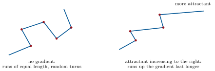
The bacterial flagellum is a rigid helical filament of the protein flagellin, thick and several body lengths long, turned by a rotary motor set in the envelope. The motor is driven not by ATP but by protons flowing down the electrochemical gradient across the membrane, at some protons per turn and up to turns per second. In E. coli the several flagella bundle together when they all turn counter-clockwise and push the cell in a straight run; when one or more reverse, the bundle flies apart and the cell tumbles to a new random direction. Chemotaxis is the bias of this random walk: the cell measures whether an attractant concentration has risen over the last second and, if so, suppresses tumbling. It steers by comparing the present with the recent past — along its path — because across its own of length no gradient is measurable.
Evidence. Berg and Brown (1972) followed single E. coli cells in three dimensions with a tracking microscope: runs of about a second at , tumbles of a tenth of a second, and in a gradient of serine the runs up the gradient were longer while the runs down it were not shortened. Tethering a cell to a glass slide by one flagellum made the whole body spin, which proved the motor is rotary; cells with the motor uncoupled from ATP synthesis still swam as long as a proton gradient could be imposed, which identified the fuel. ∎
Example 1.10 (Cyanobacteria and the heterocyst)
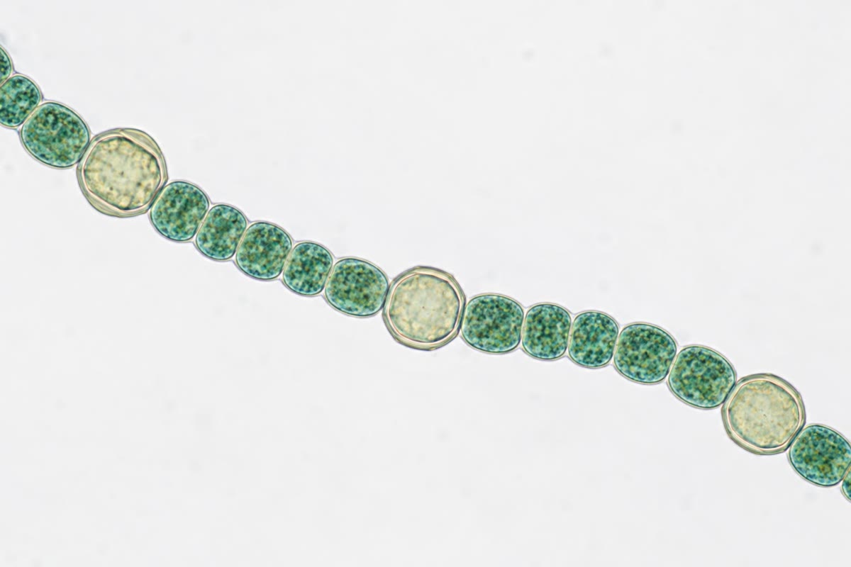
Cyanobacteria are the bacteria that photosynthesise as plants do, splitting water and releasing oxygen on stacked internal membranes (the thylakoids, which the chloroplast inherited from them). Many grow as filaments of cells sharing a sheath. In Anabaena, when combined nitrogen runs out, every tenth cell or so becomes a heterocyst: it dismantles its oxygen-producing photosystem, thickens its wall against oxygen, and fixes atmospheric nitrogen with an enzyme that oxygen would destroy, exporting glutamine to its neighbours in exchange for sugar. A filament of Anabaena is a first sketch of a division of labour between cells.
Remark 1.11 (Dormancy)
Some Gram-positive rods (Bacillus, Clostridium) answer starvation by building an endospore: a copy of the chromosome wrapped in several dehydrated coats, metabolically inert, resistant to boiling, radiation and centuries of drought, which germinates when conditions return. Many protists do the same with a cyst. Dormancy is the other way of surviving a bad season, alongside motility.
1.3 Eukaryotic unicellular organisms
Definition 1.12 (Protists: a grade, not a group)
Protists are the eukaryotes that are neither animals, nor land plants, nor fungi — mostly unicellular, some colonial, a few (the kelps) multicellular. They share the eukaryotic cell of the Year 1 volume (nucleus, mitochondria, endomembranes, cytoskeleton, cilia) and nothing else in particular: the lineages that contain them are as distant from one another as animals are from plants. Green algae belong with the land plants; diatoms and brown algae form a lineage of their own; ciliates, dinoflagellates and the malaria parasite form another; amoebae and slime moulds are closer to animals and fungi than to any of these. The word names a level of organisation — the eukaryotic cell living alone — and not a branch of the tree.
Example 1.13 (Four ways to be a eukaryotic cell alone)
Chlamydomonas: an ovoid green alga with a cellulose-free glycoprotein wall, one cup-shaped chloroplast, an orange eyespot that shades a light sensor so that the cell knows which way the light is as it rotates, and two anterior flagella that beat in a breaststroke; it is a phototroph. Paramecium: a ciliate, its whole surface beating with cilia in coordinated waves, a mouth-like oral groove that sweeps bacteria into food vacuoles, two contractile vacuoles, and two kinds of nucleus — a small germinal micronucleus and a large working macronucleus with hundreds of copies of each gene; it is a phagotroph. Amoeba: no wall, no fixed shape, moving and feeding by pseudopods that actin polymerisation pushes out. The diatom: a photosynthetic cell enclosed in two overlapping valves of silica like a box and its lid, so ornamented with pores that the shells were once used to test microscope lenses; when it divides, each daughter keeps one valve and makes a new, smaller, inner one.
Definition 1.14 (Modes of nutrition)
A unicellular eukaryote feeds in one of three ways, or in two at once. Phototrophy: it has chloroplasts and makes its own organic matter from light and (green algae, diatoms, dinoflagellates). Phagotrophy: it engulfs particles — bacteria, other protists — into a food vacuole where lysosomal enzymes digest them (amoebae, ciliates, choanoflagellates). Osmotrophy: it absorbs dissolved molecules across its membrane, often after secreting enzymes outside (yeasts, many parasites). Mixotrophs such as Euglena photosynthesise in the light and eat in the dark. Prokaryotes cannot phagocytose: the wall forbids it, which is why a prokaryote never became a predator by engulfing.
Proposition 1.15 (Osmoregulation in fresh water)
A freshwater protist is far saltier inside (some of solutes) than the pond (about ), and its membrane lets water through. Water therefore enters continuously by osmosis, at a rate proportional to the surface area and to the osmotic pressure difference — about for the figures above. A walled cell resists by turgor; a naked cell would swell and burst within minutes. The contractile vacuole is the answer: a reservoir fed by radial canals that fills with water pumped in by proton-driven transporters and then contracts, expelling the water through a pore, every few seconds to every few minutes. A Paramecium passes its own volume of water every half hour; marine relatives, in a medium as salty as themselves, have no contractile vacuole at all.
1.4 The physics of a small swimmer
Proposition 1.16 (Life at low Reynolds number)
The Reynolds number compares inertial to viscous forces for a body of size moving at speed in a fluid of density and viscosity . For a human swimmer it is about ; for Paramecium (, ) it is 0.2, and for a bacterium . Below inertia is irrelevant: a cell that stops beating stops moving within a nanometre, water feels like thick syrup, and a reciprocal motion (the same stroke forwards and backwards) produces no net displacement. Small swimmers therefore use non-reciprocal strokes: the rotating helix of the bacterial flagellum, the asymmetric beat of a cilium (stiff power stroke, curled recovery stroke), the breaststroke of Chlamydomonas.
Theorem 1.17 (Stokes’ law and sinking)
A sphere of radius and density moving slowly through a fluid of viscosity feels a drag . Left to itself in water of density it sinks at the terminal speed
proportional to the square of its radius: a cell ten times larger sinks a hundred times faster. This is why the phytoplankton is small, why diatoms carry spines and oil droplets, and why a Chlamydomonas that stops swimming sinks out of the light in a few days.
Proof. At terminal speed the drag balances the apparent weight (weight minus buoyancy): . Solving for gives the formula. The drag law itself is derived in the physics series for ; here it is taken as given. ∎
Example 1.18 (A diatom’s fall)
A centric diatom of radius , denser than sea water (), sinks at , about per day: it would leave the sunlit layer in under a month. Its populations survive because turbulence stirs the water, because oil droplets and a low density trim , and because a cell that sinks and divides on the way leaves descendants that are lifted again.
1.5 Reproduction and sex in one cell
Definition 1.19 (Modes of division)
A unicellular organism reproduces by dividing. Binary fission: the cell grows to twice its size and splits into two equal daughters (bacteria, Paramecium, Amoeba). Budding: a small daughter grows out of the mother and detaches (yeast; the mother keeps a scar and can bud some twenty times). Multiple fission: the cell grows large, then divides several times in quick succession inside the mother wall and releases 4, 8, 16 or more daughters at once (Chlamydomonas, the malaria parasite in a red cell). Under constant conditions the number of cells grows exponentially, with the generation time: twenty minutes for E. coli in rich medium, a day for many algae, a week for some ciliates. (Growth as a function of food is treated in Chapter 2.)
Proposition 1.20 (Sex without reproduction: conjugation in Paramecium)
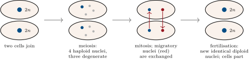
Two Paramecium of complementary mating types join by their oral surfaces. In each, the diploid micronucleus undergoes meiosis to four haploid nuclei, three of which degenerate; the survivor divides by mitosis into a stationary and a migratory nucleus, and the two cells exchange their migratory nuclei across the junction. Each cell then fuses its stationary nucleus with the one it received, and the two partners separate, each with a new diploid micronucleus of the same genotype as its partner’s. The old macronucleus breaks down and a new one is built from the new micronucleus. No new cell was made: conjugation is genetic mixing — meiosis and fertilisation, Chapter 4 — decoupled from multiplication. Bacterial conjugation, which transfers a plasmid one way from donor to recipient, is a different process with the same name (Chapter 3).
1.6 Toward multicellularity
Proposition 1.21 (The volvocine series)
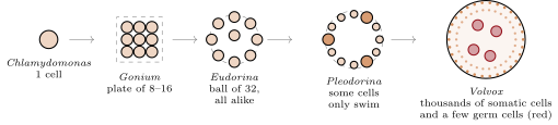
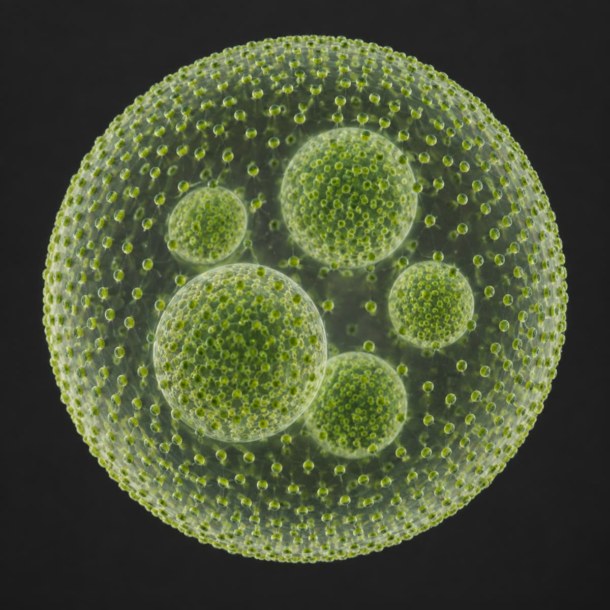
Among green algae one lineage displays, in living species, the steps from one cell to a body. Chlamydomonas lives alone. Gonium is a flat plate of 4 to 16 identical cells that swim together. Pandorina and Eudorina are hollow balls of 16 to 32 cells, still all alike, each able to found a new colony. Pleodorina has some small cells that only swim and never divide. Volvox is a sphere of to small somatic cells, flagellated, which will die with the colony, around a handful of large germ cells (gonidia) that divide to form daughter spheres inside the mother. Somatic and germinal cells have appeared: this is the threshold of multicellularity, crossed in this lineage some million years ago and, independently, at least twenty-five times elsewhere in the tree of life — in animals, plants, fungi, brown algae, red algae and several bacterial groups.
Evidence. The molecular phylogeny of the volvocine algae, built from dozens of genes, places Chlamydomonas at the base and Volvox at the tips, with the colonial forms between and the number of cells increasing along the branches; the genome of Volvox carteri (2010) differs from that of Chlamydomonas by a few hundred genes, chiefly those of the extracellular matrix that holds the cells and of the regulators that silence division in somatic cells. The transition needed remarkably little new genetic material. ∎
Remark 1.22 (Why divide the labour?)
In these algae a cell cannot swim and divide at the same time: the basal bodies that anchor the flagella are needed to organise the spindle. A single cell alternates; a small colony can afford to stop swimming while all its cells divide; a large colony would sink out of the light (Stokes’ law, Theorem 1.17) during the hours its division takes. Beyond a certain size, keeping some cells swimming while others divide is worth the loss of their descendants — and a soma is born. The choanoflagellates, collared flagellates whose cell is a copy of the sponge’s feeding cell, show the same first steps on the branch that led to animals.
1.7 Exercises
Exercise 1.1 ★
Define unicellular, colonial and multicellular, and place E. coli, Gonium, Volvox and a yeast cell in the right category with a reason.
Solution
Solution of Exercise 1.1.
Unicellular: one cell performs all functions and lives alone; colonial: identical cells stay attached but each could live alone; multicellular: several cell types, interdependent, only the germ line reproduces. E. coli and the yeast are unicellular (a bud detaches and lives alone); Gonium is colonial (16 identical cells, each able to found a colony); Volvox is multicellular (mortal somatic cells, a few germ cells).
Exercise 1.2 ★
Compute the surface-to-volume ratio of a coccus of radius and of a spherical protist of radius . Which one respires faster per unit of cytoplasm, and why?
Solution
Solution of Exercise 1.2.
: for the coccus, for the protist, a hundredfold difference. The coccus respires faster per unit volume: every part of its cytoplasm is within a fraction of a micrometre of the surface through which oxygen and food enter, whereas the protist’s interior is fed through a hundred times less surface per unit volume.
Exercise 1.3 ★
Give three structural differences between a bacterium and a unicellular eukaryote, and one thing a bacterium cannot do that an amoeba can.
Solution
Solution of Exercise 1.3.
No nucleus (a nucleoid) versus a nuclear envelope; no mitochondria or endomembranes, the respiratory chain sitting in the plasma membrane, versus organelles; a flagellum that is a rotating rigid helix of flagellin turned by protons, versus a cilium bending by dynein and ATP; also size ( versus tens) and a peptidoglycan wall. A bacterium cannot phagocytose: its wall forbids engulfing a particle, which an amoeba does with its pseudopods.
Exercise 1.4 ★
Name the function of each of the following structures of Paramecium: cilia, oral groove, food vacuole, contractile vacuole, macronucleus, micronucleus.
Solution
Solution of Exercise 1.4.
Cilia: locomotion and sweeping food toward the mouth; oral groove: funnels particles to the gullet; food vacuole: digests engulfed bacteria with lysosomal enzymes; contractile vacuole: expels the water that enters by osmosis; macronucleus: the working nucleus, hundreds of gene copies transcribed for daily life; micronucleus: the germinal diploid nucleus, used in meiosis and conjugation.
Exercise 1.5 ★★
With for a metabolite in cytoplasm, how long does diffusion take across a bacterium and across a amoeba? Comment on what the amoeba must do that the bacterium need not.
Solution
Solution of Exercise 1.5.
: for the bacterium; , about four minutes, for the amoeba. The amoeba cannot rely on diffusion to distribute metabolites: it needs cytoplasmic streaming and motor-driven transport along the cytoskeleton, which the bacterium does without.
Exercise 1.6 ★★
Compute the Reynolds number of a Paramecium (, ), of a bacterium (, ) and of a human swimmer (, ), with and . Why can a swimmer glide after a stroke while a bacterium cannot?
Solution
Solution of Exercise 1.6.
: Paramecium ; bacterium ; swimmer . At inertia carries the swimmer on after the stroke; at viscous drag stops the bacterium within a nanometre, so it moves only while it beats.
Exercise 1.7 ★★
A diatom of radius is denser than water. Compute its sinking speed and the time to fall through . By what factor does the time change if the cell halves its radius? If it trims its excess density to ?
Solution
Solution of Exercise 1.7.
; takes , 27 days. Halving the radius divides by 4: 108 days. Reducing to divides it by 5: 135 days.
Exercise 1.8 ★★
Model a Paramecium as an ellipsoid of half-axes , and (). It expels its own volume of water every . Compute the water influx in and in litres per second.
Solution
Solution of Exercise 1.8.
. In : ; since , this is .
Exercise 1.9 ★★
An attractant doubles in concentration over . What is the relative difference between the two ends of a bacterium? Between the start and the end of a run at ? Counting molecules has a relative error of about : explain why the bacterium compares times rather than sides.
Solution
Solution of Exercise 1.9.
A doubling over is a rise of about per micrometre (since ), so across the cell. A run covers : . To resolve a difference the cell would have to count some molecules at each end, far more than bind its few thousand receptors in a second; needs about , which is feasible. Temporal comparison converts a hopeless spatial measurement into a possible one by letting the run do the integrating.
Exercise 1.10 ★★★
Thiomargarita namibiensis is a spherical sulfur bacterium of diameter whose cytoplasm is a shell thick around a vacuole of nitrate. Compute the ratio of its surface to its cytoplasmic volume and compare with E. coli (a cylinder of radius ; ignore the ends). Explain how the vacuole lets it be so large, and what the nitrate is for.
Solution
Solution of Exercise 1.10.
Surface with : ; cytoplasmic volume surface ; ratio . For E. coli, a cylinder: . The giant is only eight times worse than E. coli, not 500 times, because every point of its cytoplasm is within of the surface. The vacuole stores nitrate, its electron acceptor, so that it can respire sulfide in the anoxic sediment between the rare stirrings that bring nitrate (Chapter 2).
Exercise 1.11 ★★★
In Volvox the somatic cells never divide and die with the colony. Using the flagellation constraint and Stokes’ law, explain why such cells are worth having in a colony of cells but not in one of 16, and why the germ cells are the largest cells of the sphere.
Solution
Solution of Exercise 1.11.
A cell cannot swim and divide at once. A 16-cell colony that stops swimming for the hours of division sinks, by Stokes’ law, a negligible distance (its radius is a few tens of micrometres, its sinking speed micrometres per second); the whole colony can divide and lose nothing. A -cell sphere of radius would sink a hundred times faster — millimetres per second, metres in an hour — out of the light: it pays to keep most cells swimming while a few divide. The germ cells are large because they must carry the resources to build a whole daughter sphere of thousands of cells by successive divisions without feeding, so they are provisioned by the soma.
Exercise 1.12 ★★★
“Protist is a grade, not a clade.” Explain the distinction with the lineages named in this chapter, and say why the same statement is true of “prokaryote” and false of “cyanobacterium”.
Solution
Solution of Exercise 1.12.
A clade is an ancestor and all its descendants; a grade is a level of organisation reached by several lineages. Protists — green algae (with the land plants), diatoms (with brown algae), ciliates (with dinoflagellates and Plasmodium), amoebae (near animals and fungi) — share only the eukaryotic cell lived alone, and their last common ancestor is that of all eukaryotes, including us: a grade. “Prokaryote” unites bacteria and archaea by what they lack; the archaea are closer to eukaryotes than to bacteria, so the term is again a grade. Cyanobacteria descend from one ancestor that invented oxygenic photosynthesis and include all its descendants (the chloroplast lineage aside): a clade.
1.8 Problem: A Drop of Pond Water
Problem 1.1
Weekend problem — a sample of pond water is counted, grown, and analysed for the physics that governs its cells, ending on the water and energy budget of a green alga
A pond of is sampled in spring. Under the microscope, a counting chamber whose grid covers under a coverslip held above it shows 24 cells of Chlamydomonas (spheres of radius , density ). Take , , , , , for in water.
Part I — Counting.
- Compute the volume of water under the grid, in microlitres.
- Deduce the density of Chlamydomonas in the pond, in cells per millilitre.
- Compute the volume and the surface-to-volume ratio of one cell.
- What fraction of the pond water’s volume is occupied by these cells?
- How many Chlamydomonas does the pond hold?
- A filamentous cyanobacterium is also present. Give two features visible under the microscope that distinguish it from the alga.
Part II — Growth. The sample is kept in the light and counted again after : cells under the grid.
- Compute the generation time .
- Compute the growth rate constant in , in .
- How long would it take the pond population to reach cells per millilitre at this rate?
- Give three reasons why it will not.
- If the cells grow only during the of daylight and not at all at night, how long does the same increase take?
- Chlamydomonas divides by multiple fission, releasing 4, 8 or 16 daughters at once. Explain why a generation time is nevertheless well defined by the count.
Part III — The physics of the cell.
- Compute the Reynolds number of a cell swimming at .
- A cell of mass that stops beating its flagella coasts a distance . Compute it.
- Compute the sinking speed of a cell that has stopped swimming.
- Compare the time to sink with the time to swim up .
- The pond holds of dissolved . Compute the maximal diffusive supply of to a cell, in molecules per second.
- A cell contains of carbon and doubles it in one generation. Compute its mean carbon demand in molecules per second, and the ratio of supply to demand.
- Redo the comparison for a hypothetical cell of radius with the same carbon density. Conclude.
Part IV — Water and energy. The cytoplasm holds of solutes, the pond . The membrane’s hydraulic conductivity is (water volume flux per unit area per unit pressure difference). Hydrolysing one ATP yields ; fixing one carbon atom costs three ATP.
- Compute the osmotic pressure difference across the membrane.
- Compute the volume of water entering the cell per second, in .
- Without a contractile vacuole, how long would the cell take to double its volume?
- Compute the minimal power needed to expel this water against the osmotic pressure.
- Convert it to ATP molecules per second and compare with the ATP the cell spends on carbon fixation (question 18).
- State the result: the cell’s water influx, the time it would take to burst, and the fraction of its energy that keeping dry costs.
Solution
Solution of Problem 1.1.
1. . 2. per cells per millilitre. 3. ; . 4. per millilitre, and : a fraction , . 5. : cells. 6. The cyanobacterium is a filament of much smaller cells (a few micrometres), blue-green and without a distinct chloroplast or nucleus, and it does not swim with flagella; the alga is a single ovoid cell with two flagella, an eyespot and one cup-shaped chloroplast. 7. : four doublings in , . 8. . 9. , about 4.3 days. 10. Nutrients (phosphate, nitrate) run out; the cells shade one another so that light per cell falls; grazers (ciliates, rotifers, Daphnia) multiply on them; at per millilitre the cells would fill some five percent of the volume and exhaust the dissolved . 11. Growth only half the time: the mean rate is halved, , 8.7 days. 12. The count measures the population, not individual cells: if a cell releases 8 daughters every , the population is multiplied by 8 in three generation times of each. The generation time is the mean doubling time of the number, whichever way the divisions are grouped. 13. . 14. ; ; : half a nanometre. 15. , . 16. Sinking : , 4.3 days; swimming up at : , 2.8 hours. Swimming wins by a factor of 37. 17. : molecules per second. 18. of carbon, doubled in : atoms per second. Supply over demand: . 19. Supply grows tenfold (), demand a thousandfold (): the ratio becomes ; such a cell could grow at only six percent of the rate, so a diffusion-fed alga of that size is impossible without stirring, internal transport or a much slower metabolism. 20. , . 21. Area ; influx . 22. , about eleven minutes. 23. flow . 24. One ATP: : ATP per second. Carbon fixation: ATP per second. The vacuole costs of that — cheap insurance against bursting. 25. Water influx (the cell’s volume in eleven minutes); without a contractile vacuole the cell would double its volume in about ; keeping dry costs at least ATP per second, under one percent of what its photosynthesis spends on carbon.