University Biology — Year 3 · Bachelor Year 3
11Cancer Biology
A tumour large enough to feel, a centimetre across, contains a billion cells and is the work of about thirty doublings from one cell that went wrong — typically a decade of growth before anyone knew. Behind that cell lie several mutations, each of which had to occur in the same lineage and each of which removed one of the controls of the two previous chapters: a brake on division, a trigger of death, a limit on the number of divisions, a requirement to stay put. Cancer is the evolution of a cell population inside a body, by mutation and selection, toward a state that serves the cells and kills the organism. This chapter treats the genes that are hit and what they normally do, the statistics that reveal how many hits are needed, the physiology a tumour must acquire to grow beyond a millimetre and to spread, the causes that can be avoided, and the therapies — from poisons that kill dividing cells to antibodies that release the immune system — that the biology has made possible, together with the evolutionary reason they so often fail.
11.1 What a cancer is
Definition 11.1 (Tumour, cancer, hallmarks)
A tumour (neoplasm) is a clone of cells that has escaped the controls on its growth and accumulates in a tissue. It is benign if it stays confined and encapsulated, malignant — a cancer — if its cells invade neighbouring tissue and seed secondary tumours elsewhere (metastasis), which is what kills. Cancers are named for their cell of origin: carcinomas from epithelia ( of human cancers), sarcomas from connective tissue, leukaemias and lymphomas from blood-forming cells, gliomas from glia. Whatever the tissue, a malignant clone has acquired the same set of capabilities, the hallmarks of cancer: it proliferates without external growth signals and ignores the signals that would stop it; it resists apoptosis; it divides without limit, having reactivated telomerase; it induces blood vessels; it invades and metastasises; and, underlying these, its genome is unstable, it reprograms its metabolism toward glycolysis even in oxygen (the Warburg effect), it recruits inflammatory cells that help it, and it evades the immune system. Each hallmark is a control of normal tissue defeated, and each maps onto genes.
11.2 The genes that are hit
Definition 11.2 (Oncogenes and tumour suppressors)
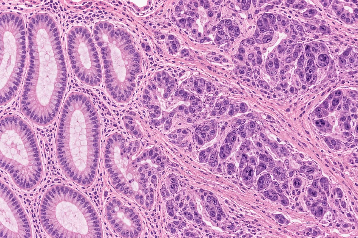
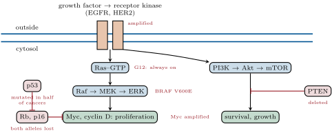
A proto-oncogene is a normal gene that promotes proliferation or survival — a growth factor, its receptor, a signalling protein, a transcription factor, a cyclin — and an oncogene is a mutant form that is active without its normal signal: a point mutation that locks the small GTPase Ras in its GTP state (glycine 12 in a quarter of all cancers), an amplification of the gene for the receptor HER2 or for the transcription factor Myc, a translocation that fuses BCR to the kinase ABL in chronic myeloid leukaemia or puts MYC next to an antibody enhancer in Burkitt lymphoma. One mutant allele suffices: oncogenes act dominantly. A tumour suppressor gene restrains proliferation or enforces death or repair — Rb and p16 at the restriction point, p53, the guardian that arrests or kills a damaged cell, APC in the Wnt pathway of the colon, PTEN, BRCA1 and BRCA2, the mismatch-repair genes — and it must lose both alleles to contribute: two hits, the first often inherited in the familial cancer syndromes, the second somatic. p53 is mutated in half of all human cancers and its pathway disabled in most of the rest.
Evidence. Rous (1911) transmitted a chicken sarcoma with a cell-free filtrate, a virus, whose single transforming gene, src, was shown by Varmus and Bishop (1976) to be a captured copy of a normal chicken gene — oncogenes are altered cellular genes. Weinberg (1982) transferred DNA from a human bladder carcinoma into cultured mouse cells, which became transformed; the responsible fragment was RAS with a single base change, and the same change was absent from the patient’s normal tissue. Knudson (1971) compared children with the eye tumour retinoblastoma: those with a family history developed several tumours in both eyes, early; those without developed one, later — consistent with the familial cases needing one somatic event, the sporadic two, and thus with a gene whose two copies must both be lost. RB was cloned in 1986 and both alleles were indeed inactivated in every tumour. ∎
Theorem 11.3 (Hits and the age curve)
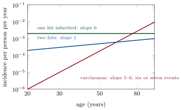
Suppose a cancer requires independent, rate-limiting events in one cell lineage, each occurring at a constant rate per cell per year, in a population of susceptible cells, with . Then the probability that a given cell has suffered all events by age is about , the cumulative risk in the tissue is about , and the incidence — new cases per year at age — is
a power of age with exponent : a straight line of slope on a log–log plot. For an individual who inherits one of the events, the same cancer has incidence , one power lower.
Proof. Each event, at rate , has occurred by time with probability ; the events are independent, so all have occurred with probability , and the order in which they occurred does not matter here. With cells and rare events, the expected number of cells that have completed the sequence is , and the incidence is its time derivative, . Inheriting one event leaves to occur: , incidence . If the order of events mattered (only of orders led to cancer) the prefactor would change, not the exponent. ∎
Example 11.4 (Reading the exponent)
The incidence of most carcinomas in adults rises as about the fifth to sixth power of age — from about in per year at thirty to in at eighty, a factor of for a factor in age, and — which suggests six or seven rate-limiting events. The estimate is crude: the expansion of a clone after each hit raises the number of cells at risk of the next, so fewer events with clonal growth between them give the same slope, and the number of driver mutations found in sequenced tumours is two to eight. Knudson’s retinoblastoma fits the theorem’s second statement exactly: the sporadic disease, needing two hits, has an incidence that rises with age (through the few years the retinoblasts exist); the hereditary disease, needing one, is present at a nearly constant rate from birth and strikes early and repeatedly.
Proposition 11.5 (The pathways that are hit)
The hundreds of cancer genes fall into a dozen pathways, each of which a tumour disables at some point. The growth-factor pathways: a receptor tyrosine kinase (EGFR, HER2), Ras, the kinase cascade Raf–MEK–ERK to the nucleus, and in parallel PI3K–Akt–mTOR toward survival and growth, restrained by PTEN; a tumour activates one of them, by whichever gene. The cell-cycle brake: cyclin D, Cdk4, p16, Rb (Chapter 10). The guardian: p53 and its regulators. Death: Bcl-2. Developmental signalling: Wnt (APC, -catenin) in the colon, Hedgehog in skin and brain, Notch in T cells. Genome maintenance: mismatch repair, BRCA, ATM. Chromatin: the writers and remodellers of Chapter 1, mutated in a third of tumours. Immortality: the telomerase promoter. Because a pathway is a series of steps, mutations in different genes are alternatives — a tumour with mutant Ras rarely also has mutant EGFR — and drugs against one step can be defeated by a mutation downstream.
11.3 A tumour evolves
Proposition 11.6 (Multistep carcinogenesis)
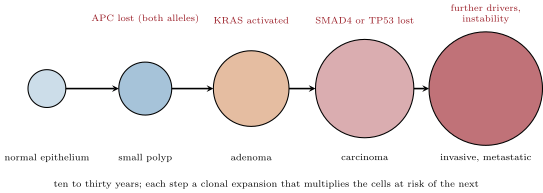
A tumour is the product of somatic evolution: mutation generates variants, and selection within the tissue favours the ones that divide more, die less or escape a constraint. The colorectal sequence worked out by Vogelstein is the paradigm: loss of APC produces a small benign polyp; a KRAS mutation lets it grow into an adenoma; loss of SMAD4 or TP53 converts it to a carcinoma; further changes allow invasion. Each step is a clonal expansion that multiplies the cells at risk of the next. A sequenced tumour carries thousands of mutations, of which a handful — typically two to eight — are drivers that conferred selective advantage; the rest are passengers, carried along in the clone, and their spectrum is a record of what caused them: CT at adjacent pyrimidines from ultraviolet light in melanoma, GT from tobacco smoke in lung cancer, the fingerprints of the APOBEC enzymes, of defective mismatch repair, of aflatoxin — mutational signatures. Tumours are heterogeneous, a branching tree of subclones, and the subclone that kills is often a minority at diagnosis.
Method 11.7 (The Ames test)
To test whether a chemical is mutagenic, and hence a probable carcinogen: (1) take a strain of Salmonella with a point mutation in a histidine biosynthesis gene, which cannot grow without histidine; (2) mix the chemical with a liver extract, whose enzymes convert many compounds into their active mutagenic forms, as the body would; (3) plate some bacteria on histidine-free agar with the mixture; (4) count the colonies that grow — each is a revertant in which a new mutation restored the gene; (5) compare with the spontaneous rate. A dose-dependent increase in revertants means the compound causes point mutations. Most known carcinogens are positive; the test is cheap, takes two days, and a compound that fails it is examined further before it is made.
Definition 11.8 (Immortality and instability)
Normal somatic cells lack telomerase and count their divisions in telomere length (Chapter 24): after some fifty they senesce, and a cell that bypasses senescence by losing p53 and Rb goes on until its telomeres are exhausted and its chromosomes fuse and break — a crisis that kills most clones. The rare survivor has reactivated telomerase, usually by a promoter mutation, and is immortal; of cancers express the enzyme. The crisis leaves scars: fused and broken chromosomes, amplifications, deletions, translocations — chromosomal instability, which, with the loss of the spindle checkpoint and of p53’s response to missegregation, makes most solid tumours aneuploid and keeps them generating variants. A minority of tumours instead have a mutator phenotype from lost mismatch repair, with a normal karyotype and a thousandfold point-mutation rate.
11.4 The tumour as a tissue
Proposition 11.9 (Oxygen sets the size of a vessel-free tumour)
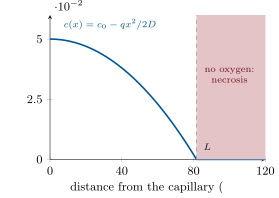
Consider tissue consuming oxygen at a constant rate per unit volume, supplied by diffusion (coefficient ) from a vessel at whose wall the concentration is . In one dimension at steady state, , so and oxygen runs out at the distance
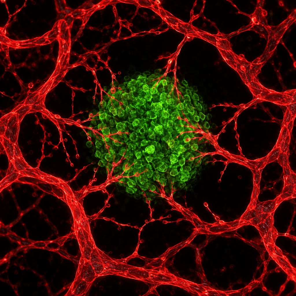
With , and (a respiring tumour), : no cell can live more than about a hundred micrometres from a capillary. A clone with no vessels of its own therefore stops at a diameter of one to two millimetres, its centre necrotic and its rim alive. To grow further it must induce angiogenesis — its hypoxic cells stabilise the transcription factor HIF, which induces VEGF, which makes nearby endothelial cells sprout toward it. Judah Folkman proposed in 1971 that blocking this step would starve tumours; antibodies against VEGF are now standard in several cancers, though the tumours adapt.
Proof. At steady state the diffusive flux must change with exactly as fast as oxygen is consumed: , that is . Integrating twice with and at the depth where nothing is left (no flux beyond it) gives after using to fix . The numbers follow. ∎
Definition 11.10 (Invasion and metastasis)
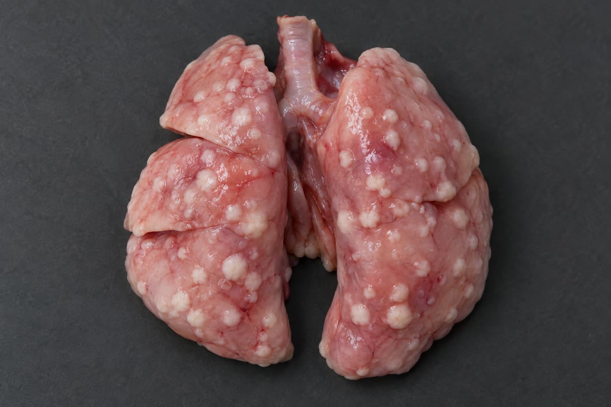
To metastasise, a carcinoma cell must leave the epithelium — lose its cell junctions and polarity and acquire motility, an epithelial–mesenchymal transition driven by transcription factors normally used in embryonic development — degrade the basement membrane and matrix with secreted proteases, cross the wall of a blood or lymph vessel, survive in the circulation (fewer than one in ten thousand do, killed by shear, by the absence of attachment, and by natural killer cells), lodge in a capillary bed of another organ, cross out, and grow there. Where it grows is not random: breast cancer goes to bone, lung, liver and brain, colon cancer to the liver, prostate cancer to bone — Paget’s seed and soil of 1889, explained partly by blood flow and partly by the fit between the cell and the tissue’s signals. A disseminated cell may lie dormant for years before it grows, which is why cancers recur a decade after apparent cure. Metastasis causes nine deaths in ten from solid tumours, and it is the step least understood.
Proposition 11.11 (Escaping the immune system)
A tumour’s mutations create neoantigens, peptides no normal cell presents, and cytotoxic T cells recognise and kill tumour cells — the reason immunosuppressed patients have more cancers and the reason tumours with many mutations (melanoma, lung, mismatch-repair-deficient colon) respond best to immunotherapy. A clinically evident tumour is one that has escaped: by losing the MHC molecules that present peptides, by secreting suppressive factors, by recruiting regulatory T cells, and above all by expressing PD-L1, which engages the inhibitory receptor PD-1 on T cells and switches them off, the same brake that normally ends an immune response (Chapter 16). Antibodies that block PD-1 or PD-L1, or the earlier brake CTLA-4, release the T cells; in a fraction of patients — a fifth to a half depending on the tumour — they produce lasting remissions of cancers that were untreatable, and their side-effects are autoimmunity, the brake’s other function.
11.5 Causes and treatments
Proposition 11.12 (Causes)
Cancer is caused by whatever raises the rate of the events in the theorem or the number of cells at risk. Chemical carcinogens, most of them mutagens after activation by the liver: the seventy carcinogens of tobacco smoke, which cause a third of cancer deaths and raise the risk of lung cancer fifteen- to twenty-five-fold; aflatoxin from mouldy grain; asbestos, which is not a mutagen but a chronic irritant. Radiation: ultraviolet light for skin (the dimers of Chapter 3), ionising radiation for leukaemia, thyroid and breast. Infections, a sixth of cancers worldwide: the papillomaviruses, whose proteins E6 and E7 destroy p53 and inactivate Rb and cause nearly all cervical cancers (a vaccine now prevents them); hepatitis B and C viruses in liver cancer; Epstein–Barr virus in lymphomas; Helicobacter pylori in stomach cancer, through decades of inflammation. Inherited mutations in a tumour suppressor, the first hit at birth, in some of cancers. And chance: most of the mutations in a tumour arose from the ordinary errors of replication, so tissues that divide most — colon, skin, blood — have the most cancers, and the risk rises with age whatever one does.
Theorem 11.13 (Why one drug fails and two may not)
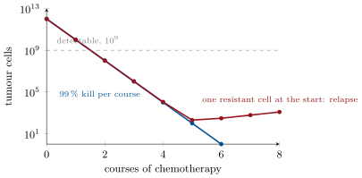
Let a tumour of cells have been produced by successive divisions from one cell, and let a mutation conferring resistance to a given drug arise at rate per cell division. Then the probability that the tumour contains no resistant cell when treatment begins is about
so that for resistance pre-exists with near certainty. For two drugs with independent resistance mechanisms, a cell resistant to both arises at rate about per division, and : with and , one drug faces resistant clones on average, two drugs a probability of that even one doubly resistant cell exists.
Partial proof. Growing from to cells takes about divisions in all (a tree with leaves has internal nodes). If a resistant cell is produced at each division with probability and, to a first approximation, its descendants neither die nor outgrow the rest, the number of resistance events is Poisson with mean , and the probability of none is . Independent mechanisms multiply the per-division probabilities. The approximation neglects that early mutants leave larger resistant clones — the full distribution is that of Luria and Delbrück, treated in the Year 2 volume — but the probability of no mutant, which is what decides whether the tumour can be cured, is exactly the Poisson term. ∎
Example 11.14 (Therapies and their logic)
Surgery and radiotherapy remove or kill a localised tumour; radiation kills by double-strand breaks, and its fractionation into daily doses lets normal tissue repair between them (Chapter 3). Cytotoxic chemotherapy — alkylating agents, antimetabolites, microtubule poisons, topoisomerase inhibitors — kills dividing cells of any kind, tumour cells slightly more, and follows a log-kill law: each course kills a constant fraction, say , so that six courses reduce cells to one, and the same six courses are needed whether the tumour is large or small; the patient’s hair, gut and marrow, which also divide, set the dose. Targeted therapy attacks a lesion the tumour depends on: imatinib blocks the BCR–ABL kinase and turned chronic myeloid leukaemia from a fatal disease into a chronic one; trastuzumab binds HER2; the PARP inhibitors, the Cdk4/6 inhibitors and venetoclax of earlier chapters each exploit one defect. Immunotherapy releases the T cells. And the theorem says how they should be used: in combination, early, when is smallest — which is what adjuvant chemotherapy after surgery does — and with drugs whose resistance mechanisms do not overlap. The tumour is an evolving population, and treatment is selection.
Remark 11.15 (What has changed)
Half of all patients diagnosed with cancer in a rich country now survive ten years, against a quarter in 1970. The gain came from prevention (tobacco, hepatitis and papillomavirus vaccines, screening for polyps and cervical lesions), from earlier detection, and from treatments that follow from the biology of this chapter — the combination chemotherapy that cures most childhood leukaemias and testicular cancers, the targeted drugs, and the immunotherapies. What has not changed is metastatic disease, which the biology explains but does not yet cure; the next gains will come from treating the tumour as what it is, a population evolving under the selection that treatment imposes.
11.6 Exercises
Exercise 11.1 ★
Distinguish an oncogene from a tumour suppressor gene in mechanism, in the number of alleles that must change, and with two examples each.
Solution
Solution of Exercise 11.1.
An oncogene is a gain-of-function version of a gene that promotes proliferation or survival; one mutant allele suffices (dominant): RAS (point mutation), MYC (translocation, amplification), HER2, BCR–ABL. A tumour suppressor restrains proliferation or enforces arrest, death or repair; both alleles must be lost (recessive at the cell level): RB, TP53, APC, BRCA1.
Exercise 11.2 ★
List the hallmarks of cancer and, for each, name one gene of this or an earlier chapter whose alteration provides it.
Solution
Solution of Exercise 11.2.
Sustained proliferation: RAS, MYC. Insensitivity to growth suppressors: RB, CDKN2A (p16). Resisting death: BCL2, TP53. Replicative immortality: the telomerase promoter. Angiogenesis: VEGF induced through HIF (VHL loss). Invasion and metastasis: E-cadherin loss, the epithelial–mesenchymal factors. Genome instability: mismatch-repair genes, BRCA. Deregulated metabolism: MYC, PI3K pathway. Immune evasion: PD-L1, MHC loss. Tumour-promoting inflammation: NF-B signalling.
Exercise 11.3 ★
What did Knudson observe about hereditary and sporadic retinoblastoma, and what did he infer?
Solution
Solution of Exercise 11.3.
Children with a family history developed several tumours in both eyes, early; sporadic cases developed a single tumour, later. He inferred that two events were needed in a cell: the familial patients had inherited one and needed only a second, somatic event (frequent enough to occur in several retinal cells), while sporadic patients needed both to occur somatically in one cell — rare, late, single. Hence a gene whose two copies must both be lost: the first tumour suppressor.
Exercise 11.4 ★
Explain the Ames test and why a liver extract is added.
Solution
Solution of Exercise 11.4.
A histidine-requiring Salmonella mutant is plated without histidine with the test compound; colonies arise only from cells in which a new mutation has reverted the defect, so their number measures mutagenicity. Liver extract is added because many carcinogens are harmless until liver enzymes convert them into reactive metabolites, as happens in the body; the bacteria lack those enzymes.
Exercise 11.5 ★★
The incidence of a cancer is per year at age and at age . Estimate the exponent of the age-power law and the number of rate-limiting events it suggests.
Solution
Solution of Exercise 11.5.
Incidence rises -fold while age doubles: , , so about seven or eight rate-limiting events on the naive reading — fewer if clonal expansion between hits is allowed.
Exercise 11.6 ★★
Using Proposition 11.9 with and , compute for tissue consuming and for a tumour consuming three times as much. Why does a fast-growing tumour become necrotic sooner?
Solution
Solution of Exercise 11.6.
: for , ; for , . A fast-growing tumour respires faster, exhausts the oxygen within a shorter distance, and its centre dies at a smaller size.
Exercise 11.7 ★★
A tumour of cells is treated with a drug that kills per course. How many courses to reduce it below one cell? Why is the tumour undetectable after two courses although cells remain, and what does that imply for stopping treatment?
Solution
Solution of Exercise 11.7.
needs : four courses. After two courses cells remain — a tenth of a cubic millimetre, far below the that can be detected — so the tumour is “gone” by every test while a hundred thousand cells remain; treatment must continue for the planned number of courses, not until the tumour disappears.
Exercise 11.8 ★★
Explain why a mutation in EGFR and a mutation in KRAS are rarely found in the same lung tumour, and why a tumour treated with an EGFR inhibitor often relapses with a KRAS mutation.
Solution
Solution of Exercise 11.8.
Both lesions activate the same pathway (receptor Ras ERK); once one is present the other confers no further advantage and is not selected. An EGFR inhibitor blocks the receptor; a cell that acquires a KRAS mutation downstream restores the signal with the receptor still blocked, and is selected during treatment — resistance by bypass.
Exercise 11.9 ★★
The papillomavirus proteins E6 and E7 inactivate p53 and Rb. Explain how this drives cervical cancer, why it takes decades, and why a vaccine given before sexual activity prevents it.
Solution
Solution of Exercise 11.9.
E7 frees E2F from Rb, driving the infected epithelial cell through the restriction point without growth factors; E6 destroys p53, so the inappropriate proliferation and the damage it causes trigger neither arrest nor apoptosis. Two hallmarks are supplied at once, but the remaining ones — immortality, invasion, instability — need further somatic mutations, which take decades to accumulate in the persistently infected cells. The vaccine raises neutralising antibodies against the capsid and prevents the infection; given before exposure, there is never an infected cell to transform.
Exercise 11.10 ★★★
Extend the Armitage–Doll argument to allow each hit to expand its clone by a factor before the next: show that the incidence has the same power of but a prefactor multiplied by , and discuss why the fitted from an age curve is an upper bound on the number of drivers.
Solution
Solution of Exercise 11.10.
After the th hit the clone grows by , so times as many cells are at risk of the next hit; the probability of the full sequence in the lineage descended from one original cell is — for constant per hit the cumulative risk becomes up to order-dependent factors, and the incidence keeps the power with a prefactor larger. If the expansions themselves grow with time (exponentially growing clones), the curve steepens further, so a slope of is produced by fewer than seven drivers: the Armitage–Doll is an upper bound on the number of rate-limiting drivers, and sequenced tumours show two to eight.
Exercise 11.11 ★★★
With Theorem 11.13, compare (a) treating a -cell tumour with drug A for six courses then drug B for six, and (b) giving A and B together from the start, when . Compute for each strategy and explain why early combination is the rule.
Solution
Solution of Exercise 11.11.
(a) Drug A alone faces pre-existing resistant cells: ; the A-resistant clone survives and regrows during the eighteen weeks of A, and by the time B starts it is again a large population, so B faces as well — the sequence fails twice. (b) Together: a doubly resistant cell arises at per division, , . Combination from the start, when is smallest, is the rule because the product of two small rates is what makes resistance unlikely.
Exercise 11.12 ★★★
Peto’s paradox: an elephant has a hundred times as many cells as a human and lives as long, yet has no more cancer. Using the theorem of this chapter, say what the naive expectation is, and propose two mechanisms by which large animals could have solved the problem (the elephant has twenty copies of TP53).
Solution
Solution of Exercise 11.12.
With times more cells and the same and , cumulative risk would be times higher — every elephant should die of cancer young. It does not, so or must differ: twenty copies of TP53 make the apoptotic response to damage stronger and remove mutant cells before they expand (lowering the effective for that step); large animals also have lower per-cell mutation rates (better repair, fewer divisions per cell per year because their cells turn over more slowly), and may require more hits (an extra suppressor layer). Cancer suppression evolved with body size.
11.7 Problem: The Arithmetic of a Tumour
Problem 11.1
Weekend problem — a tumour grown from one cell and timed to detection, its age curve read for the number of hits, its oxygen supply and necrotic core computed, and its treatment planned against the certainty of resistance, ending on the years to detection, the depth of living tumour around a vessel and the probability that a combination cures
Data: a tumour cell has volume ; doubling time ; detection at ; lethal burden . Age curve: incidence per year at and at . Oxygen: , , in tumour; capillaries in a vascularised tumour are apart. Therapy: each course kills ; resistance mutation rate per division per drug; a course every three weeks; the tumour regrows between courses with the same doubling time.
Part I — Growth.
- How many cells in ? How many doublings from one cell?
- How many years from the founding cell to detection?
- How many doublings, and years, from detection to the lethal burden? Compare the two intervals.
- Many tumours are detected at but were growing for only five years. What must have been true of the doubling time early on, or of the growth law?
- Only a fraction of cells in a tumour are dividing at any time (the growth fraction), and a fraction dies. If cells cycle in but the tumour doubles in , what does that say about the balance of birth and death?
- Why does the same log-kill chemotherapy spare a slowly growing tumour more than a fast one, and yet the fast one regrow faster between courses?
Part II — Hits.
- From the two incidences, compute the exponent of the age curve and the implied number of events .
- If each event occurs at per cell per year in susceptible cells, what does the theorem predict for the cumulative risk by age with your ? Is it plausible? What is missing?
- Repeat with clonal expansion by a factor after each of the first hits, using Exercise 11.10.
- A carrier inherits one hit. By what factor is her incidence at age raised, if the other rates are unchanged? (Compare with at .)
- Smoking multiplies for one of the events by . By what factor does the incidence rise if that event is one of ; if it is two of them?
- Explain why stopping smoking at fifty lowers the risk of lung cancer below that of a continuing smoker, but never to that of a never-smoker.
Part III — Supply.
- Compute the oxygen penetration depth in the tumour.
- A vessel-free spheroid: what is the maximum diameter at which every cell is oxygenated? What is the thickness of the living rim of a spheroid, and what fraction of its volume is alive?
- In a vascularised tumour with capillaries apart, is every cell oxygenated? Which cells are hypoxic, and why does that matter for radiotherapy (oxygen fixes radiation damage) and for selection of p53-mutant cells?
- A drug halves the tumour’s capillary density. Recompute the spacing and say what happens to the cells midway between vessels.
- Hypoxic cells switch to glycolysis, making per glucose instead of . By what factor must they raise glucose uptake to keep their ATP supply, and how is this exploited in imaging tumours?
- Explain why anti-angiogenic therapy alone rarely cures but can help chemotherapy reach the tumour.
Part IV — Treatment.
- A tumour is treated with one drug. How many courses reduce it below one cell, ignoring regrowth and resistance?
- Between courses (three weeks) the survivors regrow. By what factor? Recompute the net kill per cycle and the number of cycles needed.
- Compute the expected number of cells resistant to the drug at the start of treatment and the probability that there is none.
- Two drugs with independent mechanisms are given together. Probability that no doubly resistant cell exists at the start? At a tumour of cells?
- Adjuvant therapy is given after surgery, when perhaps cells remain undetected. Recompute the probabilities for one and two drugs, and state the lesson.
- An immunotherapy induces T cells that kill any cell presenting a neoantigen. Why is resistance to it a different problem from resistance to a kinase inhibitor, and which tumours does the theorem suggest it should work best on?
- Summarise: the years from founding cell to detection (question 2), the depth of living tumour around a vessel (question 13), and the probability that the two-drug combination faces no resistant cell in a tumour (question 22).
Solution
Solution of Problem 11.1.
1. cells; doublings. 2. days, about years. 3. cells: ten more doublings, days, under three years — a third of the time to detection. 4. The early doubling time must have been about days, or the tumour grew faster when small and slowed as it grew (the usual pattern, as supply and space fail). 5. A cycle of days would double the population every days; a doubling time of means the net growth is of the birth rate: nearly every cell born is matched by one dying, or few are cycling — birth and death almost balanced, growth the small difference. 6. The drugs kill cycling cells, so a tumour with a small fraction in cycle loses a smaller fraction per course; but the fast tumour, having lost more, also regrows between courses. On balance the fast tumours (leukaemias, lymphomas, testicular cancer) are the ones chemotherapy cures. 7. Ratio over an age ratio : , . 8. : absurd against a lifetime risk near in . Missing: clonal expansion multiplying the cells at risk after each hit, mutation rates raised by instability, many more divisions (stem cells cycling for decades), and fewer true drivers. 9. Multiplying by gives — still tiny with seven events; with four events and expansions, , the right order. The true picture is few drivers and large expansions. 10. : a carrier’s incidence at forty is tens of thousands of times the sporadic one — hereditary cancers strike young. 11. One event tenfold faster: incidence ; two: . The observed fifteen- to twenty-five-fold risk suggests smoke acts on more than one step. 12. On stopping, falls back for the events still to come, so the incidence stops climbing as steeply; but the hits already accumulated in the cells remain, so the ex-smoker’s cells are nearer to cancer than a never-smoker’s and the excess risk persists, frozen rather than erased. 13. . 14. Fully oxygenated up to a diameter . A spheroid has a living rim of : alive fraction . 15. Midway cells are from a vessel, just within — oxygenated in principle, but tumour vessels carry less oxygen and flow intermittently, so the midway cells are hypoxic. Hypoxic cells are two to three times more resistant to radiation (oxygen is needed to make the damage permanent), and hypoxia triggers p53-dependent apoptosis, so it selects the cells that have lost p53. 16. Halving the density spreads vessels by : apart, midway cells away, beyond : they die or, surviving hypoxic, become more aggressive and angiogenic. 17. times more glucose. Tumours therefore concentrate glucose analogues, and a positron-emitting fluorodeoxyglucose scan lights them up. 18. Starved tumours shrink to the diffusion-limited size and persist, dormant and hypoxic, or co-opt existing vessels; hypoxia selects aggressive clones. But pruning the chaotic tumour vasculature lowers the interstitial pressure and evens the flow, so chemotherapy penetrates better — the combination works where either alone does not. 19. : , five courses. 20. days doublings, regrowth ; net factor per cycle ; : still five cycles. 21. resistant cells expected; : resistance is certain. 22. ; . At cells, , . 23. cells: one drug , ; two drugs , . Treat when the population is small, and treat with combinations. 24. The T cells attack many neoantigens at once, so no single mutation escapes them; escape requires losing antigen presentation itself (MHC, -microglobulin) or suppressing the T cells, a different and rarer class of event. The same high mutation load that makes a tumour certain to resist a kinase inhibitor gives it many neoantigens: mismatch-repair-deficient, ultraviolet- and smoke-induced tumours respond best. 25. About years to detection; living tumour deep around a vessel; that a two-drug combination faces no resistant cell in a tumour.