University Biology — Year 3 · Bachelor Year 3
16Adaptive Immunity and Vaccination
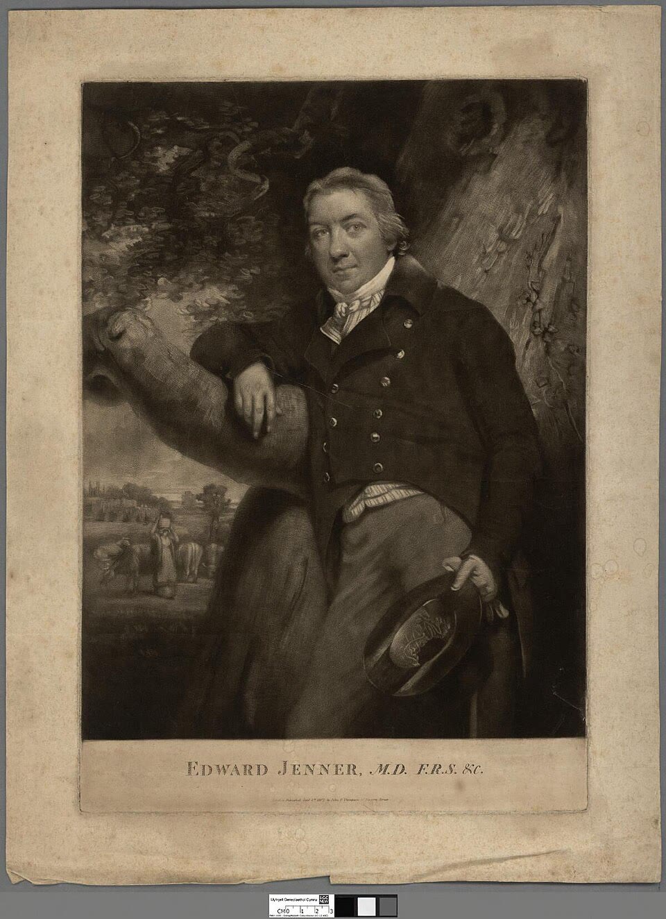
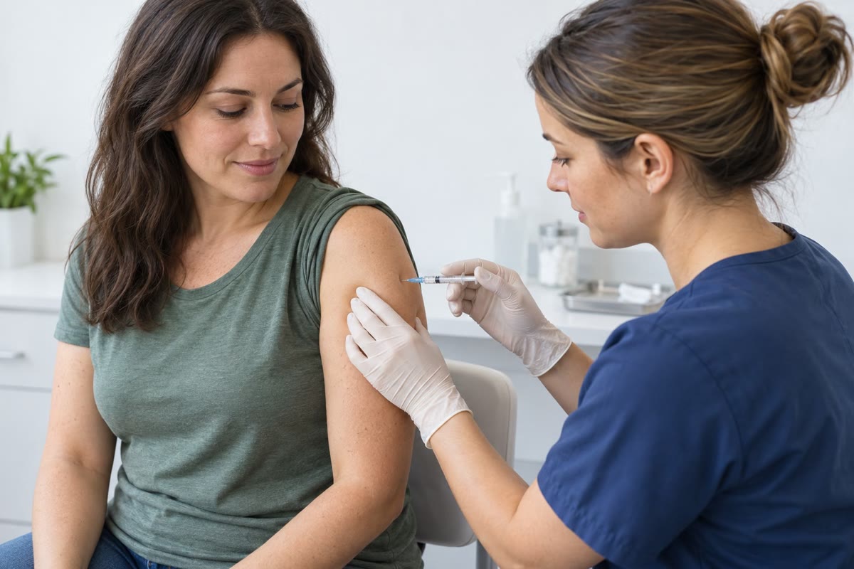
In 1796 a country doctor scratched pus from a milkmaid’s cowpox sore into the arm of a boy, and six weeks later inoculated him with smallpox; the boy did not fall ill. In 1980 the World Health Organization declared smallpox — which had killed three hundred million people in the twentieth century — eradicated from the Earth, by that method. The system that Jenner had recruited, without knowing it existed, can recognise any molecule it has never met, choose among a hundred billion different receptors the few that fit, multiply the cells that carry them a millionfold in a week, improve their fit by mutation and selection while it does so, and remember the result for a lifetime; it also learns not to attack the body that made it, and its failures to learn that are the autoimmune diseases. This chapter treats the lymphocytes and how they make their receptors, how a response is mounted and remembered, how tolerance is enforced and lost, and the arithmetic by which vaccinating enough people protects those who are not.
16.1 Lymphocytes and clonal selection
Definition 16.1 (Lymphocytes and their receptors)
Adaptive immunity is carried by lymphocytes: B cells, which mature in the bone marrow and whose antigen receptor is a membrane-bound antibody that they later secrete; and T cells, which mature in the thymus and whose receptor recognises peptide fragments displayed on the surface of other cells. An antigen is whatever a receptor binds; the part actually contacted is an epitope. The founding fact is clonal selection (Burnet, 1957): each lymphocyte carries receptors of one specificity only, fixed before it meets any antigen; the body holds a repertoire of some such cells; an antigen selects the few whose receptors fit and drives them to divide into a clone of effector and memory cells; and lymphocytes whose receptors fit the body’s own molecules are deleted or silenced while they mature. The cells circulate continuously between the blood and the lymph nodes, spleen and mucosal lymphoid tissue, where antigen carried by dendritic cells (Chapter 15) is displayed, so that the rare matching cell — one in a hundred thousand for a given epitope — finds it within a day or two.
Evidence. Nossal and Lederberg (1958) placed single lymphocytes from immunised rats in microdroplets and tested the antibody each secreted: every cell made antibody against one of the two antigens, never both. Burnet had predicted it the year before. Tonegawa (1976) compared the DNA of an antibody gene in embryonic cells and in an antibody-secreting tumour: in the embryo the variable and constant parts lay far apart, in the tumour they were joined — the gene had been cut and spliced in the somatic cell, the first demonstration of a genome rearranged on purpose. ∎
16.2 Making a hundred billion receptors
Definition 16.2 (Antibody structure and V(D)J recombination)
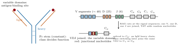
An antibody (immunoglobulin) is two identical heavy chains and two identical light chains, each with a variable domain at its end and constant domains behind; the two arms each bind an epitope through three hypervariable loops of the heavy and three of the light variable domain, and the stem (Fc) is what phagocytes, complement and mast cells recognise. Five classes differ in their heavy-chain constant region: IgM, the first made, a pentamer that activates complement well; IgG, the workhorse of blood and tissues, which crosses the placenta; IgA, a dimer secreted across mucosal surfaces into gut, airways and milk; IgE, bound by mast cells, against worms — and the cause of allergy; IgD. The variable domains are not encoded as such. The heavy-chain locus holds about functional V segments, D and J; a developing B cell chooses one of each and joins them by V(D)J recombination: the enzymes RAG1 and RAG2 cut at recombination signal sequences flanking the segments, and the ends are joined by the non-homologous end-joining machinery of Chapter 3, with nucleotides trimmed and added at random (by the enzyme TdT) at each junction — junctional diversity, which falls exactly in the third hypervariable loop. The light chains do the same with V and J. After the response begins the same variable domain is joined to a different constant region by a second rearrangement, class switching, changing the antibody’s function but not its specificity.
Theorem 16.3 (The size of the repertoire)
With , , heavy segments, and light chains from two loci with and , the number of distinct heavy–light combinations obtainable by segment choice alone is
Junctional diversity at the two heavy junctions and one light junction multiplies this by a factor of order –, giving a potential repertoire above from fewer than gene segments — more than the number of B cells in a body, so that the actual repertoire is a sample of the possible one. Somatic hypermutation during the response (below) then generates variants of each selected receptor.
Proof. Each heavy chain is one V, one D and one J; each light chain one V and one J from either locus; any heavy pairs with any light, so the counts multiply (multiplication principle), and the two light loci add. The junctional factor is the number of distinct sequences the random trimming and addition can produce at a junction — a few dozen per junction at least — raised to the number of junctions; its exact value is not defined, but three junctions each with – outcomes give to . The count for T cell receptors, with two chains rearranged in the same way and a larger junctional contribution, is of the same order. ∎
Definition 16.4 (T cell receptors and MHC)
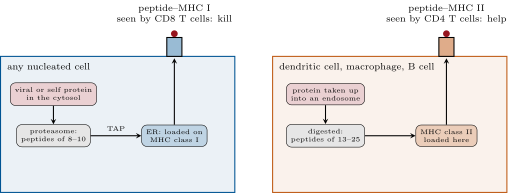
The T cell receptor is a two-chain molecule built by the same recombination, never secreted, and it does not bind antigen free in solution: it recognises a short peptide held in the groove of a major histocompatibility complex (MHC) molecule on another cell’s surface. MHC class I, on every nucleated cell, displays peptides of eight to ten residues cut by the proteasome from the cell’s own cytosolic proteins — a sample of what the cell is making, including any virus — to CD8 T cells, which kill the cell if the peptide is foreign. MHC class II, on dendritic cells, macrophages and B cells, displays peptides of thirteen to twenty-five residues from proteins the cell has taken up and digested in endosomes, to CD4 T cells, which respond by helping. A T cell is restricted: it recognises its peptide only on the MHC allele it was selected on. The MHC genes (HLA in humans) are the most polymorphic in the genome, with thousands of alleles each differing in the groove, so that each person displays a different sample of each protein’s peptides and no pathogen can escape presentation in everyone — and so that a transplanted organ from an unmatched donor is recognised as foreign by a large fraction of the recipient’s T cells.
Evidence. Zinkernagel and Doherty (1974) infected mice of two inbred strains with a virus and tested their cytotoxic T cells on infected target cells: the T cells killed infected cells of their own strain but not of the other, although the virus was the same. The T cell saw the virus only together with a self MHC molecule — MHC restriction — which was later explained when the crystal structure of an MHC molecule (Bjorkman, 1987) showed a groove with a peptide in it, and the T cell receptor was shown to bind the two together. ∎
16.3 The response
Definition 16.5 (Activation, help and killing)
A naive T cell is activated when its receptor binds peptide–MHC on a dendritic cell that also provides costimulation (Chapter 15); it then divides every six to eight hours for a week and differentiates. CD4 helper T cells differentiate, according to the dendritic cell’s cytokines, into subsets: Th1, which activate macrophages to kill what they have eaten (tuberculosis); Th2, which drive IgE and eosinophils against worms; Th17, which recruit neutrophils against extracellular bacteria and fungi; follicular helpers, which help B cells; and regulatory T cells, which suppress. CD8 cytotoxic T cells kill cells displaying their peptide on class I, by perforin and granzymes and by the Fas ligand, triggering apoptosis (Chapter 10) — the defence against viruses and tumours. A B cell is activated by antigen binding its receptor plus help from a follicular helper T cell that recognises peptides of the same antigen on the B cell’s class II; it then becomes a plasma cell secreting thousands of antibodies a second, for days (short-lived, in the node) or years (long-lived, in the marrow), or enters a germinal centre. Memory B and T cells — long-lived, more numerous than the naive precursors and quicker to respond — are the residue of every response and the basis of vaccination.
Definition 16.6 (The germinal centre)
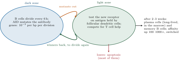
In a germinal centre, a structure that forms in a lymph node about a week into a response, activated B cells run an evolutionary algorithm. In its dark zone they divide every six hours and their antibody variable genes are mutated by the enzyme AID at about one change per thousand base pairs per division — somatic hypermutation, a million times the genomic rate, aimed at one kilobase. In the light zone each cell tests its new receptor against antigen held on follicular dendritic cells and competes for the help of T cells: cells that bind better take up more antigen, present more, receive more help and return to the dark zone to divide again; cells that bind worse, or have lost binding, die by apoptosis. Over two to three weeks of cycles the affinity of the surviving antibodies rises a hundred- to a thousandfold — affinity maturation — and the winners leave as plasma cells and memory cells, having also switched class. The secondary response is faster, larger and made of better antibody because it starts from this selected, expanded, switched population.
Example 16.7 (Kinetics of a response)
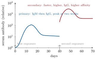
About one B cell in binds a given epitope, so a body’s B cells hold some precursors, of which perhaps a hundred meet the antigen in the draining node. Dividing every eight hours, a hundred cells become in a week. Antibody appears in the serum after four to six days, IgM first, peaks at about two weeks and declines with a half-life of three weeks for IgG as the short-lived plasma cells die, settling at the level the long-lived marrow plasma cells maintain for years. A second exposure starts from – memory cells of high affinity already switched to IgG: antibody rises within two days, ten- to a hundredfold higher, and neutralises the pathogen before it can establish itself — which is what a vaccine buys.
16.4 Tolerance and its failures
Definition 16.8 (Tolerance)
The repertoire is made at random and therefore contains receptors for self. Central tolerance removes them where the cells mature: in the thymus, T cells whose receptors bind self peptide–MHC too strongly are killed (negative selection), those that cannot bind MHC at all are also killed (they would be useless), and only the intermediate few per cent leave; a transcription factor, AIRE, makes thymic cells express proteins from every tissue — insulin, thyroglobulin — so that T cells specific for them can be deleted there. B cells that bind self in the marrow are deleted or edit their receptors. Peripheral tolerance handles the rest: a T cell that meets its antigen without costimulation is made unresponsive (anergy) or deleted; and regulatory T cells, defined by the factor FoxP3, actively suppress responses against self and against harmless antigens such as food and commensals. Autoimmunity is the failure of these controls: type 1 diabetes (T cells destroy the cells), multiple sclerosis (myelin), rheumatoid arthritis (joints), lupus (antibodies against nuclear components), thyroid disease; each is associated with particular HLA alleles, which present the relevant self peptides, and some are triggered by infection with a microbe whose peptides resemble a self protein (rheumatic fever after streptococcal infection).
Evidence. Medawar (1953) injected cells from one inbred mouse strain into newborn mice of another; as adults the recipients accepted skin grafts from the donor strain that they would otherwise have rejected, while rejecting grafts from third strains. Tolerance was acquired, specific, and set by what the immune system met early — the experimental basis for Burnet’s deletion of self-reactive clones. Children born without a functional AIRE gene develop autoimmunity against many organs at once; those without FoxP3 (a rare X-linked syndrome) develop fatal autoimmunity in infancy — the two arms of tolerance, each shown by its absence. ∎
Example 16.9 (Hypersensitivity, deficiency, transplantation)
Allergy is a Th2 response to a harmless antigen — pollen, peanut, penicillin — that produces IgE; on re-exposure the antigen cross-links IgE on mast cells, which release histamine within minutes, and if the antigen is in the blood the systemic release is anaphylaxis, treated with adrenaline. Immunodeficiency: children lacking RAG or the enzyme ADA make no lymphocytes (severe combined immunodeficiency) and die of infection unless given marrow or gene therapy; HIV destroys CD4 T cells and with them the help every response needs. Transplantation: the recipient’s T cells see the donor’s HLA molecules as foreign, and up to a tenth of all T cells respond — far more than to any pathogen — so grafts are matched at the HLA loci and the recipient is immunosuppressed for life; a graft of marrow can attack its new host instead. And the deliberate uses: the monoclonal antibodies of Chapter 6, the checkpoint inhibitors that release T cells against tumours (Chapter 11), and T cells engineered with a chimaeric receptor for a tumour antigen (CAR-T), which have cured leukaemias that nothing else touched.
16.5 Vaccination
Definition 16.10 (Vaccines and herd immunity)
A vaccine presents antigens of a pathogen, with an adjuvant or in a form that triggers innate receptors, so that the recipient acquires memory cells and antibodies without the disease (Chapter 13 lists the kinds). Its efficacy is the fraction of vaccinated people protected. Protection extends beyond the vaccinated: a pathogen spreading in a population where each case infects others on average when all are susceptible — the basic reproduction number — infects only times the susceptible fraction when some are immune, and dies out when that number falls below one. This is herd immunity: above a threshold of immune people the chain of transmission breaks and the unvaccinated — infants, the immunocompromised, those in whom the vaccine failed — are protected by the others.
Theorem 16.11 (The herd immunity threshold)
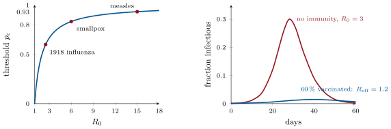
In a population where a fraction is immune and the rest susceptible, each case infects on average others. An epidemic cannot start if , that is if
With a vaccine of efficacy , the coverage needed is . If an epidemic does run in a susceptible population (the SIR model, with infection rate and recovery rate , ), it peaks when the susceptible fraction has fallen to , and the fraction never infected, , satisfies .
Proof. Each case makes contacts that would infect people if all were susceptible; a fraction of them are, so , and the condition gives ; a vaccine that protects a fraction of those who receive it makes a fraction of the population immune, so . In the SIR model, and , so grows while and falls after: the peak is at . Dividing the two equations, , so is constant; evaluating it at the start (, ) and at the end (, ) gives , the final-size relation. ∎
Example 16.12 (Measles and smallpox)
Measles has : , and with a vaccine of efficacy after two doses the coverage needed is — which is why measles returns wherever vaccination slips a few per cent, and why an unvaccinated child in a well-vaccinated country is nonetheless safe. Smallpox had –, a threshold near –, no animal reservoir, no asymptomatic carriers and a vaccine that worked in one dose: eradicable, and eradicated. Polio is nearly there. Influenza and the coronaviruses, whose antigens drift (Chapter 13) and whose immunity wanes, are not. In an unvaccinated population an epidemic with infects, by the final-size relation, of people; with , .
Remark 16.13 (What immunity remembers)
The adaptive immune system is a Darwinian machine inside each of us: random variation (V(D)J, hypermutation), selection (by antigen, in the germinal centre, against self in the thymus) and inheritance (memory cells, clonal expansion). It solves, in weeks, the problem that evolution solves in millennia — a molecule that binds a target never seen before — and its answers are among the best-studied cases of evolution observed directly. Its cost is autoimmunity, its dependence on the innate system’s judgement is absolute, and its memory, which Jenner exploited and which vaccines write deliberately, is the reason a species of long-lived, slow-breeding animals can survive in a world of fast-breeding microbes.
16.6 Exercises
Exercise 16.1 ★
State the four tenets of clonal selection and the experiment that supports each of the first two.
Solution
Solution of Exercise 16.1.
One lymphocyte, one specificity (Nossal and Lederberg: each cell secreted antibody to one antigen only); the receptor is made before antigen is met (Tonegawa: the gene is rearranged in the B cell, not in response to antigen); antigen selects and expands the matching clones into effector and memory cells; self-reactive clones are deleted during development.
Exercise 16.2 ★
Describe an antibody’s structure and give the function of each of the five classes.
Solution
Solution of Exercise 16.2.
Two heavy and two light chains, each with a variable domain at the tip and constant domains behind; two identical binding sites formed by six hypervariable loops; an Fc stem that other cells and complement read. IgM: first antibody, pentamer, complement activation. IgG: main blood and tissue antibody, opsonisation, neutralisation, crosses the placenta. IgA: dimer secreted onto mucosal surfaces and into milk. IgE: bound to mast cells, against parasites, the mediator of allergy. IgD: on naive B cells, of minor known function.
Exercise 16.3 ★
Contrast MHC class I and class II in cell distribution, source of peptide, peptide length and the T cell that reads each.
Solution
Solution of Exercise 16.3.
Class I: all nucleated cells; peptides from cytosolic proteins cut by the proteasome; 8–10 residues; read by CD8 cytotoxic T cells. Class II: dendritic cells, macrophages, B cells; peptides from proteins taken up and digested in endosomes; 13–25 residues; read by CD4 helper T cells.
Exercise 16.4 ★
Define and the herd immunity threshold, and compute the threshold for mumps (), pertussis () and the 1918 influenza ().
Solution
Solution of Exercise 16.4.
is the mean number of cases one case produces in a wholly susceptible population; the threshold is . Mumps ; pertussis ; 1918 influenza .
Exercise 16.5 ★★
A species has , , and a single light locus with , . Compute the combinatorial repertoire, then the total with a junctional factor of . How does it compare with the B cells the animal has?
Solution
Solution of Exercise 16.5.
heavy chains, light: combinations; with junctional diversity — ten times the number of B cells, so the animal expresses at most a tenth of what it could make, and almost every cell carries a unique receptor.
Exercise 16.6 ★★
A germinal-centre B cell’s variable region is ; AID mutates at per base pair per division. Over divisions, how many mutations per cell, and among cells how many distinct single-base variants of the antibody are generated? Compare with the possible single substitutions.
Solution
Solution of Exercise 16.6.
mutation per cell per division; in twenty divisions. Among cells, mutation events fall on possible single substitutions: each is sampled hundreds of times, and the double and triple combinations besides — the germinal centre explores the neighbourhood of the receptor exhaustively.
Exercise 16.7 ★★
Explain why a T cell from one person does not recognise viral peptides presented by another person’s cells, and why this same fact makes transplants difficult and the HLA system polymorphic.
Solution
Solution of Exercise 16.7.
T cells are selected in the thymus to recognise peptides on the host’s own MHC alleles; another person’s alleles have differently shaped grooves that hold different peptides in different orientations, so the same viral peptide is not seen. A graft’s foreign MHC molecules are themselves recognised by a large fraction of T cells as “self MHC with a strange peptide”, hence rejection. The polymorphism persists because a population with many alleles presents a broader sample of any pathogen’s peptides and no pathogen can escape everyone; heterozygotes present twice the range and are favoured.
Exercise 16.8 ★★
Predict the immune phenotype of a child lacking (a) RAG1, (b) AIRE, (c) FoxP3, (d) the class-switch enzyme AID, and (e) CD4 T cells.
Solution
Solution of Exercise 16.8.
(a) No V(D)J recombination: no B or T cells, severe combined immunodeficiency. (b) No AIRE: thymic cells fail to display tissue antigens, self-reactive T cells escape, autoimmunity against many organs. (c) No FoxP3: no regulatory T cells, fatal early-onset autoimmunity and allergy. (d) No AID: no class switching or hypermutation — abundant IgM, almost no IgG or IgA, low-affinity antibodies, recurrent infections. (e) No CD4 T cells: no help for B cells or macrophages, poor antibody responses and susceptibility to opportunistic infections — the immunodeficiency of AIDS.
Exercise 16.9 ★★
A vaccine has efficacy and the disease . What coverage stops transmission? If coverage is , what is , and what fraction of the population does an epidemic infect (use the final-size relation with replaced by among the susceptibles — or argue why that is approximately right)?
Solution
Solution of Exercise 16.9.
; coverage . At coverage the immune fraction is , . Among the susceptibles the epidemic spreads as if were (the immune merely dilute the contacts), and the final-size relation gives : about of the susceptibles, of the population, are infected.
Exercise 16.10 ★★★
Derive the final-size relation of the theorem from the SIR equations and solve it numerically for , , and . Why does an epidemic never infect everyone even without any intervention?
Solution
Solution of Exercise 16.10.
From and , , so is conserved; with , at the start and , at the end, . Solutions: , ( infected); , (); , (); , (). Not everyone: as the susceptibles are used up the effective reproduction number falls below one while some remain, and the epidemic burns out before reaching them.
Exercise 16.11 ★★★
Affinity maturation raises binding a thousandfold in three weeks. Treat the germinal centre as a population under selection: with a mutation rate of one per cell per division, a fraction of mutations improving affinity by a factor and the rest neutral or harmful, how many rounds of selection are needed for a thousandfold gain, and how many cells must each round screen? Comment on why the germinal centre is organised in cycles.
Solution
Solution of Exercise 16.11.
: successive improvements. With one mutation per cell per division and one in a hundred improving, at least a hundred cells must be screened per round to find one; in practice thousands, since half the mutations destroy binding and selection is noisy. Seventeen rounds of two divisions each take about nine days — the observed two to three weeks, given the inefficiencies. Cycles separate mutation (dark zone) from testing (light zone) so that each variant is judged against antigen before it is allowed to multiply, exactly as a genetic algorithm alternates variation and selection.
Exercise 16.12 ★★★
Autoimmune diseases are commoner in women and associated with HLA alleles; type 1 diabetes has risen fivefold in fifty years in rich countries. Propose, with the mechanisms of this chapter and of Chapter 14, explanations for each observation and one test of each.
Solution
Solution of Exercise 16.12.
Women: several immune genes lie on the X chromosome, some (TLR7) escape inactivation and are expressed from both copies, and oestrogens enhance B cell survival — test: men with an extra X (Klinefelter) have a female-like risk of lupus, which they do. HLA: the associated alleles present the relevant self peptides well, or fail to present them in the thymus so the clones are not deleted — test: mice transgenic for the human allele develop the disease when given the antigen. The rise: less early microbial exposure and altered microbiomes (antibiotics, diet, caesarean birth) give fewer regulatory T cells and more inflammation — tests: diabetes-prone mice raised germ-free or on antibiotics develop diabetes more, and children of immigrants acquire the incidence of the new country within a generation.
16.7 Problem: A Vaccine Campaign
Problem 16.1
Weekend problem — a repertoire counted, a response timed from a hundred cells to a millilitre of serum, a germinal centre run as an evolutionary algorithm, and a measles campaign planned against the herd threshold, ending on the size of the repertoire, the antibody a plasma cell makes in a day and the coverage a country needs
Data: , , ; light loci and ; junctional factor . A body has B cells; one in binds a given epitope; precursors are recruited; division every for days; of the clone becomes plasma cells secreting IgG molecules a second each; IgG is ; plasma volume ; IgG half-life . Germinal centre: variable region, mutations per bp per division, one in mutations improves affinity by . Measles: , vaccine efficacy after one dose and after two; a country of people with births a year.
Part I — The repertoire.
- Compute the combinatorial repertoire and the total with junctional diversity.
- Compare with the number of B cells. What fraction of the possible repertoire does one body express at a time, and what follows for two individuals’ repertoires?
- How many B cells in the body bind the epitope? How many, on average, in a lymph node holding B cells?
- A newborn has B cells. How many bind the epitope, and why is a newborn’s response weak even so?
- An antibody’s third heavy-chain loop is made by junctional diversity. Explain why this loop is the most variable part of the molecule and usually at the centre of the binding site.
- Somatic hypermutation adds diversity after selection, V(D)J before. Why is it advantageous to have both, in that order?
Part II — The response.
- From cells dividing every , how many after days? How many plasma cells?
- How many IgG molecules do they secrete per day, and what mass? What serum concentration does one day’s output give in ?
- After ten days of secretion at that rate, ignoring decay, what is the concentration in g/L? Compare with the normal total IgG of .
- The plasma cells die after two weeks; the antibody then decays with half-life . How long until the concentration of question 9 falls a hundredfold? What maintains antibody for years?
- A secondary response starts from memory cells. How many cells after days, and how does that compare with the primary response at day 3 and day 7?
- Explain why IgM comes first and IgG later in a primary response, and why the secondary response is IgG from the start.
Part III — The germinal centre.
- How many mutations does a B cell acquire in its variable region per division? Over divisions?
- What fraction of the daughters of one division carry an improving mutation? In a germinal centre of dividing cells, how many improved cells appear per division?
- How many successive improvements of give a thousandfold gain in affinity? If each round of selection takes two divisions (), how long is that?
- Most mutations damage the antibody. If of mutations destroy binding, what fraction of cells survive each division’s mutation, and why must the light zone kill most cells?
- A vaccine given as two doses six weeks apart yields antibodies of higher affinity than one dose. Explain with the germinal centre.
- Some viruses (HIV, influenza) escape antibodies by mutating their surface. Explain why the germinal centre is nevertheless the basis of hope for a broadly protective vaccine.
Part IV — The campaign.
- Herd threshold for measles, and coverage required with one dose; with two doses.
- The country vaccinates of children with two doses. Fraction immune, , and the verdict: does measles circulate?
- How many susceptible children accumulate per year of births at coverage (unvaccinated plus vaccine failures)? After five years without measles, how many susceptibles are there, and why does an importation then cause an outbreak?
- Use the final-size relation with the effective reproduction number among susceptibles to estimate what fraction of those susceptibles an outbreak infects. (Take from question 20 as the reproduction number in the whole population, and note that among the susceptible pool the disease spreads as if were .)
- Coverage is raised to with two doses. Fraction immune and ; is the threshold met?
- Infants under one year cannot be vaccinated against measles and are the most likely to die of it. Explain how the campaign protects them, and what happens to their protection if coverage falls to .
- Summarise: the total repertoire (question 1), the IgG one day’s plasma cells make (question 8), and the two-dose coverage the country needs (question 19).
Solution
Solution of Problem 16.1.
1. ; . 2. cells against possible: a body could hold every receptor about twice, but clones are unequal and most sequences never happen to be made, so each body expresses a large but incomplete sample — and two people share only a small fraction of their receptors. 3. cells; in a node of , about . 4. precursors — a hundred times fewer; the newborn also has immature follicles and dendritic cells, no memory, and limited T cell help, so responses are slow and small, and maternal IgG bridges the gap. 5. It spans the V–D and D–J junctions, where nucleotides are removed and added at random: its sequence is not encoded anywhere in the genome. Sitting between the two chains at the centre of the binding site, it is the loop most often in contact with the epitope. 6. Before antigen, diversity must be broad so that something binds whatever comes; after antigen, effort is better spent varying the few receptors already known to bind. A coarse search then a fine one is far more efficient than either alone. 7. Seven days is divisions: cells; , plasma cells. 8. molecules a day; g ; in , per day. 9. in : , a thousandth of the total IgG — the antibody to any one antigen is a small fraction of the whole. 10. A hundredfold is half-lives, about days. Years of antibody come from long-lived plasma cells in the marrow, which secrete continuously, and from memory cells re-stimulated on re-exposure. 11. Nine divisions: cells at day 3 — a thousand times the primary’s at day 3, and a quarter of what the primary reached only at day 7. 12. The rearranged VDJ lies next to C, so the first transcript is IgM; switching to C needs T cell help and AID, which take days. Memory cells have already switched, so their progeny make IgG at once. 13. per division; over twenty. 14. One in a hundred; improved cells per division. 15. : ; days. 16. Half the daughters lose binding: without selection, of the lineage would still bind after twenty divisions. The light zone must remove the losers at every cycle so that only functional and improved receptors go on. 17. The first dose leaves memory B cells of raised affinity and switched class; the second dose seeds new germinal centres with them, and hypermutation and selection resume from a higher starting point, so the antibodies that emerge are better still. 18. Some infected people eventually make antibodies against conserved sites that the virus cannot change (broadly neutralising antibodies), after years of maturation. The germinal centre can be steered: a sequence of immunogens that first engages the right germline precursors and then rewards binding to the conserved site could guide maturation along that path in months rather than years. 19. . One dose: — unattainable; two doses: . 20. Immune ; : measles circulates. 21. susceptibles per birth cohort; after five years — a pool in which an imported case finds enough susceptibles in its contacts to sustain chains. 22. Among the susceptibles, gives : about of them, some cases. 23. Immune ; — still above one; the threshold needs with two doses, and measles is the disease that punishes the last few per cent. 24. Infants are protected only by the immunity of those around them (and by maternal antibody for a few months): if chains of transmission cannot form, no case reaches them. At coverage the immune fraction is and : outbreaks run, and the infants, unvaccinated and most vulnerable, are infected. 25. Repertoire about ; of IgG a day from one response’s plasma cells; two-dose coverage.