University Biology — Year 3 · Bachelor Year 3
13Virology
There are some virus particles on Earth, ten for every cell, and in the sea they kill a fifth of all bacteria every day. A virus is not a cell: it has no metabolism, no ribosomes, no way to make anything by itself. It is a genome — as short as two genes, as long as two thousand — wrapped in a protein shell, which enters a cell and turns the cell’s machinery to making copies of itself. The influenza of 1918 killed fifty million people; smallpox, which killed three hundred million in the twentieth century alone, was eradicated in 1980 by a vaccine first tried in 1796; and a coronavirus that crossed into humans in 2019 had its genome sequenced within weeks, a vaccine designed within days of that, and drugs against its protease within two years. This chapter treats what viruses are and how they are classified by the chemistry of their genomes, how they multiply, the kinetics of an infection inside one host — which a pair of differential equations describes well enough to have changed the treatment of AIDS — their evolution, and how they are fought.
13.1 What a virus is
Definition 13.1 (Virus)
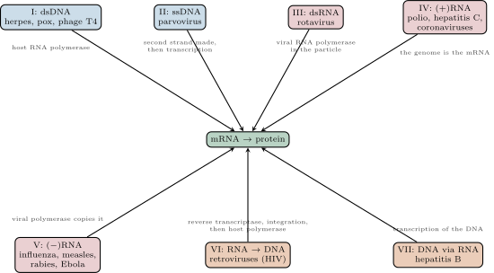
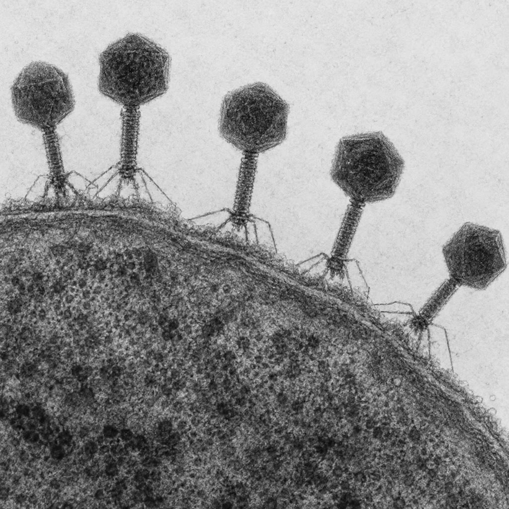
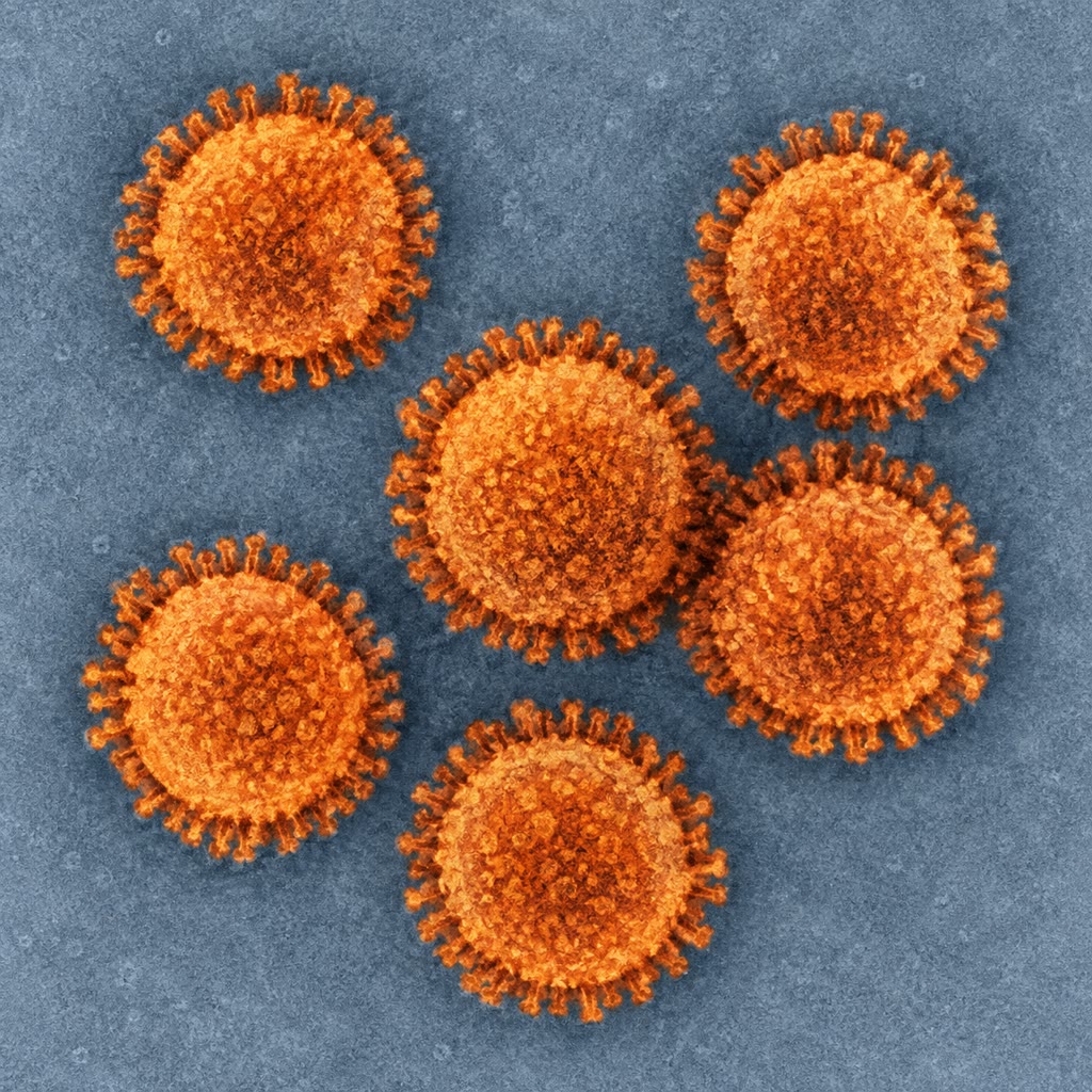
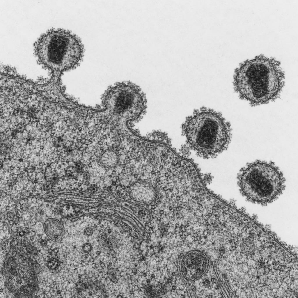
A virus is an obligate intracellular parasite consisting of a nucleic acid genome — DNA or RNA, single- or double-stranded, linear or circular, one molecule or several — packaged in a protein capsid, and in many cases wrapped in an envelope taken from a host membrane and studded with viral glycoproteins. The complete infectious particle is the virion, across for most (a few giant viruses reach a micrometre). Capsids are built from many copies of one or a few proteins arranged with helical symmetry (a rod, as in tobacco mosaic virus) or icosahedral symmetry (a shell of subunits, ). The Baltimore classification groups viruses by the route from genome to messenger RNA: I, double-stranded DNA (herpes, pox, adenovirus, most phages); II, single-stranded DNA (parvovirus); III, double-stranded RNA (rotavirus); IV, positive-strand RNA, itself a messenger (polio, hepatitis C, the coronaviruses); V, negative-strand RNA, complementary to messenger (influenza, measles, rabies, Ebola); VI, RNA reverse-transcribed into DNA (the retroviruses, HIV); VII, DNA replicated through an RNA intermediate (hepatitis B). Every class but I must bring or encode an enzyme the cell lacks — an RNA-dependent RNA polymerase, a reverse transcriptase — and those enzymes are the targets of most antiviral drugs.
Evidence. Ivanovsky (1892) and Beijerinck (1898) showed that the mosaic disease of tobacco was transmitted by sap passed through filters that retained all bacteria — a contagium vivum fluidum, a living infectious fluid. Stanley (1935) crystallised the agent, tobacco mosaic virus, and showed it to be protein with (as Bawden and Pirie found) RNA. Hershey and Chase (1952) labelled a bacteriophage’s protein with S and its DNA with P, let it infect E. coli, sheared off the empty coats in a blender, and found the P inside the cells and the S outside: the DNA enters and the protein stays behind, so the DNA carries the instructions. Fraenkel-Conrat (1955) took tobacco mosaic virus apart into protein and RNA and reassembled infectious particles from the RNA of one strain and the protein of another; the progeny were of the RNA’s strain. ∎
Proposition 13.2 (Genetic economy: why capsids are symmetric)
A capsid must enclose the genome that encodes it. A protein of mass needs about codons, that is nucleotides, a stretch of RNA weighing about : any protein is encoded by a nucleic acid nine times its own mass. A shell made of a single protein molecule enclosing a genome would have to enclose a genome nine times its own mass just to encode itself, and a shell cannot hold nine times its own mass of nucleic acid at biological density. The way out, seen by Crick and Watson (1956), is to build the shell from many copies of one small protein: tobacco mosaic virus encodes a coat protein in of its genome and uses copies of it. Identical subunits that pack identically must be arranged symmetrically, so viral capsids are helices or icosahedra, and the same economy gives them the capacity to self-assemble, since each subunit needs only to recognise its neighbours.
Proof. The mass ratio follows from a codon being three nucleotides of about each and a residue about : . A capsid of one protein would enclose of nucleic acid, a sphere of radius about at nucleic-acid density, whereas the protein itself would make a shell of about thickness and the same radius — geometrically possible, but a single polypeptide of residues that folds correctly is not, and one mutation would destroy it. With identical subunits the coding cost falls by while the enclosed volume is unchanged. ∎
13.2 How a virus multiplies
Definition 13.3 (The replication cycle)
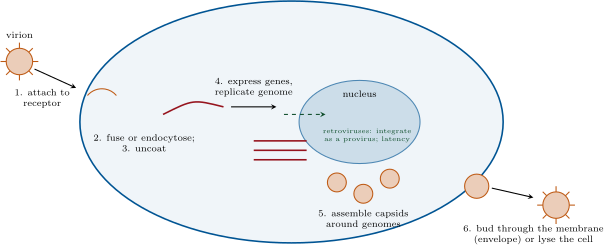
Every virus passes through the same stages. Attachment to a specific host receptor — CD4 and a chemokine receptor for HIV, ACE2 for the SARS coronaviruses, sialic acid for influenza, a lipopolysaccharide or a porin for a phage — which decides which species and which cells the virus can infect (its tropism). Entry: fusion of the envelope with the plasma membrane, or endocytosis followed by fusion from within the acid endosome, or, for a phage, injection of the genome through the wall. Uncoating, expression of the viral genes by the class-specific route, and replication of the genome, often in a factory the virus builds in the cytoplasm or nucleus. Assembly of new capsids around new genomes, driven by the self-assembly of the symmetric subunits, and release: by lysis of the cell, or by budding through a membrane, which supplies the envelope. Some viruses can instead insert their genome into the host’s and wait: the lysogeny of phage , whose repressor keeps it silent until the host is damaged; the latency of herpesviruses in neurons for a lifetime and of HIV as a provirus in resting T cells, from which it is not removed by any drug that acts on replication.
Method 13.4 (Counting infectious particles: the plaque assay)
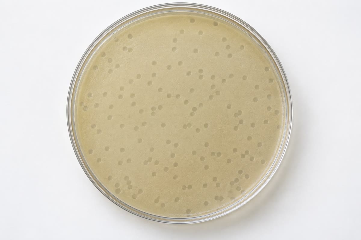
To measure a virus stock: (1) dilute it in tenfold steps; (2) mix each dilution with an excess of host cells — bacteria in soft agar for a phage, a monolayer of cultured cells for an animal virus; (3) incubate: each infectious particle infects one cell, the progeny infect the neighbours, and after a day or two a clear hole, a plaque, marks the spot; (4) count the plaques on a plate with and multiply by the dilution: the titre in plaque-forming units (PFU) per millilitre. Since each plaque is a clone of one particle, picking it yields a pure isolate. The ratio of physical particles (counted by electron microscopy or PCR) to plaque-forming units is usually far above one — ten to a thousand for many animal viruses — since most particles are defective or fail to infect.
Theorem 13.5 (Infection dynamics within a host)
Let be the density of susceptible target cells, of infected cells and of free virions. Virions infect targets at rate , infected cells produce virions at rate each and die at rate , and free virions are cleared at rate :
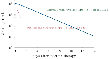
With held constant, the infection grows if the basic reproduction number exceeds , and at steady state with production balancing clearance. If a drug stops all new infections at (), the virions decay at first at the clearance rate , then, once the free virus has fallen to the level the remaining infected cells maintain, at the death rate of those cells:
The two slopes of the viral load after treatment measure and , and gives the daily production of virions before treatment.
Proof. An infected cell lives on average and makes virions; each virion lives and infects cells in that time; the product is the number of infected cells one infected cell gives rise to, . At steady state gives . After treatment , so , and is a linear equation whose solution with is the expression given (check: at it equals , and it satisfies the equation term by term). For the term is gone and follows the infected cells with slope ; for and the early slope is . ∎
Example 13.6 (HIV before and after the equations)
Until 1995 the years of clinical latency of HIV infection — a stable viral load of – copies per millilitre and a slow fall of T cells — were read as a quiescent virus. Ho, Perelson and colleagues gave patients a protease inhibitor and fitted the decline of the viral load to the theorem: the fast phase gave (a virion half-life of about six hours), the slow phase (an infected cell lives about a day and a half). A steady load of per millilitre in of body fluid, cleared at , therefore requires the production of about virions a day, every day, for years: the “latent” period is a furious steady state of infection and death, with the T cell pool replaced daily until it fails. The mutation arithmetic of the next section then follows at once, and with it the reason single drugs failed and three did not.
13.3 How viruses evolve
Definition 13.7 (Mutation, quasispecies, drift and shift)
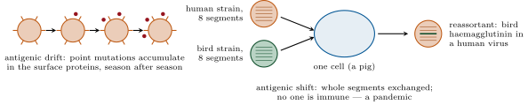
RNA-dependent polymerases and reverse transcriptases have no proofreading, and RNA viruses mutate at to per nucleotide per replication — a thousand to a million times the rate of their hosts — so that a population of an RNA virus is a cloud of related genomes, a quasispecies, in which every single point mutant is present at all times if the population is large. Selection by antibodies drives antigenic drift: the surface proteins of influenza accumulate substitutions year by year, and last year’s immunity protects less each season. Antigenic shift is a jump: a segmented genome (influenza has eight RNA segments) can be reassorted when two strains infect one cell, and a segment encoding a haemagglutinin from a bird or pig virus, to which no human has immunity, produces a pandemic — 1918, 1957, 1968, 2009. Recombination within a genome does the same for unsegmented viruses, and the coronaviruses recombine freely. Most new human viruses are zoonoses, crossing from an animal reservoir — bats for the coronaviruses, Ebola and rabies; birds and pigs for influenza; primates for HIV — and the crossing is usually a matter of a few mutations in a receptor-binding protein.
Proposition 13.8 (The error threshold)
A genome of nucleotides copied with error rate per nucleotide is copied without any error with probability . If the fittest sequence is to persist in the population against the flood of its mutants, the fraction of error-free copies must exceed about the inverse of its selective advantage : , that is , a number of order one. The product of mutation rate and genome length is therefore bounded: an RNA virus with cannot have a genome much longer than nucleotides, which is about what RNA viruses have, and the coronaviruses, at the longest RNA genomes known, achieve it only by encoding a proofreading exonuclease that cuts tenfold. The threshold is also a weapon: drugs that raise the polymerase’s error rate (ribavirin, molnupiravir) push a virus over it into error catastrophe, and the quasispecies collapses.
Proof. Independent errors at each of sites give the error-free probability , and for small . The population balance is the standard quasispecies argument (Eigen, 1971): the master sequence is replenished at rate relative to the average mutant and lost to mutation otherwise; it is maintained only if the former exceeds , whence the bound. ∎
Example 13.9 (Why HIV needed three drugs)
HIV’s reverse transcriptase errs about once in nucleotides, so each genome copied carries about mutations, and new genomes a day carry mutations among them: every one of the possible single point mutations arises many times every day, before any drug is given. A drug that a single mutation defeats — as one mutation defeats most reverse-transcriptase and protease inhibitors — is therefore defeated within weeks, which is what happened to AZT in 1987. Two independent mutations arise together at per genome, about once a day; three at , once in a thousand days — and a patient on three drugs whose load has fallen a thousandfold produces genomes a day, so the triple mutant never appears. That, and the theorem’s demonstration that the virus was replicating at full speed, made combination therapy from 1996 the treatment that turned AIDS into a chronic condition.
13.4 Hosts, defences and drugs
Proposition 13.10 (Pathogenesis and defence)
A virus harms by lysing cells (polio destroys motor neurons, HIV kills T cells), by making cells produce toxic products, by transforming them (Chapter 11), and often mainly through the host’s own response: the fever, tissue damage and, at worst, the cytokine storm of the immune reaction to influenza or the SARS coronaviruses. The host answers with innate defences that recognise the signatures of viral replication — double-stranded RNA, RNA without a cap, DNA in the cytosol — and respond with interferon, which puts every neighbouring cell into an antiviral state (Chapter 15); with natural killer cells that destroy cells that have hidden their MHC; with antibodies that neutralise virions and cytotoxic T cells that kill infected cells before they release progeny (Chapter 16); and, in bacteria, with restriction enzymes that cut foreign DNA and the CRISPR memory of Chapter 6. Viruses answer back: almost every large virus encodes proteins that block interferon, hide MHC, mimic cytokines or inhibit apoptosis, and the arms race is written in both genomes.
Definition 13.11 (Antiviral drugs and vaccines)
Antiviral drugs attack the viral enzymes or the steps a host cell does not perform. Nucleoside analogues are chain terminators: aciclovir is phosphorylated only by the herpesvirus thymidine kinase and then blocks the viral DNA polymerase, so it acts only in infected cells; AZT, tenofovir and lamivudine stop reverse transcriptase; remdesivir and molnupiravir act on the coronavirus polymerase, the second by mutagenesis. Protease inhibitors block the cleavage of viral polyproteins (HIV, hepatitis C, SARS-CoV-2’s nirmatrelvir). Integrase inhibitors stop HIV’s provirus forming; neuraminidase inhibitors stop influenza virions detaching from the cell they bud from; entry inhibitors block a receptor. The hepatitis C virus is now cured in twelve weeks by combinations of direct-acting drugs, the first chronic viral infection ever cured by chemotherapy. Vaccines present the virus’s antigens without its disease: a live attenuated virus (measles, polio by mouth, yellow fever), an inactivated virus (polio by injection, influenza), a subunit (the hepatitis B surface antigen made in yeast, the papillomavirus capsid protein), a harmless viral vector carrying a gene of the target, or, since 2020, messenger RNA encoding the antigen, delivered in lipid nanoparticles, which the recipient’s own cells translate. The immunology of why they work, and of how many must be vaccinated to protect the rest, is the matter of Chapter 16.
Remark 13.12 (Viruses in the economy of life)
The viruses that harm humans are a rounding error in the world’s virus population, most of which infects bacteria and archaea. Phages kill a fifth to a third of the ocean’s bacteria daily and return their contents to the dissolved pool — the viral shunt of the Year 2 volume’s carbon cycle. They move genes between bacteria (Chapter 12), and their toxins are the toxins of diphtheria, cholera and the worst E. coli. Eight per cent of the human genome is the remains of ancient retroviruses (Chapter 4), one of whose envelope genes now fuses the cells of the placenta. Viruses are the largest reservoir of genetic novelty on the planet, and the boundary of what counts as alive runs through them.
13.5 Exercises
Exercise 13.1 ★
Place polio, influenza, HIV, herpes simplex, hepatitis B and rotavirus in their Baltimore classes and say what enzyme each must bring or make that the cell lacks.
Solution
Solution of Exercise 13.1.
Polio: IV, positive-strand RNA — an RNA-dependent RNA polymerase. Influenza: V, negative-strand RNA — its own RNA polymerase, carried in the virion. HIV: VI — reverse transcriptase (and integrase). Herpes simplex: I, double-stranded DNA — the host could in principle supply everything, but it brings its own DNA polymerase and thymidine kinase. Hepatitis B: VII — a reverse transcriptase to copy its pregenomic RNA into DNA. Rotavirus: III, double-stranded RNA — an RNA-dependent RNA polymerase inside the particle.
Exercise 13.2 ★
What did the Hershey–Chase experiment show, and what would have been found if protein carried the instructions?
Solution
Solution of Exercise 13.2.
The phage’s DNA (P) entered the bacteria and the protein (S) stayed outside and could be sheared off; the progeny phage inherited the P. So the DNA is what the phage injects and what directs the making of new phage. Had protein carried the instructions, the S would have been found inside and passed to the progeny.
Exercise 13.3 ★
A phage stock diluted gives plaques from . What is the titre? Why may the electron microscope count ten times more particles?
Solution
Solution of Exercise 13.3.
plaque-forming units per mL. The microscope counts every particle, including empty capsids, particles with defective genomes and those that fail to attach or inject; only particles that complete an infection make plaques.
Exercise 13.4 ★
List the six stages of a replication cycle and, for each, name one antiviral drug or host defence that acts on it.
Solution
Solution of Exercise 13.4.
Attachment: entry inhibitors (maraviroc), neutralising antibodies. Entry/fusion: fusion inhibitors, endosomal-acidification blockers. Uncoating: capsid-binding drugs (pleconaril; lenacapavir). Expression and replication: nucleoside analogues, reverse-transcriptase and integrase inhibitors, interferon, RNA interference. Assembly: protease inhibitors (uncleaved polyproteins cannot assemble). Release: neuraminidase inhibitors (influenza), and cytotoxic T cells killing the cell before release.
Exercise 13.5 ★★
Using Proposition 13.2, compute the nucleic acid needed to encode the capsid of a virus made of copies of a protein, and compare with the genome such a virus might have. What fraction of the genome is the capsid gene?
Solution
Solution of Exercise 13.5.
One protein is residues, nucleotides: , of a genome. Encoding the copies separately would take , thirty-three times the whole genome; the capsid protein itself () outweighs the genome () fourfold.
Exercise 13.6 ★★
With , and per mL, compute from the theorem at , and days. How long until the load falls below per mL, the limit of detection?
Solution
Solution of Exercise 13.6.
: : ; : ; : . Below when : .
Exercise 13.7 ★★
An RNA virus has and . What fraction of its progeny genomes carry no mutation? What happens to that fraction if a mutagenic drug raises threefold? Explain why the coronaviruses needed a proofreading enzyme.
Solution
Solution of Exercise 13.7.
; error-free fraction . Threefold: , — most progeny now carry mutations and the fittest sequence is diluted toward the threshold. A coronavirus at with would have and only error-free copies, beyond the threshold; its exonuclease brings to , , three quarters error-free.
Exercise 13.8 ★★
Explain antigenic drift and shift in influenza, and why a pandemic strain has always so far carried a new haemagglutinin.
Solution
Solution of Exercise 13.8.
Drift: point mutations in the haemagglutinin and neuraminidase accumulate under antibody selection, so each season’s strain is partly new and last year’s immunity is partly obsolete. Shift: co-infection of one cell by a human and an animal strain reassorts the eight segments, and a haemagglutinin subtype from birds or pigs appears in a virus adapted to humans. No one has antibodies to the new subtype, so the whole population is susceptible at once — the condition for a pandemic, met in 1918, 1957, 1968 and 2009 by a new haemagglutinin each time.
Exercise 13.9 ★★
Aciclovir is a guanosine analogue that must be phosphorylated to act. Explain why it harms herpes-infected cells and not others, and predict the resistance mutations.
Solution
Solution of Exercise 13.9.
The herpesvirus thymidine kinase phosphorylates aciclovir, which the cellular kinases barely touch; cellular kinases then complete the triphosphate, which the viral DNA polymerase incorporates and, lacking a 3 hydroxyl, terminates the chain. Uninfected cells never make the active form, and the viral polymerase prefers it to the cellular one. Resistance: loss or alteration of the viral thymidine kinase (the commonest), or mutations in the viral polymerase that exclude the analogue.
Exercise 13.10 ★★★
Derive from the two equations of the theorem by finding the condition for a small infection (, near zero, constant) to grow, and interpret each factor.
Solution
Solution of Exercise 13.10.
With constant the equations are linear in with matrix , trace and determinant . The eigenvalues have negative trace, so one is positive exactly when the determinant is negative: , i.e. . Factors: is the number of virions an infected cell makes in its life; is the number of cells a virion infects in its life; their product is the number of infected cells one infected cell produces.
Exercise 13.11 ★★★
A patient on effective therapy still harbours latently infected resting T cells whose provirus is silent, with a half-life of months. How long would therapy have to continue for the reservoir to fall below one cell? Why does this make HIV incurable by drugs that block replication, and what would a cure require?
Solution
Solution of Exercise 13.11.
: twenty half-lives, months, about years — longer than a life. Drugs that block replication do not touch a provirus that is not being transcribed; the reservoir persists, and when therapy stops a single reactivating cell restarts the infection. A cure must remove or permanently silence the reservoir: kill the latent cells (reactivate them under therapy so they die or are killed), excise or disable the provirus, or replace the immune system with one the virus cannot infect.
Exercise 13.12 ★★★
Explain the argument of Example 13.9 with your own numbers for a virus of with producing genomes a day. How many drugs would you combine, and why might drugs against two sites of the same enzyme not count as two?
Solution
Solution of Exercise 13.12.
mutations per genome; genomes a day carry mutations among possible single mutations: each arises about times a day. A specific double mutant arises at per genome, a day — once in three years; a triple at , never. Two drugs with independent resistance mutations would suffice in principle, three give a margin for poor adherence. Two drugs binding the same enzyme may be defeated by one mutation that alters the site or the enzyme’s conformation, and their resistance mutations are not independent; they count as less than two.
13.6 Problem: A Virus in a Patient
Problem 13.1
Weekend problem — an HIV infection counted virion by virion, its dynamics fitted to a treatment curve, its mutations tallied against the drugs, and its reservoir timed, ending on the daily production of virions, the day the load becomes undetectable and the probability that a triple mutant exists
Data: viral load per mL in of body fluid; , ; each infected cell makes virions a day; genome , per nucleotide per replication; single mutations confer resistance to each of three drugs; on triple therapy production falls to genomes a day; latent reservoir cells, half-life months; a virion is a sphere of containing two RNA strands and about protein molecules of .
Part I — Counting.
- How many virions are in the body at steady state?
- How many are cleared per day, and hence produced per day?
- How many infected cells produce them, and how many die per day?
- What is the mass of one virion (RNA at per nucleotide plus protein), and the total mass of virions in the body? Compare with a grain of sand ().
- The body has CD4 T cells and replaces the infected ones. What fraction of the pool is infected at any moment, and how does a fraction so small kill the pool in ten years?
- If each infected cell dies by apoptosis and is engulfed within an hour (Chapter 10), how many are being cleared at any moment?
Part II — Treatment.
- Therapy stops new infections at . Compute at , , and days from the theorem.
- On what day does the load fall below the detection limit of per mL?
- From the two slopes, how would a clinician estimate and from measurements on days – and –?
- After the two phases the load flattens at a few copies per mL and stays there for years. Which compartment of the model is missing, and what is its decay rate?
- Compute the half-life of a free virion and of an infected cell.
- Explain why the pre-treatment plateau was misread as latency, and what the equations showed instead.
Part III — Mutation.
- Mutations per genome per replication, and mutations produced per day in the untreated patient.
- How many distinct single point mutations are possible in the genome? How many times per day does each arise?
- Rate per genome of a specific double mutant and of a specific triple mutant; how many of each arise per day untreated?
- On triple therapy, with genomes a day, expected triple mutants per year?
- The patient misses doses so that one drug’s level falls to nothing for a week while the other two hold. Which mutants can now be selected, and what is the expected number of the relevant double mutants produced in that week at genomes a day?
- Why does resistance to one nucleoside analogue often confer resistance to another, and how does that change the count of “independent” drugs?
Part IV — The reservoir and the cure.
- With a half-life of months, how long for the latent cells to fall to one? To ?
- If therapy is stopped, one latent cell reactivating suffices to restart the infection. How long after stopping does the load return to per mL, if it grows with per generation of days from one virion?
- Propose two strategies for a cure that follow from the model, and state the obstacle to each.
- The theorem’s is within one host. A population for HIV between people is about –. What fraction of transmissions must be prevented to end the epidemic ()?
- Treatment lowers a patient’s transmissibility essentially to zero. Explain how “treatment as prevention” follows from the within-host theorem.
- A vaccine of efficacy (the fraction of vaccinated people protected) given to a fraction of the population protects of it. What coverage is needed to reach the threshold of the previous question with ? With ? What does the first answer mean?
- Summarise: virions produced per day (question 2), the day the load becomes undetectable (question 8), and the expected number of triple mutants per year on therapy (question 16).
Solution
Solution of Problem 13.1.
1. virions. 2. Cleared a day; produced the same. 3. infected cells; dying a day. 4. RNA Da; protein Da; total Da g. All virions together: g, a third of a microgram — three thousand times less than a grain of sand. 5. of the pool. Ten years of deaths a day is cells, three per cent of the pool: the pool fails not by arithmetic subtraction but because a decade of forced regeneration and chronic activation exhausts the capacity to replace them. 6. corpses being cleared at any moment. 7. : : ; : ; : ; : . 8. : days. 9. Days 3–10 lie on the slow phase: the slope of there is (from to in seven days, ). The fast phase lasts only hours (), so needs samples every few hours on the first day, fitted to the two-exponential formula. 10. The latently infected cells, which release virus as they reactivate; their decay rate is per month, about per day. 11. Virion , about ; infected cell . 12. A constant load looked like a virus doing nothing; the equations show it as a steady state in which virions are made and cleared and T cells infected and killed every day. 13. mutations per genome; mutations a day. 14. possible single mutations; each arises times a day. 15. Specific double: per genome, about one a day untreated; specific triple: , a day — once in three centuries. 16. a day, a year. 17. With one drug gone, mutants resistant to the other two suffice: a specific double at ; genomes in the week give expected — still unlikely, unless the load rebounds toward a day during the lapse, when it becomes one a day and the escape is probable. Missed doses are how resistance arrives. 18. The analogues share the reverse transcriptase active site, and one mutation there (or a set of thymidine-analogue mutations) alters the enzyme’s handling of several of them: cross-resistance. Two nucleosides count as less than two independent drugs, so regimens combine classes — a nucleoside, an integrase inhibitor, a protease inhibitor or a non-nucleoside. 19. half-lives, months, years; to , half-lives, months, years. 20. From one virion to at per generation: generations, three weeks. 21. Shock and kill: reactivate the latent cells under therapy so that they die or are killed — obstacle: no agent reactivates them all, and the reactivated cells are not reliably killed. Gene editing: excise the provirus or destroy the receptor CCR5 in the patient’s cells — obstacle: reaching every cell. (Replacing the marrow with CCR5-deficient donor cells has cured a handful of patients at a risk acceptable only when a transplant was needed anyway.) 22. : for , for . 23. Therapy sets inside the patient, the load falls by the theorem to almost nothing, and transmission, which is proportional to the load, stops; treating every infected person drives the population’s below one. 24. : with and , — more than everyone: the vaccine alone cannot reach the threshold; with , . 25. About virions produced a day; undetectable on about day ; some triple mutants a year on therapy.