University Biology — Year 3 · Bachelor Year 3
21Endocrinology and Homeostasis
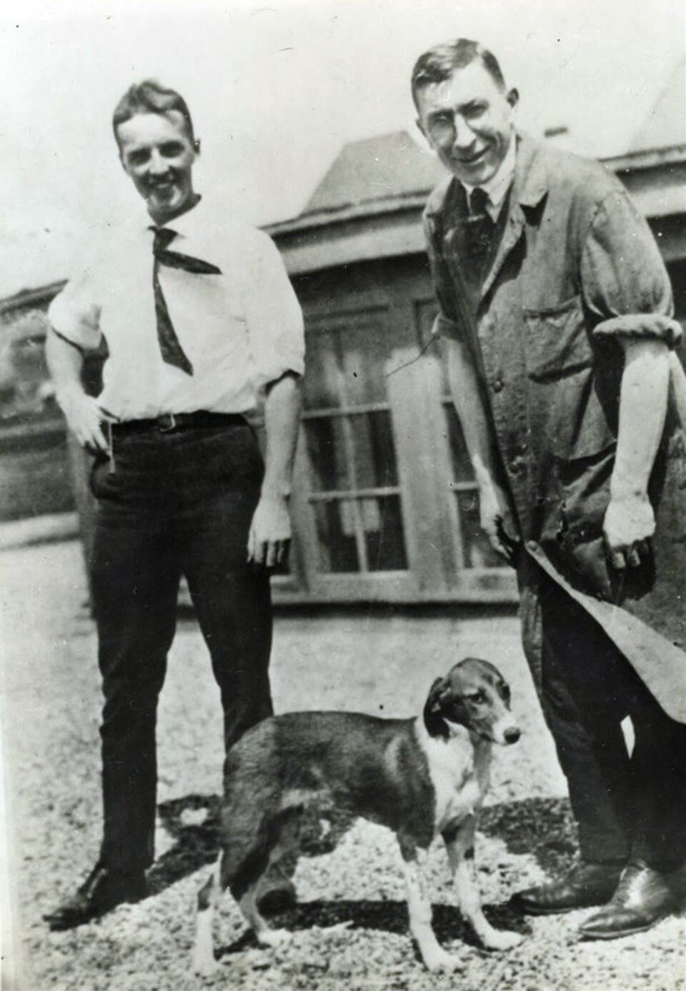
In the summer of 1921 two young men in a hot Toronto laboratory tied off the pancreatic ducts of dogs, waited for the digestive tissue to wither, ground up what was left, and injected the extract into a dog made diabetic by removing its pancreas. Its blood sugar fell. By January a fourteen-year-old boy dying of diabetes in Toronto General Hospital was receiving the extract, and within weeks he was eating, walking and gaining weight; he lived another thirteen years. The substance, insulin, is a peptide of fifty-one amino acids secreted by a per cent of the pancreas into the blood, where at a concentration of a few hundred picomolar it tells every muscle and fat cell in the body what to do with the sugar of the last meal. This chapter is about such messengers: what they are, how a cell that has never met the gland knows what the message means, how the glands are themselves governed in a hierarchy of feedback loops, and how the whole system holds the blood’s glucose, salt, calcium and temperature within a few per cent for a lifetime — and what happens, in the clinic, when one of the loops fails.
21.1 Hormones and their receptors
Definition 21.1 (Hormone)
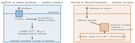
A hormone is a substance released by a cell into the blood that acts on distant cells carrying its receptor; a paracrine signal acts on neighbours, an autocrine one on the cell that made it, and a neurohormone is released by a neuron into the blood. The classical endocrine glands — pituitary, thyroid, parathyroids, adrenals, pancreatic islets, gonads — have been joined by the heart (natriuretic peptide), the kidney (erythropoietin), fat (leptin), the gut (a dozen peptides) and bone. Chemically hormones are of three kinds, and the chemistry fixes the mechanism. Peptide and protein hormones (insulin, growth hormone, the pituitary hormones) are made on ribosomes, stored in granules, released by exocytosis, travel free in plasma, cannot cross membranes, and act on receptors at the cell surface — G-protein-coupled or kinase-linked — through second messengers, within seconds to minutes; their half-lives are minutes. Steroids (cortisol, aldosterone, oestrogens, testosterone, and the secosteroid vitamin D) are made from cholesterol as needed, not stored, travel bound to carrier proteins, cross membranes, and bind nuclear receptors that are themselves transcription factors: their effects take hours and last days. Amines straddle the two: adrenaline acts at the surface within a second; the thyroid hormones, though amino-acid derivatives, act on nuclear receptors like steroids. A hormone means nothing in itself: the same adrenaline dilates one vessel and constricts another because their smooth muscles carry different receptor subtypes, and the same cortisol raises blood glucose in the liver and suppresses a lymphocyte because the two cells’ chromatin offers its receptor different genes.
Proposition 21.2 (Dose and response)
A cell with receptors of dissociation constant in a hormone concentration has of them occupied, and the response usually rises with occupancy along a sigmoid on a logarithmic dose axis, half-maximal at an that is often far below : a few per cent occupancy gives a full response, because the signal is amplified downstream (one receptor activates many G proteins, one kinase phosphorylates many substrates), and the surplus spare receptors shift the curve to the left and make the cell sensitive to low doses. The cell tunes its own sensitivity: prolonged exposure to a hormone removes its receptors from the surface (down-regulation), the mechanism by which a persistently high insulin blunts insulin’s own effect; deprivation increases them. Hormones circulate at to molar — a thousand to a million times below most metabolites — which is why receptors must bind them with nanomolar affinity, and why measuring them needed a new method.
Proof. Occupancy is the mass-action isotherm of a single binding site. If the response saturates when receptors are occupied, then it is half-maximal when , i.e. : the EC falls in proportion to the excess of receptors. Losing receptors raises it, which is down-regulation’s effect on the curve. ∎
Method 21.3 (Radioimmunoassay)
To measure a hormone at picomolar concentration (Yalow and Berson, 1959): (1) raise an antibody against it; (2) mix a fixed small amount of antibody with a fixed amount of radioactively labelled hormone, and add the sample; (3) the unlabelled hormone of the sample competes with the labelled one for the antibody’s sites, so the more hormone the sample holds, the less label is bound; (4) separate bound from free label and count; (5) read the sample’s concentration off a standard curve made with known amounts. Its descendants replace the isotope with an enzyme or a fluorophore and use two antibodies (a “sandwich”) to gain specificity; they measure every hormone in this chapter from a drop of blood, and made endocrinology a quantitative science.
21.2 The hierarchy: hypothalamus and pituitary
Definition 21.4 (The axes)
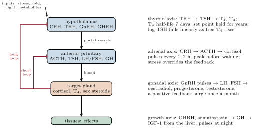
The hypothalamus, a few grams of brain above the pituitary, turns neural information — stress, cold, day length, the blood’s osmolarity and glucose — into hormonal commands. Its neurosecretory cells release releasing hormones (peptides: CRH, TRH, GnRH, GHRH, and the inhibitor somatostatin; dopamine for prolactin) into a private portal system of vessels that carries them, undiluted, a centimetre down to the anterior pituitary, whose cells respond with tropic hormones: ACTH to the adrenal cortex, TSH to the thyroid, LH and FSH to the gonads, growth hormone to the liver and tissues, prolactin to the breast. The target glands’ hormones — cortisol, thyroxine, sex steroids — feed back on both pituitary and hypothalamus to shut off their own commands: negative feedback, in three tiers. The posterior pituitary is not a gland but the axon terminals of hypothalamic neurons, releasing vasopressin and oxytocin straight into the blood. Each axis has its own dynamics: the thyroid axis is slow and steady, holding a set point for years; the adrenal axis is pulsatile and circadian, cortisol peaking before dawn and secreted in hourly bursts; the gonadal axis of a woman runs a monthly cycle on a positive feedback that the others lack.
Evidence. Berthold (1849) castrated cockerels, which lost their comb, crow and combativeness; re-implanting a testis anywhere in the abdomen, without its nerves, restored them — the organ acted through the blood. Harris (1950s) cut the portal vessels between hypothalamus and pituitary in rats: the pituitary, its blood supply restored from elsewhere, stopped responding to stress and the ovaries stopped cycling; transplanting a pituitary under the kidney left it inert, but under the hypothalamus, where portal vessels regrew into it, it worked — the brain governs the gland by blood-borne factors, and Guillemin and Schally then isolated the first of them, TRH, from millions of sheep and pig hypothalami (1969): three amino acids. ∎
Proposition 21.5 (Feedback and the log-linear thyroid)
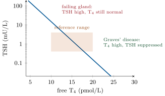
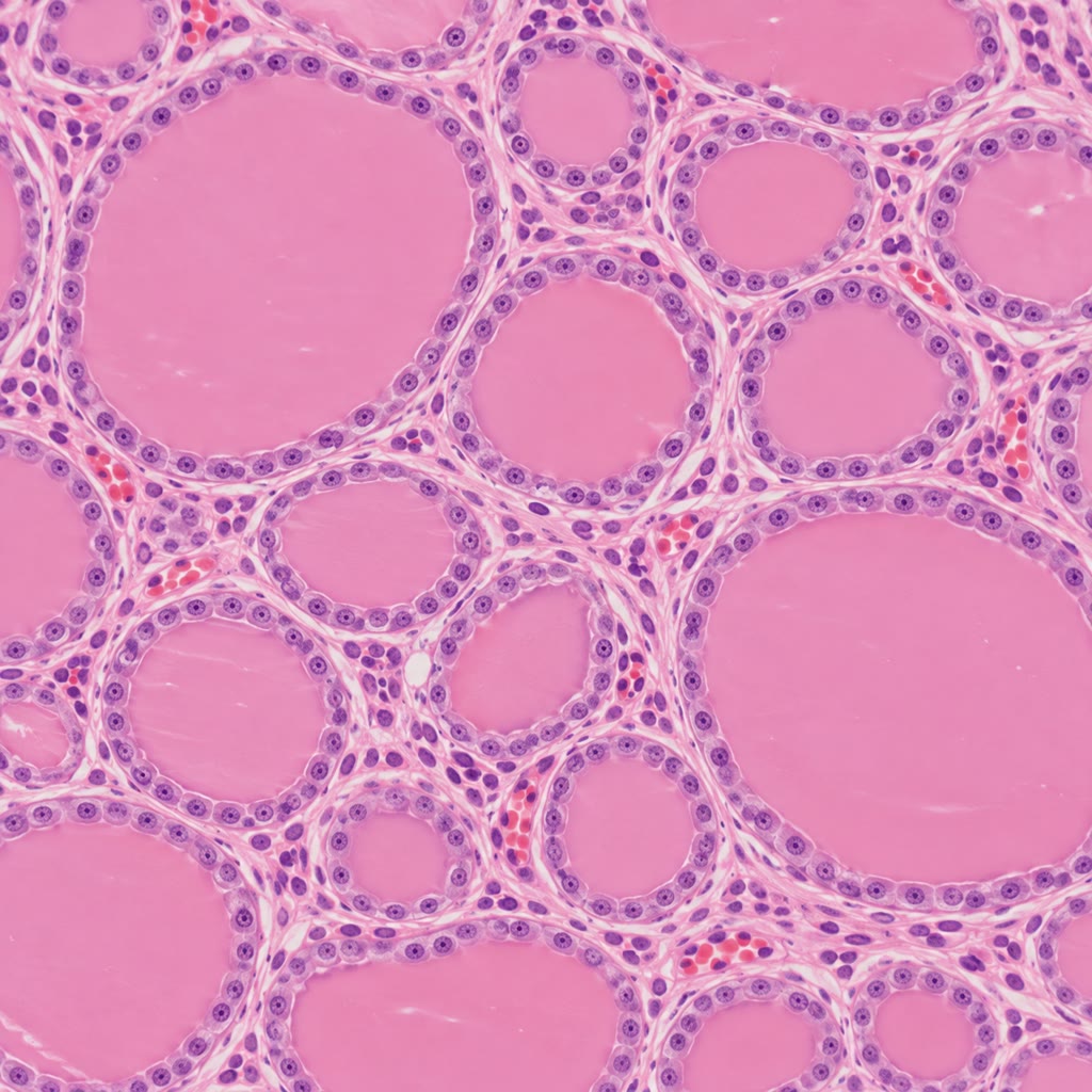
The thyroid takes up iodide, and makes thyroxine T (four iodines), which the tissues convert to the active T; both raise the metabolic rate of nearly every cell, set the heart rate, and are needed for brain development before and after birth. TSH from the pituitary drives both the synthesis and the growth of the gland; T suppresses TSH. The relation at steady state is log-linear: the logarithm of TSH falls in proportion to the free T,
so that a fall of free T by a third raises TSH about tenfold — which is why TSH, and not T, is the sensitive test: a gland that is beginning to fail shows a normal T held up by a TSH already several times normal. Failure of the gland (autoimmune destruction, iodine deficiency) gives hypothyroidism — cold, slow, tired, with a high TSH and, in iodine deficiency, a gland enlarged into a goitre by the TSH that cannot make it produce; an antibody that mimics TSH gives hyperthyroidism (Graves’ disease) with a hot, fast, thin patient and a TSH suppressed to nothing, since the stimulation escapes the feedback. A newborn without thyroid hormone develops irreversible intellectual disability within months, which is why every newborn’s TSH is measured on a spot of blood.
Proof. The pituitary cell’s TSH output responds to the receptor occupancy by T, itself proportional to T, and the response of a transcriptional repression to a change in occupancy is multiplicative — each increment of hormone represses the same fraction of what remains — so , whence the exponential. With fitted so that falling from to raises TSH from to , : TSH doubles for each of T lost, which is the amplification that makes it the sensitive test. ∎
Example 21.6 (Cortisol: the stress axis)
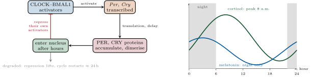
Cortisol, from the adrenal cortex under ACTH, mobilises fuel (glucose from the liver, amino acids from muscle, fatty acids from fat), sensitises vessels to adrenaline, and suppresses inflammation and the immune response — the reason its synthetic relatives are the commonest anti-inflammatory drugs. Its rhythm is set by the clock: at 8 a.m., a fifth of that at midnight, and pulses every hour or two throughout, since the hypothalamic CRH neurons fire in bursts. Stress — haemorrhage, infection, surgery, fear — raises it tenfold within minutes, overriding the feedback. Too little (Addison’s disease, destruction of the adrenal cortex) gives weakness, low blood pressure, low glucose, and, under stress, collapse and death unless replaced; too much (Cushing’s syndrome: a pituitary tumour making ACTH, an adrenal tumour, or, most often, prescribed steroids) gives the round face, thin skin, wasted muscle, high glucose, high pressure and brittle bones of prolonged exposure. Stopping a long course of steroids abruptly is dangerous for the reason the axis predicts: the suppressed hypothalamus and pituitary take weeks to recover, and the adrenal, unstimulated, has shrunk.
21.3 Glucose: the tightest loop
Definition 21.7 (Insulin and glucagon)
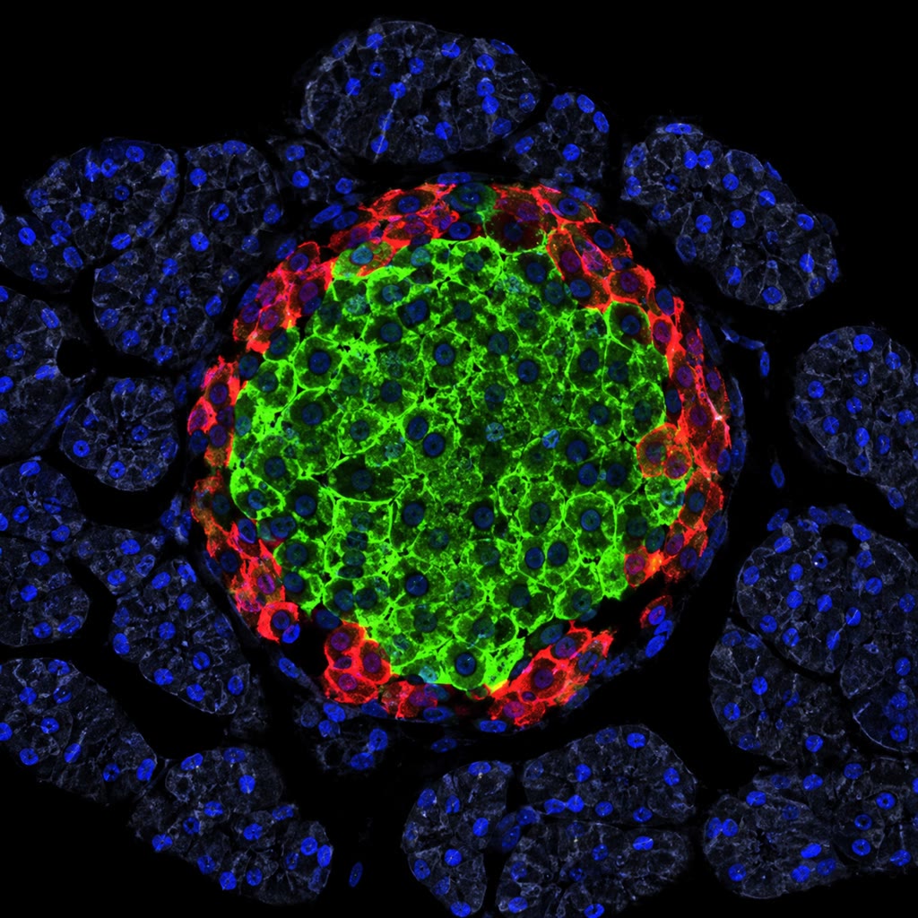
The blood holds about of glucose, at (), and the brain burns a day and can burn nothing else; a meal delivers in an hour. The islets of Langerhans, a million clusters of a few thousand cells scattered through the pancreas, hold the loop. cells sense glucose by metabolising it: more glucose, more ATP, closure of an ATP-sensitive potassium channel, depolarisation, calcium entry and exocytosis of insulin, a peptide hormone cleaved from a single precursor. Insulin tells muscle and fat to move the transporter GLUT4 to their surfaces and take glucose up, the liver to store glucose as glycogen and stop making it, and fat to stop releasing fatty acids: it is the hormone of the fed state, the only one that lowers blood glucose. cells release glucagon when glucose falls: the liver breaks down glycogen and makes new glucose from amino acids and lactate. Adrenaline, cortisol and growth hormone raise glucose too, and the redundancy is not symmetric: a body can defend against a fall by four routes and against a rise by one. Diabetes mellitus is the failure of that one: type 1, the autoimmune destruction of the cells, needs insulin from the first day; type 2, nine cases in ten, begins as resistance of the tissues to insulin, met for years by more insulin, until the cells fail to keep up and glucose rises.
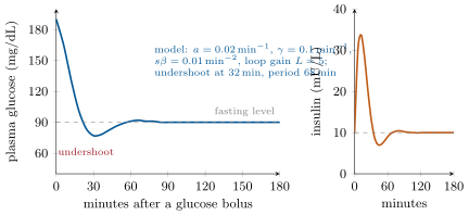
Theorem 21.8 (The insulin–glucose loop)
Write and for the deviations of plasma glucose and insulin from their fasting values. Glucose is cleared at a rate proportional to its own excess (the glucose effectiveness ) and to the insulin excess (sensitivity ); insulin is secreted in proportion to the glucose excess () and cleared at rate ; a load enters:
Under a steady load the glucose settles at
so the feedback divides the disturbance an unregulated body would suffer by , the loop gain plus one. After a bolus the return is governed by the eigenvalues : monotone if , and otherwise a damped oscillation of angular frequency decaying as — glucose undershoots its fasting value before settling, which is the mild hypoglycaemia two or three hours after a sugary meal. Insulin resistance lowers : the fasting steady state under the body’s own hepatic output rises, and the loop compensates with a higher until the cells’ falls too, when the compensation fails.
Proof. Steady state: gives ; putting it in : , whence . Dynamics: the system matrix is , with trace and determinant ; its characteristic equation has the roots stated. Both have negative real part, so the fasting state is stable; the roots are complex when the discriminant is negative, the condition given, and then , which crosses zero. Resistance: with the liver’s basal output and reduced, falls and rises for the same ; rises with it (hyperinsulinaemia); if then falls, falls further and climbs toward . ∎
Evidence. Von Mering and Minkowski (1889) removed a dog’s pancreas and produced diabetes — the flies gathering on its urine gave the sugar away; tying the ducts, which destroyed the digestive tissue but spared the islets, did not. Banting and Best (1921), with Collip’s purification, extracted the islet principle and lowered a diabetic dog’s blood sugar, then Leonard Thompson’s (January 1922). Sanger sequenced insulin (1955), the first protein sequence; Yalow and Berson measured it in blood (1959) and found that adult-onset diabetics had, at first, more of it than the healthy — resistance, not deficiency. Genentech’s bacteria made human insulin in 1978, the first recombinant drug. ∎
Method 21.9 (Reading the glucose loop in a patient)
(1) Fasting plasma glucose: normal below , diabetes at or above on two occasions. (2) Oral glucose tolerance test: of glucose drunk; plasma glucose at two hours normal below , diabetes at or above — the two-hour value reads the loop’s gain, the fasting value its set point. (3) Glycated haemoglobin, HbA1c: glucose attaches irreversibly to haemoglobin at a rate proportional to its concentration, and red cells live 120 days, so the fraction glycated integrates the mean glucose of the past three months without a fast — normal below , diabetes from . (4) To separate resistance from deficiency, measure insulin (or its by-product C-peptide) alongside: high insulin with high glucose is resistance; absent C-peptide is type 1.
21.4 Calcium, and the clock
Definition 21.10 (Calcium homeostasis)
Ionised calcium in plasma is held at within a few per cent, because it sets the excitability of every nerve and muscle: too low and they fire spontaneously (tetany), too high and they are sluggish. The four parathyroid glands sense it with a surface receptor and secrete parathyroid hormone (PTH) along a steep inverse sigmoid: PTH mobilises calcium from bone, makes the kidney reabsorb it and excrete phosphate, and activates vitamin D — made in skin by ultraviolet light or eaten, hydroxylated in liver, then in kidney to calcitriol — which makes the gut absorb calcium. Bone is the reservoir, a kilogram of calcium, remodelled continuously by osteoclasts and osteoblasts under PTH, calcitriol, oestrogen and load. Deficiency of vitamin D gives rickets in children and soft bones in adults; the fall of oestrogen at the menopause tilts remodelling toward resorption and gives osteoporosis; a parathyroid adenoma raises calcium, dissolves bone and forms kidney stones — “bones, stones, groans and moans.”
Definition 21.11 (The circadian clock)
Nearly every cell carries a circadian clock: a transcription–translation loop in which the proteins CLOCK and BMAL1 switch on the genes Per and Cry, whose proteins accumulate, enter the nucleus after a delay of hours and repress their own transcription, then are degraded, releasing the repression — one cycle in about twenty-four hours, set by the rates of the delay and degradation. The clocks of the body are synchronised by a master clock of twenty thousand neurons in the suprachiasmatic nucleus of the hypothalamus, which is itself set by light through a dedicated class of retinal ganglion cells (containing melanopsin, not the rods’ or cones’ pigments), and which signals night to the body through the pineal’s melatonin, secreted only in darkness. The clock schedules cortisol before waking, body temperature lowest at 4 a.m., growth hormone in the first sleep, insulin sensitivity higher in the morning; food, exercise and temperature are secondary zeitgebers that can pull the peripheral clocks away from the central one, which is the malaise of the shift worker and of jet lag — a liver clock on one time and a brain on another.
Evidence. Konopka and Benzer (1971) screened mutant flies for altered rhythms of emergence and found three alleles of one gene, period: a short day (19 h), a long day (29 h), and no rhythm at all — a clock with a genetic period. Hardin, Hall and Rosbash (1990) found that period mRNA and protein oscillate with a lag between them, and that the protein represses its own gene: the loop. Ralph and Menaker (1990) transplanted the suprachiasmatic nucleus of a mutant hamster with a 20-hour rhythm into a normal hamster whose own nucleus had been destroyed: the recipient ran on 20 hours — the period belonged to the transplanted tissue. In humans kept in a bunker without time cues the rhythm free-ran at a little over 24 hours; a bright light in the evening delayed it, in the morning advanced it. ∎
Remark 21.12 (Homeostasis as control)
Every loop in this chapter has the same parts: a sensor (the cell’s metabolism, the parathyroid’s calcium receptor, the pituitary’s thyroid-hormone receptor), a controller that compares with a set point, an effector hormone with its own half-life, and a feedback that reduces the error by a factor but never to zero. What differs is the timing — seconds for adrenaline, an hour for insulin, days for thyroxine, a lifetime for bone — and the price of the compromise: a fast loop with a delayed effector oscillates, a slow loop lets a disturbance persist. The clinic sees the loops from outside, through the pairs the feedback couples: a high TSH with a low T is a failed gland, a high TSH with a high T a failed pituitary; a high insulin with a high glucose is resistance, a low insulin with a high glucose is destruction. To read a hormone’s level one must always ask what its commander was doing.
21.5 Exercises
Exercise 21.1 ★
Classify insulin, cortisol, adrenaline, thyroxine and vasopressin by chemistry, receptor location, speed of action and half-life.
Solution
Solution of Exercise 21.1.
Insulin: peptide, surface receptor (a tyrosine kinase), acts in minutes, half-life about . Cortisol: steroid, nuclear receptor, hours, half-life (mostly bound to a carrier). Adrenaline: amine, surface G-protein-coupled receptors, seconds, half-life about . Thyroxine: iodinated amino acid, nuclear receptor, days, half-life (bound to carriers). Vasopressin: peptide, surface G-protein-coupled receptor (cAMP in the collecting duct), minutes, half-life about .
Exercise 21.2 ★
Draw the thyroid axis with its feedback and predict TSH and T in: a destroyed thyroid; a TSH-secreting pituitary tumour; a patient taking too much thyroxine; iodine deficiency.
Solution
Solution of Exercise 21.2.
Hypothalamus (TRH) pituitary (TSH) thyroid (T), with T inhibiting both upper tiers. Destroyed thyroid: T low, TSH high. TSH-secreting tumour: TSH high and T high (the feedback fails to suppress the tumour). Excess thyroxine tablets: T high, TSH suppressed. Iodine deficiency: T low or low-normal, TSH high, and the gland grows under it into a goitre.
Exercise 21.3 ★
Why does a body have four hormones that raise blood glucose and only one that lowers it? What does this predict about the relative dangers of too much and too little insulin?
Solution
Solution of Exercise 21.3.
The brain dies of a low glucose in minutes and is harmed by a high one only over years; and evolution met famine far more often than feasts. So the defence against a fall is redundant (glucagon, adrenaline, cortisol, growth hormone) and the defence against a rise is single. Prediction: too much insulin is acutely lethal (hypoglycaemic coma), too little is a chronic disease — which is what the clinic sees.
Exercise 21.4 ★
What is a zeitgeber? Give three, say which the master clock uses, and explain what happens to a traveller who crosses eight time zones.
Solution
Solution of Exercise 21.4.
A zeitgeber is an environmental cue that entrains a clock: light, food timing, temperature, exercise, social schedule. The master clock uses light, through the melanopsin ganglion cells of the retina. After eight time zones the clock shifts only about an hour a day, so for a week sleep, cortisol, temperature and appetite run on the old time while the peripheral clocks, reset by meals, drift at their own pace: jet lag, worse eastward, since advancing the clock is harder than delaying it.
Exercise 21.5 ★★
A cell has receptors with and responds maximally when are occupied. Find the EC. After a week of high hormone the cell has receptors: new EC? Interpret for insulin resistance.
Solution
Solution of Exercise 21.5.
Half-maximal response at receptors occupied: , eighty times below . With receptors: , eleven times less sensitive. Prolonged high insulin down-regulates its receptors, so a given effect needs more insulin: one component of insulin resistance, which feeds on itself.
Exercise 21.6 ★★
With and a normal TSH of at free T of , compute TSH when T is , , and . At which of these would T still be within a reference range of while TSH is outside ?
Solution
Solution of Exercise 21.6.
: ; : ; : ; : . At the T is still within while TSH is more than double its upper limit (subclinical hypothyroidism); at TSH is just within range; at both are abnormal; at both are abnormal the other way.
Exercise 21.7 ★★
In the loop of Theorem 21.8 with , , per mg/dL and per mU/L, compute the loop gain, the steady glucose excess under a constant load of , the same without feedback, and the steady insulin excess.
Exercise 21.8 ★★
Same parameters: is the return after a bolus oscillatory? Compute the period and the time for the amplitude to fall by . Now halve (insulin resistance): recompute both and the new loop gain.
Solution
Solution of Exercise 21.8.
Discriminant : oscillatory. , period ; amplitude falls by in . Halving : , discriminant , still oscillatory, , period ; the decay time is unchanged; . A weaker loop returns more slowly and holds a larger steady error.
Exercise 21.9 ★★
Cortisol’s half-life is . A patient on of prednisolone daily for a year stops abruptly. Explain, tier by tier, why they may collapse under a minor infection three days later, and what the axis would show if measured (ACTH, cortisol, response to injected ACTH).
Solution
Solution of Exercise 21.9.
For a year the prednisolone suppressed CRH and ACTH; the adrenal cortex, unstimulated, atrophied. On stopping there is no exogenous steroid, the hypothalamus and pituitary take weeks to resume, and the shrunken adrenal could not respond to ACTH if it came. An infection demands ten times the normal cortisol; with none, vessels dilate, pressure and glucose fall: adrenal crisis. Measured: ACTH low (the lesion is central), cortisol low, and a poor cortisol rise to injected ACTH (the gland has wasted) — unlike Addison’s disease, where ACTH is high. Hence the slow taper.
Exercise 21.10 ★★★
Show that a one-variable negative feedback with cannot oscillate, while the two-variable loop of the theorem can, and explain in words why the clock needs a delay of hours between transcription and repression to produce a 24-hour period. What would happen to the period if PER were degraded twice as fast?
Solution
Solution of Exercise 21.10.
One variable: with has a single fixed point ; for , and decreases monotonically toward , never crossing it (uniqueness of solutions), and symmetrically below: no oscillation. Two variables: the matrix can have complex eigenvalues, as in the theorem, when the effector acts through a second variable with its own time scale. An instantaneous repression would settle at a steady level; the delay between transcription and nuclear repression (translation, dimerisation, phosphorylation, import) lets the protein overshoot before the gene is turned off and undershoot before it is turned on again, and the period is roughly twice the sum of the delay and the time to degrade the protein. Faster PER degradation shortens the period: a destabilising mutation of human PER2 gives a clock of about 20 hours and a family that falls asleep at 7 p.m.
Exercise 21.11 ★★★
Parathyroid hormone follows with and . Compute PTH as a fraction of maximum at Ca , , , and , the slope at the set point, and explain why such steepness is wanted for calcium but would be dangerous for glucose.
Solution
Solution of Exercise 21.11.
Exponent : at , , PTH ; : ; : ; : ; : . Slope at the set point per mmol/L: of maximum for each . Calcium must be held within a few per cent and its effector acts on a huge buffer (bone) without overshoot, so a steep response is safe and needed. A glucose loop that steep, acting through insulin with its own clearance time, would have a large loop gain and oscillate deeply into hypoglycaemia after every meal.
Exercise 21.12 ★★★
Glucose attaches to haemoglobin at a rate per unit time, with such that a mean glucose of gives glycation over a red cell’s 120-day life. Derive HbA1c as a function of mean glucose, the value at , and explain why a patient with a haemolytic anaemia (red cells living 60 days) has a misleadingly low HbA1c.
Solution
Solution of Exercise 21.12.
Glycation is irreversible and slow, so a cell of age has a fraction glycated; averaged over cells uniformly aged days, HbA1c , proportional to the mean glucose. Calibration: gives per (mmol/L)(day); at : . With cells living 60 days the mean age is 30 days and HbA1c is halved for the same glucose: a patient at would read , normal.
21.6 Problem: The Sugar in the Blood
Problem 21.1
Weekend problem — a glucose tolerance test read through the linear insulin–glucose loop: the distribution of a dose, the loop gain and the damped return, what resistance and -cell failure do to the fasting set point, a diagnosis from the numbers, and the thyroid axis beside it, ending on the loop gain, the return time and the fasting glucose of the failing loop
Data: a healthy adult, glucose distribution volume , fasting glucose (), fasting insulin . Loop parameters: , , (mU/L per min per mg/dL), (mg/dL per min per mU/L). Basal hepatic glucose output (already balanced at the fasting state by the fasting disposal). An oral dose of is absorbed over an hour. Thyroid: mU/L. Glucose molar mass .
Part I — The dose.
- Express the fasting glucose in mmol/L and check the conversion from mg/dL. How many grams of glucose are in the distribution volume at fasting?
- If the whole appeared at once in , by how much would glucose rise (mg/dL and mmol/L)? Why does the real peak, about above fasting, fall so far short?
- Absorbed over , the dose enters at what rate in mg/dL per minute? Compare with .
- Without any insulin response (), glucose would obey . Steady excess under the absorption rate, and the time constant of the approach. Where would glucose be after an hour?
- With the loop, compute and the steady excess under the same rate. Compare with question 4.
- Explain why the brain is protected on both sides: what it needs, and what too much glucose does to tissues over years.
Part II — The return.
- Write the characteristic equation of the loop and compute the trace, the determinant and the discriminant.
- Compute and the period of the damped oscillation, and the decay time .
- Starting from an excess of with insulin at its fasting value, the solution is . Find from .
- When does glucose first return to fasting (first zero of )? Estimate the undershoot at the first minimum.
- Insulin: has the same exponential and frequency. Argue from that its peak comes after glucose has started falling and before glucose reaches fasting.
- Why does a real tolerance test, with absorption over an hour rather than a bolus, show a peak at 30–60 minutes and a return by two hours, and only a mild dip at three?
Part III — When the loop fails.
- Insulin resistance halves . New ? The liver’s is now met by a new fasting steady state: using as the excess over an idealised unregulated baseline, find by how much the fasting glucose rises relative to the healthy state (compute both values and subtract).
- Fasting insulin in the resistant state: compute for both states and the ratio. What does the clinic call this?
- Now the cells fail: falls to a fifth as well. New , new fasting excess. Convert to mmol/L and compare with the diabetic threshold of .
- Is the return still oscillatory in the state of question 15? Compute the discriminant and the time constant of the slowest mode.
- A patient has fasting glucose , fasting insulin , two-hour value . Diagnose type and mechanism from the pairs.
- Another has fasting glucose , C-peptide undetectable, and lost in two months. Diagnose, and explain the weight loss in terms of what the tissues do without insulin.
- HbA1c: if the mean glucose of the first patient is , and gives , estimate their HbA1c assuming proportionality.
Part IV — The gland next door.
- Free T is . Compute TSH. Is the T in the range ? Is the TSH in ? What is the state called?
- T is : TSH? A patient with this T but a TSH of : where is the lesion?
- Thyroxine’s half-life is . A patient on replacement forgets a week of tablets. To what fraction does the level fall, and why is the missed week less dangerous than a missed day of cortisol replacement?
- Graves’ disease: an antibody occupies the TSH receptor. Predict T, TSH, and the effect of the log-linear relation on the measured TSH.
- Explain in one paragraph why a hormone level is meaningless without its commander’s level, using the four pairs of the last remark of the chapter.
- Summarise: the healthy loop gain (question 5), the period and decay time of the return (question 8), and the fasting glucose of the failing loop (question 15).
Solution
Solution of Problem 21.1.
1. ; in the distribution volume. 2. : a rise of , . The real peak is far lower because absorption is spread over an hour while disposal removes glucose as it arrives, and the liver takes up a large share on first pass. 3. ; over : , four times . 4. Steady excess , time constant ; after an hour above fasting: about , . 5. ; steady excess , six times smaller than without insulin. 6. The brain burns of glucose a day, stores none and can use little else: a low glucose gives confusion and coma within minutes. A high glucose glycates proteins and, over years, damages the small vessels of retina, kidney and nerves and the large arteries. 7. : trace , determinant , discriminant . 8. ; period ; decay time . 9. , so , . 10. when , , . At the first minimum, near : , glucose . 11. At , : insulin rises while , and peaks when , which requires still positive — so the peak comes before glucose reaches fasting, and after glucose has begun to fall (it falls from ). In the model the peak is near . 12. With input spread over an hour the peak occurs when absorption and disposal balance, lower and later (30–60 min); the return waits for absorption to end, hence two hours; the insulin overshoot is smaller for a gradual input, so the dip is mild. 13. . Healthy ; resistant : a rise of about (), fasting glucose near , — the threshold of impaired fasting glucose. 14. : healthy , resistant , ratio : compensated insulin resistance, hyperinsulinaemia with near-normal glucose. 15. ; , above the healthy state: , fasting glucose (), above the diabetic threshold. 16. Discriminant : monotone return. : and per minute; slowest time constant , against for the healthy decay: the failing loop returns more slowly and never undershoots. 17. High glucose with high insulin: resistance with partial compensation — type 2; the two-hour value of exceeds , so diabetes, not merely impaired tolerance. 18. No C-peptide means no endogenous insulin: type 1. Without insulin muscle and fat cannot take up glucose, fat is broken down to fatty acids and ketones, muscle protein is catabolised for gluconeogenesis, and glucose is lost in the urine with its calories: the patient starves in the midst of plenty. 19. . 20. . T within range, TSH above it: subclinical hypothyroidism, the gland failing and the pituitary compensating. 21. . A low T with a TSH of only means the pituitary is not responding: the lesion is central (pituitary or hypothalamus), not in the thyroid. 22. One half-life: the level falls to , and the symptoms creep in over weeks. Cortisol, with a half-life of , is gone within hours of a missed dose, and a stress on that day meets no defence: the missed week of thyroxine is uncomfortable, the missed day of cortisol can kill. 23. T high (the antibody drives the gland regardless of feedback); TSH suppressed by the high T; the log-linear relation drives it to undetectable — at , — so an undetectable TSH is the first sign. 24. A hormone’s level reports the balance of its command and its gland: high TSH with low T is a failed gland and high TSH with high T a runaway pituitary; high insulin with high glucose is a resistant body and low insulin with high glucose a destroyed islet. Read alone, a T of or an insulin of does not say where the fault lies; read with its commander, it does. 25. Loop gain ; period and decay time ; fasting glucose of the failing loop .