University Biology — Year 3 · Bachelor Year 3
3Genome Stability: DNA Damage, Repair and Recombination
An afternoon on a beach puts some hundred thousand chemical lesions into the DNA of every skin cell exposed to the sun: adjacent thymines welded into dimers by ultraviolet light, bases oxidised, strands nicked. Without any sun at all, the warm water of the cell knocks about ten thousand purines off their sugars every day and turns hundreds of cytosines into uracil. Yet the mutation rate of a human cell is about one change in ten billion base pairs per division — in a genome of six billion, less than one new mutation per division. The gap between the damage a genome suffers and the damage it keeps is closed by repair: a dozen enzyme systems that patrol the double helix, recognise what is not supposed to be there, cut it out and rebuild from the undamaged strand, and, when both strands are broken, rejoin them or copy the missing piece from a sister. The children who lack one of these systems — burning at the first touch of sunlight, or developing colon cancer in their thirties — show what the systems are worth. This chapter is about damage, the repair pathways, the recombination machinery that repairs the worst breaks and that meiosis borrows, and the cell’s decision, when repair fails, to stop dividing or to die.
3.1 What goes wrong with DNA
Definition 3.1 (DNA lesions)
A lesion is any chemical alteration of DNA away from the four normal bases correctly paired on an intact backbone. Spontaneous lesions arise from the chemistry of water and oxygen: depurination, hydrolysis of the bond between a purine and its sugar, leaving an abasic site (about per cell per day); deamination, which turns cytosine into uracil, 5-methylcytosine into thymine, and adenine into hypoxanthine (a few hundred per day); oxidation by reactive oxygen species, chiefly to 8-oxoguanine, which pairs with adenine as readily as with cytosine; and replication errors, a misincorporated or slipped base. Environmental lesions include the cyclobutane pyrimidine dimer and the 6-4 photoproduct made by ultraviolet light between two adjacent pyrimidines; alkylated bases from alkylating agents (O-methyl- guanine pairs with thymine); bulky adducts from aromatic hydrocarbons and aflatoxin; interstrand cross-links from cisplatin and nitrogen mustards; and the single- and double-strand breaks made by ionising radiation, about single-strand and double-strand breaks per cell per gray.
Proposition 3.2 (The fidelity ladder)
Replication achieves its error rate of about per base pair in three multiplicative steps. Base selection by the polymerase, on geometry and hydrogen bonding, errs about once in ; proofreading by the polymerase’s 35 exonuclease, which excises a mispaired terminal base before extension, removes of those; mismatch repair, acting behind the fork on the newly made strand, removes of what remains. The product is the observed rate, and a defect in either correction step raises it a hundred- or a thousand-fold: such cells are mutators.
Example 3.3 (Lesions against mutations)
A human cell of base pairs dividing once a day accumulates on the order of – lesions a day and about new mutations per division: fewer than one lesion in ten thousand survives to become a permanent change. The rest are repaired before replication reads them, and the rate of survival to mutation — not the rate of damage — is what selection has minimised. The same arithmetic shows why the number of mutations in a tumour or in the sperm of an older father counts divisions: the mutations a cell carries are, to first order, the divisions it has undergone times the error per division.
3.2 Repair of one damaged strand
Definition 3.4 (Excision repair)
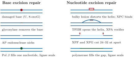
Most lesions affect one strand and are repaired by cutting out the damaged piece and copying the other strand. Base excision repair (BER) handles small, non-distorting lesions: a DNA glycosylase specific for one kind of wrong base (uracil-DNA glycosylase, 8-oxoG glycosylase, and several others) flips the base out of the helix and cuts it off its sugar; an AP endonuclease nicks the backbone at the abasic site; a polymerase (Pol ) removes the sugar and inserts one nucleotide; a ligase seals. Nucleotide excision repair (NER) handles bulky, helix-distorting lesions — pyrimidine dimers, chemical adducts — without recognising the chemistry of the lesion itself: the distortion is detected (by XPC scanning the genome, or by a stalled RNA polymerase in the transcription-coupled branch), the helix is opened by the helicase subunits of TFIIH, the damage is verified (XPA), two endonucleases (XPF, XPG) cut the damaged strand nucleotides apart, the oligonucleotide is released, and polymerase and ligase fill the gap. Loss of any of the seven XP proteins causes xeroderma pigmentosum: extreme sensitivity to sunlight, freckling, and a thousand-fold increased rate of skin cancer.
Evidence. Cleaver (1968) cultured skin fibroblasts from xeroderma pigmentosum patients, irradiated them with ultraviolet light and gave them radioactive thymidine outside S phase. Normal cells incorporated label at many small sites — the “unscheduled DNA synthesis” of repair patches — while the patients’ cells incorporated almost none, and died at doses normal cells survived: the disease is a failure to excise the dimers. Fusing cells from two patients often restored repair in the hybrid, each cell supplying what the other lacked; by this test the patients were sorted into complementation groups A to G, one per gene of the pathway, years before the genes were cloned. ∎
Definition 3.5 (Mismatch repair)
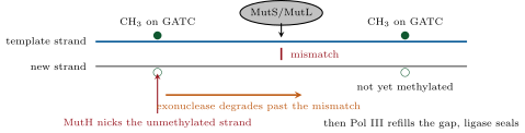
Mismatch repair (MMR) corrects the mispairs and small insertion–deletion loops that escape proofreading. The MutS protein (MSH2–MSH6 in humans) recognises the distortion of a mismatch and, with MutL (MLH1–PMS2), licenses an exonuclease to degrade the newly synthesised strand from a nearby nick back past the mismatch; the gap is refilled by polymerase. The difficulty is strand discrimination: a mismatch has two bases and only one is wrong. In Escherichia coli the new strand is identified because it is not yet methylated at GATC sites, which the Dam methylase marks a few minutes after synthesis; MutH nicks the unmethylated strand. In eukaryotes the new strand is identified by the nicks between Okazaki fragments and the free 3 end of the leading strand. Loss of MMR raises the mutation rate a hundred- to a thousand-fold, most visibly at microsatellites — runs of a short repeat where the polymerase slips — whose lengths change from cell to cell: microsatellite instability. Inherited loss of one MMR allele is Lynch syndrome, with colorectal cancer in some of carriers, often before fifty.
Method 3.6 (Sorting a repair disease into genes)
To find how many genes a recessive phenotype involves, before any gene is known: (1) take cell lines from unrelated patients, each mutant in some gene of the pathway; (2) fuse pairs of lines into hybrid cells; (3) test the hybrid for the function (here, unscheduled DNA synthesis after ultraviolet light); (4) if the hybrid is repaired, the two patients carry mutations in different genes (each supplies the other’s missing protein: they complement); if not, in the same gene; (5) group the patients into complementation groups — one per gene. The count of groups is a lower bound on the number of genes.
3.3 Repair of a broken chromosome
Definition 3.7 (Double-strand break repair)
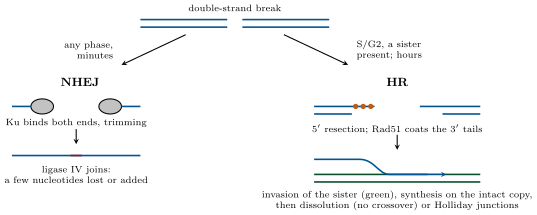
A double-strand break severs the chromosome and has no intact strand to copy. Two pathways repair it. Non-homologous end joining (NHEJ): the Ku70–Ku80 ring binds each end, recruits the kinase DNA-PKcs, nucleases trim overhangs, and ligase IV joins the ends; it works at any stage of the cycle and in minutes, but loses or adds a few nucleotides at the junction and can join the wrong ends. Homologous recombination (HR): the MRN complex and nucleases resect the 5 strands to leave long 3 single-stranded tails, which are coated by RPA and then, with the help of BRCA2, by the recombinase Rad51; the Rad51 filament searches for a homologous duplex — the sister chromatid, in S and G2 — invades it, and the invading 3 end primes synthesis on the intact copy. The joint molecules may be dissolved after a short synthesis (most mitotic repair, no exchange of flanking DNA) or mature into two four-way Holliday junctions, whose resolution by endonucleases yields either a crossover or a non-crossover product. HR is accurate because it copies a sister, but is available only when a sister exists.
Theorem 3.8 (Damage load, repair rate and the fork)
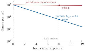
Let lesions of one kind arise at a constant rate per cell per hour and be removed by first-order repair with rate constant (half-life ). The number of lesions obeys , so that at steady state
and a pulse of lesions delivered at leaves lesions when a replication fork arrives at time . If two pathways compete for the same lesion with rate constants and , each lesion is repaired by the first with probability and the overall half-life is . The number of lesions that reach the fork is Poisson-distributed with mean , so the probability that none does is .
Proof. The steady state is where . For the pulse, and the equation integrates to . With two pathways the loss rate is ; in any short interval a given lesion is removed by pathway 1 with probability and by pathway 2 with , so the conditional probability that its removal, when it happens, is by pathway 1 is . Independent lesions each surviving with probability give a binomial count, Poisson in the limit of many lesions and small survival, and the Poisson probability of zero is . ∎
Example 3.9 (Why xeroderma is a disease of the fork)
A dose of sun leaves pyrimidine dimers in a keratinocyte. With a nucleotide excision half-life of () and a fork arriving after , dimers are still there to be copied; with the near-absent repair of an XP cell (), are. Each dimer met by the fork is bypassed by translesion synthesis (below), which is error-prone: the patient’s mutation load per division is fifteen times the normal one, and the skin cancers follow in childhood. The cure that works is to keep small — total avoidance of ultraviolet light.
Evidence. Holliday (1964) proposed the four-way junction to explain a puzzle of fungal genetics: in a cross, a tetrad of spores sometimes showed a instead of the Mendelian segregation of a marker, always for markers close to a crossover. His model — the exchange of single strands between two homologous duplexes, forming a heteroduplex that mismatch repair then “corrects” in one direction or the other (gene conversion) — explained both the odd ratios and their association with crossing over. The junction itself was later seen in the electron microscope in recombining plasmids and in the structure of the resolvase RuvC bound to it. ∎
3.4 Tolerance, alarm and the decision to stop
Definition 3.10 (Translesion synthesis)
When a fork meets a lesion that has not been repaired, the replicative polymerase stalls. Translesion synthesis (TLS) calls in a specialised polymerase with a roomier active site and no proofreading — Pol for pyrimidine dimers, Pol , , and Rev1 for other lesions — which inserts a few nucleotides opposite the lesion and steps aside. Pol puts two adenines opposite a thymine dimer, which is usually right; the others are often wrong. TLS trades accuracy for the survival of the fork, and is the source of most ultraviolet-induced mutations. The variant form of xeroderma pigmentosum (XP-V) has intact excision repair and lacks Pol : the dimers that escape excision are bypassed by the more error-prone polymerases, and the cancers follow.
Proposition 3.11 (The bacterial SOS response)
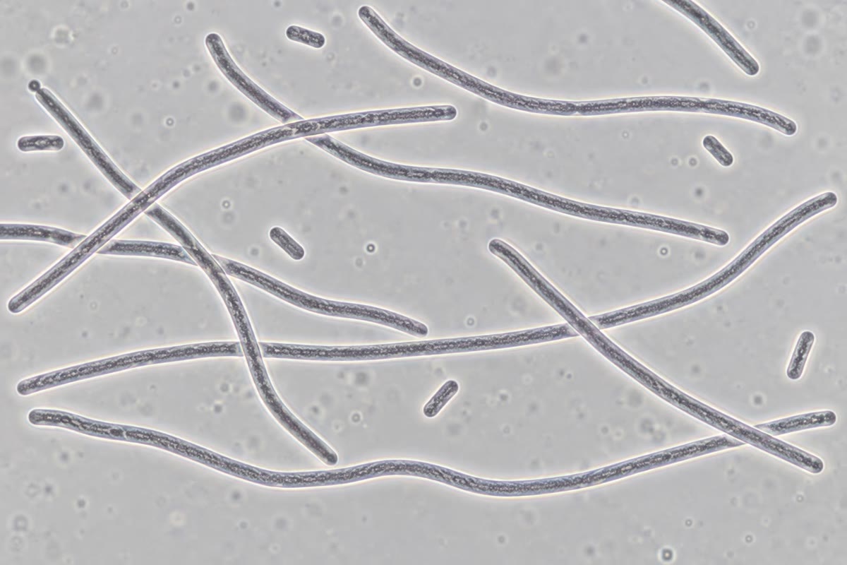
In E. coli the recombinase RecA, bound to the single-stranded DNA that accumulates at stalled forks and resected breaks, becomes a co-protease that induces the repressor LexA to cleave itself. LexA represses some forty genes; as it falls, they are switched on in order of their operators’ affinity: first the excision-repair genes uvrA and uvrB, then recA itself and the cell-division inhibitor sulA (the cells stop dividing and grow into filaments), and last the error-prone polymerases Pol IV and Pol V, which bypass lesions at the cost of mutations. The graded response tries accurate repair first and mutagenic tolerance only when the damage persists; the mutations it induces are the raw material on which antibiotic resistance evolves under treatment.
Definition 3.12 (The DNA damage response)
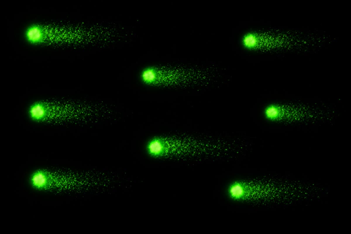
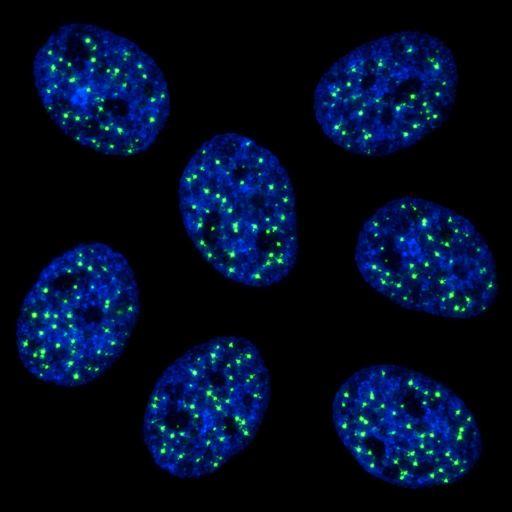
Eukaryotes signal damage through two kinases: ATM, recruited by the MRN complex to double-strand breaks, and ATR, recruited to the single-stranded DNA of stalled forks. They phosphorylate hundreds of targets. Within minutes histone H2AX is phosphorylated (-H2AX) over megabases around each break, forming a focus that recruits repair proteins and can be counted under the microscope, one focus per break. The kinases Chk1 and Chk2 halt the cell cycle — the checkpoints of Chapter 10 — by inactivating the phosphatases that would drive entry into S or M; and the transcription factor p53, stabilised by phosphorylation, induces the cyclin-dependent kinase inhibitor p21 (a durable arrest in G1), repair genes, and, if the damage is heavy or persists, the apoptotic genes that kill the cell. The response buys time for repair, and when repair fails removes the cell rather than let it divide with a broken genome.
Example 3.13 (Synthetic lethality: BRCA and PARP)
Women who inherit one defective copy of BRCA1 or BRCA2 have a lifetime risk of breast cancer of ; the tumours arise in cells that have lost the second copy and can no longer do homologous recombination. Such cells repair their double-strand breaks by end joining alone and accumulate rearrangements. They also acquire a specific weakness. Single-strand breaks, some a day, are repaired by a route that needs the enzyme PARP; when PARP is inhibited by a drug, single-strand breaks persist to S phase, where a fork converts each into a double-strand break with only one end — repairable only by recombination. A normal cell, with one good BRCA allele, copes; the tumour cell dies. Two defects, each harmless alone, are lethal together: the drug kills by synthetic lethality, and spares the patient’s other cells.
Proposition 3.14 (Recombination as a tool)
Site-specific recombinases cut and rejoin DNA at short defined sequences without homology search or synthesis: the phage P1 enzyme Cre acts on loxP sites, the yeast enzyme Flp on FRT sites. Two sites in the same orientation on one molecule excise the DNA between them as a circle; in opposite orientations they invert it; sites on two molecules exchange their flanks. A gene flanked by loxP sites (“floxed”) is deleted only in the cells where Cre is expressed, from a promoter of the experimenter’s choice, or at a time the experimenter chooses if Cre is made active by a drug: this is how a gene essential to the embryo is studied in the adult liver, or a neuron’s, alone (Chapter 6). The cell’s own homologous recombination is what gene targeting and CRISPR-directed editing borrow to write a chosen sequence into a chosen place.
Remark 3.15 (Repair and ageing)
Cells that fail to repair accumulate mutations, senesce or die; the inherited defects of repair (Werner syndrome, a helicase; Cockayne syndrome, transcription-coupled repair; ataxia telangiectasia, ATM) cause features of premature ageing, and the somatic mutation count of normal tissues rises linearly with age at a rate of some tens of mutations per cell per year. Whether accumulated damage causes ordinary ageing, or merely accompanies it, is not settled; that the capacity to repair is a determinant of lifespan across species — longer-lived species repair more — is well supported.
3.5 Exercises
Exercise 3.1 ★
Classify these lesions as spontaneous or environmental and name the repair pathway for each: a uracil in DNA; a thymine dimer; an 8-oxoguanine; a G–T mismatch behind the fork; a double-strand break in G1.
Solution
Solution of Exercise 3.1.
Uracil: spontaneous (deamination of C), base excision repair by uracil-DNA glycosylase. Thymine dimer: environmental (UV), nucleotide excision repair. 8-oxoguanine: spontaneous (oxidation), base excision repair by the 8-oxoG glycosylase. G–T mismatch behind the fork: replication error, mismatch repair. Double-strand break in G1: environmental (radiation) or spontaneous; non-homologous end joining, since no sister chromatid is available.
Exercise 3.2 ★
Why does nucleotide excision repair need no enzyme specific to each kind of lesion, whereas base excision repair needs a glycosylase for each?
Solution
Solution of Exercise 3.2.
Nucleotide excision recognises a physical property shared by all bulky lesions — the distortion of the helix, or a stalled RNA polymerase — and removes a patch containing whatever caused it. Small lesions (uracil, 8-oxoG, alkylated bases) hardly distort the helix, so they must be recognised by their chemistry, one glycosylase per kind of wrong base.
Exercise 3.3 ★
State the strand-discrimination problem of mismatch repair and how E. coli solves it. What would happen in a dam mutant, which methylates no GATC sites?
Solution
Solution of Exercise 3.3.
A mismatch consists of two normal bases, and only the one on the new strand is wrong; the system must know which strand is new. E. coli marks the parental strand with methyl groups at GATC, which the new strand lacks for a few minutes; MutH nicks the unmethylated strand. In a dam mutant neither strand is methylated: MutH nicks either, the repair removes the right base as often as the wrong one — fixing half the errors as mutations (a mutator) — and nicks on both strands at nearby GATC sites make double-strand breaks.
Exercise 3.4 ★
A cell receives of X-rays. How many double-strand breaks does it suffer, roughly, and how many -H2AX foci would you expect to count an hour later? Why fewer after six hours?
Solution
Solution of Exercise 3.4.
About double-strand breaks. After an hour most are joined: with a half-life near , about foci. After six hours nearly all have been repaired (), leaving only a few persistent, complex breaks.
Exercise 3.5 ★★
Using Proposition 3.2, compute the mutation rate per base pair and the number of new mutations per diploid genome per division in (a) a normal cell, (b) a cell lacking mismatch repair, (c) a cell whose polymerase has lost its exonuclease. Which is a more dangerous mutator?
Solution
Solution of Exercise 3.5.
(a) ; mutations per division. (b) Without mismatch repair ; per division. (c) Without proofreading ; per division. The mismatch-repair mutant is the more dangerous, a thousand-fold over normal.
Exercise 3.6 ★★
Abasic sites arise at per cell per day and are repaired with a half-life of . Use Theorem 3.8 to find the steady-state number of abasic sites in a cell. If a drug slows repair tenfold, what is the new steady state, and how many sites would a fork meet if it copies the genome in ?
Solution
Solution of Exercise 3.6.
; ; abasic sites present at any moment. Tenfold slower repair: . The fork passes every locus once, meeting the lesions present there at that moment; averaged over the genome it meets about of them — some normally, with the drug.
Exercise 3.7 ★★
In G2 a break can be repaired by NHEJ (half-life ) or HR (half-life ), both available. What fraction of breaks goes by HR? What is the half-life of the breaks? Why is HR nevertheless the pathway that matters at collapsed replication forks?
Solution
Solution of Exercise 3.7.
, ; HR fraction ; half-life . A collapsed fork gives a one-ended break: there is no second end for NHEJ to join, so only recombination with the sister can restore the fork — and it is the only accurate pathway in any case.
Exercise 3.8 ★★
Predict the microsatellite behaviour, the tumour spectrum and the sunlight sensitivity of a patient with (a) Lynch syndrome, (b) xeroderma pigmentosum group A, (c) the XP-V variant lacking Pol .
Solution
Solution of Exercise 3.8.
(a) Lynch: microsatellites unstable (lengths differ between tumour cells), colorectal, endometrial, gastric and ovarian cancers, normal response to sunlight. (b) XP-A: microsatellites stable, extreme sunburn on minimal exposure, skin and eye cancers in childhood, and in this group progressive neurological degeneration. (c) XP-V: microsatellites stable, excision repair normal so sunburn is milder, but the dimers that escape excision are bypassed by error-prone polymerases — skin cancers, of later onset than in group A.
Exercise 3.9 ★★
Two loxP sites in the same orientation flank exon 3 of a gene; Cre is expressed from a promoter active only in liver from birth. Describe the genotype of liver cells and of neurons in the adult, and what happens if the sites are placed in opposite orientations instead.
Solution
Solution of Exercise 3.9.
Liver cells: Cre has excised exon 3 from both alleles (as a circle that is lost), so the gene is inactive in every hepatocyte from birth — a liver-specific knockout. Neurons: no Cre, the floxed alleles are intact and functional. Opposite orientations: Cre inverts the exon and inverts it back indefinitely, so at any time about half the cells carry the inverted, non-functional exon — a mosaic, not a clean deletion.
Exercise 3.10 ★★★
Explain why the SOS response switches on repair genes before the error-prone polymerases, using the affinities of LexA for their operators, and why a bacterium that lacked the error-prone polymerases altogether might nonetheless be at a disadvantage in a patient treated with antibiotics.
Solution
Solution of Exercise 3.10.
As LexA is cleaved its concentration falls gradually. Operators that bind LexA weakly are vacated at a small fall — uvrA, uvrB, recA — so accurate repair starts first; the operators of sulA and of the error-prone polymerases bind LexA tightly and are vacated only when it is nearly gone, that is, when damage has persisted for a long time. A bacterium without the error-prone polymerases would die at forks blocked by lesions it could not bypass, and, with no induced mutagenesis, would evolve resistance to the antibiotic far more slowly — a disadvantage to the bacterium, an advantage to the patient; inhibitors of the SOS response are studied for that reason.
Exercise 3.11 ★★★
A PARP inhibitor kills BRCA-deficient tumour cells but not the patient’s normal cells. Set out the causal chain, then predict two ways a tumour could become resistant to the drug and how you would detect each.
Solution
Solution of Exercise 3.11.
Chain: PARP inhibited single-strand breaks persist a replication fork converts each into a one-ended double-strand break repair requires homologous recombination the tumour cell, lacking BRCA, cannot mis-joining, chromosome loss, death. Normal cells, with one functional allele, recombine and survive. Resistance: (1) a second mutation in BRCA that restores the reading frame and a working protein — detect by sequencing the tumour or the tumour DNA circulating in blood, and by the return of Rad51 foci after irradiation; (2) loss of the proteins that block end resection (53BP1–shieldin), which partly restores recombination without BRCA1 — detect by their absence on staining and by restored Rad51 foci; also mutations of PARP1 that prevent its trapping on DNA, or drug efflux pumps, detected by sequencing and expression respectively.
Exercise 3.12 ★★★
Gene conversion at a crossover gives tetrads. Draw (or describe) a Holliday junction with a heteroduplex spanning a marker, and show how mismatch repair of the heteroduplex in one direction yields , in the other , and no repair gives a spore that is itself heterozygous (a post-meiotic segregation). Why is conversion confined to markers near the crossover?
Solution
Solution of Exercise 3.12.
Two homologous duplexes, one carrying and the other , exchange single strands across the marker: each now has one strand from each parent over the tract — a heteroduplex with an mismatch. If mismatch repair converts the mismatch on the invaded duplex to , three chromatids carry and one : ; converted to : ; left unrepaired, the chromatid segregates and at the first mitosis after meiosis, giving two different daughter spores (post-meiotic segregation, in an eight-spored ascus). The heteroduplex tract is only a few hundred to a few thousand base pairs around the initiating break, so only markers within it can be converted, and they are, by construction, next to the crossover.
3.6 Problem: A Day in the Life of a Genome
Problem 3.1
Weekend problem — the daily damage of one human cell counted, the fidelity ladder multiplied, a dose of sunlight followed to the fork in a normal and a repair-deficient cell, a dose of X-rays divided between two repair pathways, and a tumour’s weakness turned into a drug, ending on the number of dimers the fork meets, the mutation rate per base and the fraction of breaks that recombination repairs
Data: a diploid human cell has bp, of them protein-coding. Spontaneous damage per day: depurinations, cytosine deaminations, single-strand breaks. Fidelity: polymerase , proofreading leaves of errors, mismatch repair . A sunbath leaves pyrimidine dimers per skin cell; excision repair has half-life normally and in xeroderma pigmentosum; a fork arrives later; translesion bypass of a dimer causes a mutation with probability . X-rays produce double-strand breaks per gray; NHEJ half-life , HR half-life .
Part I — Ordinary days.
- How many spontaneous lesions does the cell suffer per day, and per year? Compare with the size of the genome.
- Compute the mutation rate per base pair per division, and the expected number of new mutations per division.
- How many of those fall in protein-coding sequence? Over the roughly divisions from zygote to an adult skin cell, how many coding mutations does a skin cell carry from replication alone?
- A cell lacking mismatch repair: mutation rate and mutations per division? A microsatellite slips once in divisions without mismatch repair and once in with it. In a tumour of cells derived through divisions, how many cells carry a slipped allele at a given microsatellite in each case? (Estimate with rate divisions cells.)
- Deamination of cytosine gives uracil, of 5-methylcytosine gives thymine. Which is repaired, by what, and which becomes a mutation? Connect to the CpG deficit of Chapter 1.
- An oxidised guanine (8-oxoG) pairs with A during replication. The cell has a glycosylase for 8-oxoG opposite C, another for A opposite 8-oxoG, and a hydrolase that destroys oxidised dGTP. Explain what each prevents and what mutation results when all three fail.
Part II — A day in the sun.
- Compute the excision rate constants for the normal and the XP cell.
- How many dimers remain at the fork in each?
- How many mutations does the sunbath cause per cell in each case, and how many of them are in coding sequence?
- Over a childhood of such exposures, how many coding mutations per skin cell in each case? Compare with the replication-only number of question 3.
- About genes, totalling of coding sequence, can drive a skin cancer when mutated. What is the probability, per cell, that at least one sunbath-induced mutation lands in one of them, in the XP child? (Use a Poisson with the right mean.) With exposed cells, how many are hit?
- Explain why the XP cells’ problem is one of the fork, not of the lesion itself, and why the cancers appear in the skin only.
Part III — A dose of X-rays.
- A dose: how many double-strand breaks, and how many -H2AX foci after one hour if NHEJ is the only pathway (G1 cell)?
- In a G2 cell both pathways operate. Compute the rate constants and the fraction of breaks repaired by HR.
- What is the half-life of the breaks in G2, and how many remain after one hour?
- Each NHEJ event alters a few base pairs at the junction. If a junction falls in coding sequence with probability and the alteration then disrupts the gene with probability , how many genes does the dose disrupt per G1 cell?
- With simultaneous breaks, the ends can be mis-joined into a translocation. Suppose each of the pairs of breaks is joined wrongly with probability . Expected translocations per cell at ? At ? Why does the risk scale with the square of the dose while the break number scales linearly?
- The same given over instead of in a flash: how many breaks are present at any one time (steady state, NHEJ)? What does this imply for translocations, and for the practice of fractionating radiotherapy?
- A cell survives a dose with probability , with for a cell with intact repair and for one lacking NHEJ. Compute the survival of each at and at , and interpret .
Part IV — The tumour’s weakness.
- A BRCA2-deficient tumour cell repairs breaks by NHEJ only. Using question 14, what fraction of its S/G2 breaks is now repaired inaccurately that a normal cell would have repaired accurately? What does this do to its genome over years?
- PARP inhibition leaves the daily single-strand breaks unrepaired; of them are converted into one-ended double-strand breaks by forks. How many such breaks per day per cell, and why can NHEJ not repair a one-ended break correctly?
- Explain why the patient’s normal cells, heterozygous for BRCA2, survive the drug.
- A tumour becomes resistant through a second mutation that restores the BRCA2 reading frame. Explain, and say how you would detect it.
- Chemotherapy with an alkylating agent kills tumour cells through mismatch repair: the enzyme tries to repair the alkylated bases and triggers cell death. Predict the effect of the drug on a Lynch-syndrome tumour, which lacks mismatch repair.
- State the three results: the dimers met by the fork in the XP cell (question 8), the normal mutation rate per base pair (question 2), and the fraction of G2 breaks that HR repairs (question 14).
Solution
Solution of Problem 3.1.
1. lesions a day, a year — roughly one per kilobase of the genome per year. 2. per bp; mutations per division. 3. coding mutations per division; over divisions, about . 4. per bp, mutations per division. Slipped cells: without repair ; with it — a thousand-fold difference, visible as microsatellite instability. 5. Uracil is foreign to DNA, is recognised by uracil-DNA glycosylase and excised: repaired. Thymine from 5-methylcytosine is a normal base opposite a G; nothing marks it as wrong outside replication, and it becomes a CT mutation — the mechanism that has depleted CpG from the genome. 6. The 8-oxoG glycosylase removes the oxidised base while it is still opposite C (before replication); the second glycosylase removes an A misincorporated opposite 8-oxoG (after replication, restoring a chance to repair the G); the hydrolase destroys oxidised dGTP so it is not incorporated opposite A. When all fail, 8-oxoG pairs with A and the next round fixes a GCTA transversion. 7. Normal ; XP . 8. Normal ; XP . 9. Mutations and per cell; coding and . 10. exposures: coding mutations (normal) and (XP), against from replication — sunlight dominates the mutation load of skin even in a normal person. 11. Coding sequence bp; the driver genes are of it. Mean hits ; . Of cells, carry a driver mutation (normal child: mean , ). 12. A dimer is not a mutation; it becomes one only when a fork copies it by error-prone bypass, so dividing cells with many unrepaired dimers are the ones that mutate. Ultraviolet light reaches only the skin and the eye; internal tissues receive no dimers and, in XP, are almost normal. 13. breaks; after with NHEJ alone, foci. 14. , ; HR fraction . 15. Half-life ; after breaks. 16. genes disrupted per cell. 17. : pairs translocations. : . The break number is but the number of pairs of simultaneous breaks is . 18. , breaks present at a time: pairs, translocations — two hundred times fewer than the same dose in a flash. Fractionation lets normal tissue repair between fractions and keeps simultaneous breaks few. 19. Intact repair: at , at . No NHEJ: and . is the dose that leaves on average one lethal unrepaired lesion per cell (survival ). 20. The of two-ended G2 breaks that HR would have repaired accurately now go through NHEJ, and every one-ended fork break — which NHEJ cannot repair correctly at all — is mis-joined: over years the genome accumulates deletions, translocations and copy-number changes, the characteristic scars of BRCA tumours. 21. one-ended breaks per day. A one-ended break has no partner end; NHEJ can only leave it or join it to some unrelated end, producing a translocation or a loss. 22. One functional BRCA2 allele makes enough protein for recombination: the normal cells repair their fork breaks and survive the inhibitor. 23. A second frameshift downstream of the first restores the reading frame and yields a shortened but partly functional BRCA2, so recombination returns and the drug no longer kills. Detect by sequencing BRCA2 in the resistant tumour or in circulating tumour DNA, and functionally by the reappearance of Rad51 foci after irradiation. 24. The killing needs mismatch repair to engage the alkylated bases; a Lynch tumour lacking it is tolerant — the drug fails to kill it (such tumours are instead treated by other means, including immunotherapy, to which their heavy mutation load makes them sensitive). 25. About dimers at the fork in the XP cell; mutations per base pair per division; HR repairs of G2 breaks.