University Biology — Year 3 · Bachelor Year 3
15Innate Immunity and Inflammation
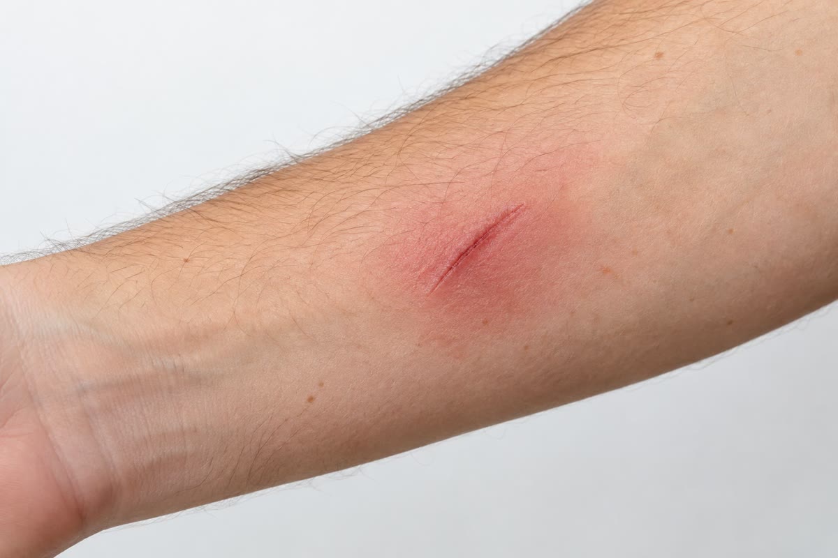
A splinter under the skin carries in ten thousand bacteria that will double every half hour. Within minutes the vessels around it dilate and leak; within an hour the first neutrophils have squeezed out of the blood and are crawling toward the intruders along a chemical trail; within a day the site is red, hot, swollen and painful — the four signs of inflammation that Celsus listed two thousand years ago — and the bacteria are dead, eaten alive by cells that recognised them as foreign without ever having met them before. This is innate immunity: the defence every animal has, ready before infection, encoded in the genome rather than learned. It is fast, it is what kills most invaders, and it is what tells the slower, learned immunity of the next chapter that there is something to learn. Its excesses — septic shock, chronic inflammation, the fevers of autoinflammatory disease — are as instructive as its successes.
15.1 Layers and cells
Definition 15.1 (Innate immunity)
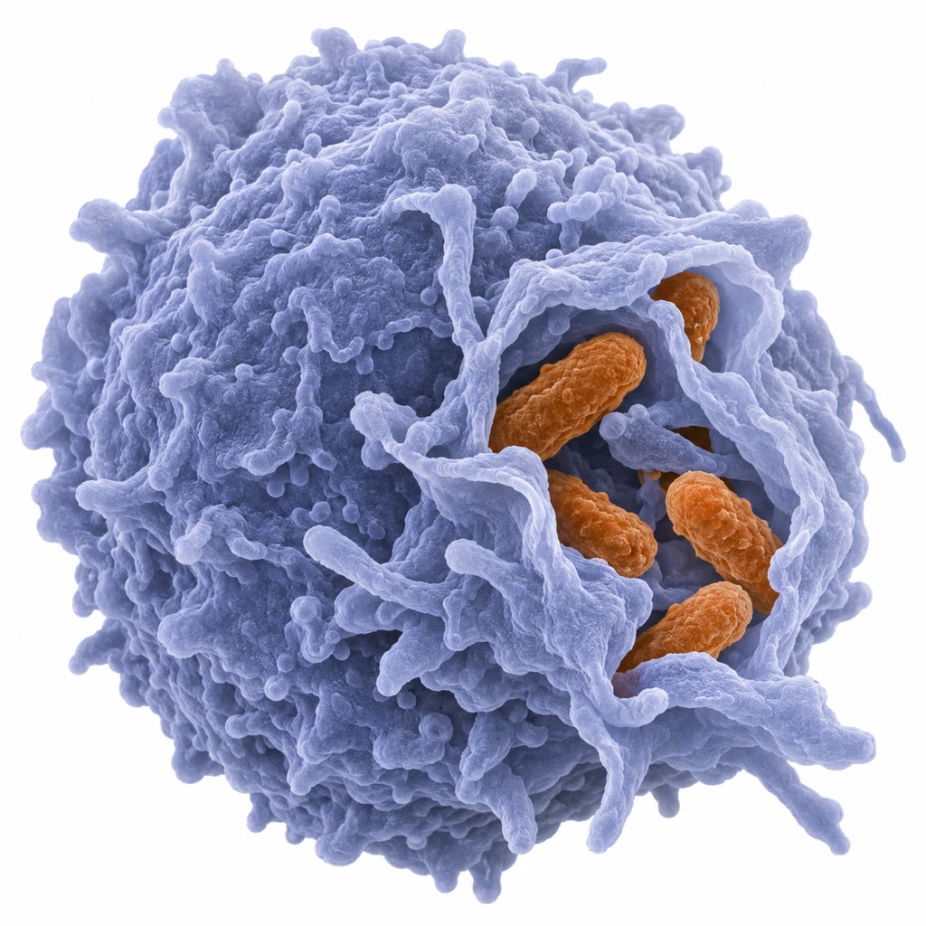
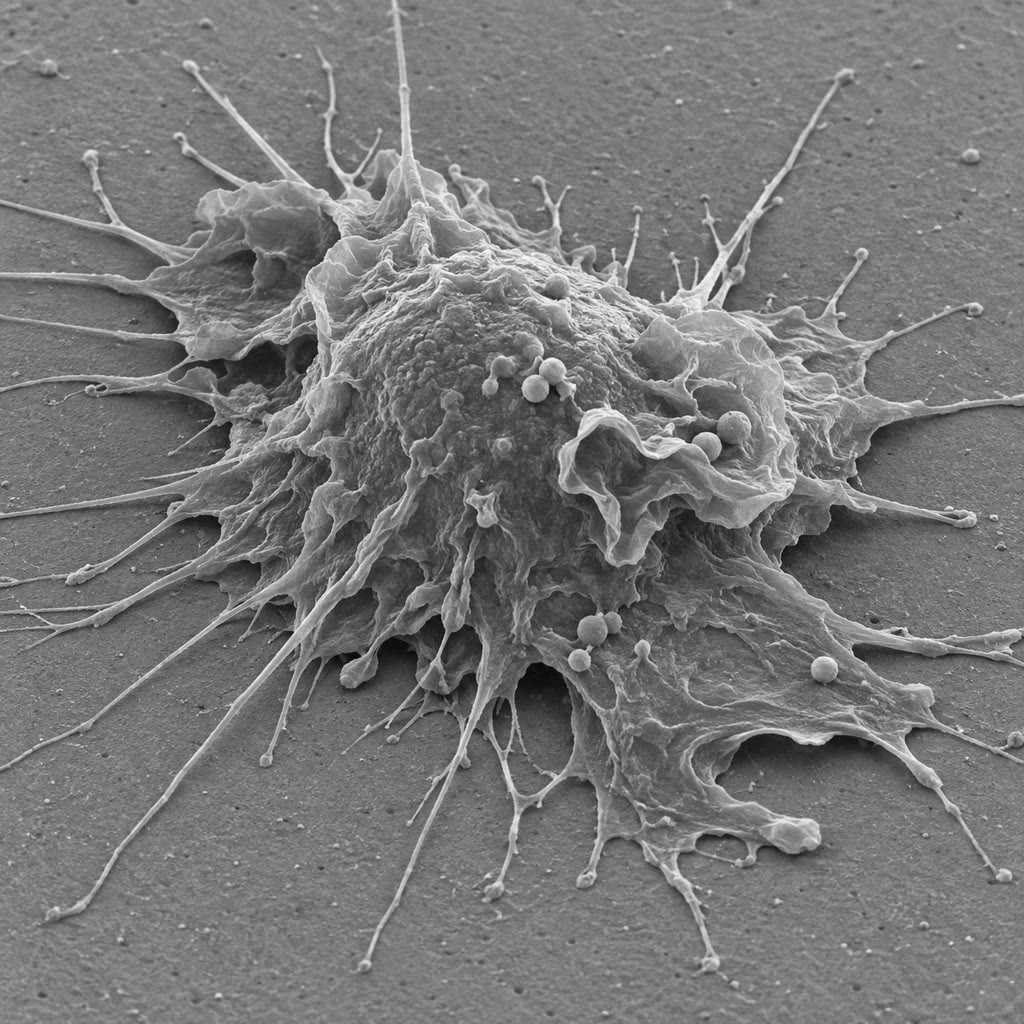
Innate immunity is the set of defences present before any exposure, encoded by germline genes that do not rearrange, recognising conserved features of microbes rather than specific individuals, acting within minutes to hours, and (with qualifications) without memory. Its first layer is the barriers: the skin’s keratin, the mucus and cilia of the airways, the acid of the stomach, the flushing of urine and tears, the enzyme lysozyme that cuts peptidoglycan, the antimicrobial peptides (defensins, cathelicidins) that puncture microbial membranes, and the commensal microbiome that occupies the niches (Chapter 14). Its cells, all from the myeloid lineage except the last: neutrophils, of the blood’s leukocytes, made a day, living a day, the first to arrive and the main killers of bacteria; macrophages, long-lived residents of every tissue (Kupffer cells in the liver, microglia in the brain, alveolar macrophages in the lung) that eat, clean up and raise the alarm; dendritic cells, which sample tissues and carry what they find to the lymph nodes; mast cells, basophils and eosinophils, armed against parasites and responsible for allergy; and natural killer (NK) cells, lymphocytes that kill infected or transformed cells on sight.
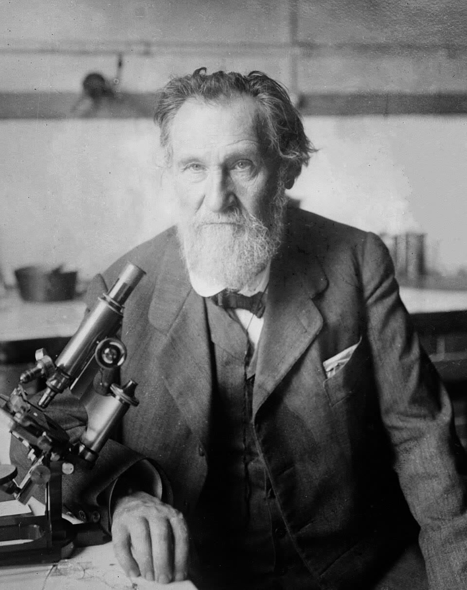
Evidence. Metchnikoff (1882), in Messina, pushed a rose thorn into a transparent starfish larva and watched, the next morning, motile cells crowding round it; he had seen the same cells engulf food particles and dye grains, and proposed that they engulfed microbes too — phagocytes, the agents of a host defence he then traced in every animal he could find, from amoebae to man. He shared the Nobel prize of 1908 with Ehrlich, whose antibodies represented the opposite, humoral view; both were right, and a century later the two halves of immunity were shown to be one system, the innate half instructing the adaptive. ∎
15.2 Recognising the enemy
Definition 15.2 (Pattern recognition)
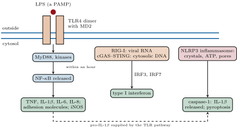
Innate cells recognise pathogen-associated molecular patterns (PAMPs): molecules common to whole classes of microbes, essential to them, and absent from the host — lipopolysaccharide of Gram-negative walls, peptidoglycan and lipoteichoic acid of Gram-positives, flagellin, fungal -glucans, double-stranded RNA, RNA without a 5 cap, DNA with unmethylated CpG, DNA in the cytosol. The receptors are pattern recognition receptors (PRRs), a few dozen in all, each encoded by one gene: the Toll-like receptors (TLRs) on the surface and in endosomes (TLR4 for lipopolysaccharide, TLR5 for flagellin, TLR3 for double-stranded RNA, TLR9 for CpG DNA); the cytosolic NOD proteins for peptidoglycan fragments, RIG-I for viral RNA, cGAS–STING for cytosolic DNA; C-type lectins for fungal sugars. The same receptors, or others of the same families, recognise damage-associated molecular patterns (DAMPs) released by injured cells — ATP, uric acid, the nuclear protein HMGB1, mitochondrial DNA — so that sterile injury also inflames. Binding activates the transcription factors NF-B and the interferon regulatory factors, which switch on cytokines, chemokines, adhesion molecules and antimicrobial genes within an hour; a subset of the cytosolic sensors assemble the inflammasome, a platform that activates caspase-1 to cut interleukin-1 into its active form and to trigger the inflammatory death called pyroptosis (Chapter 10).
Evidence. Janeway proposed in 1989 that the adaptive immune system does not respond to antigen alone but needs a signal from innate receptors that have recognised microbial patterns — the explanation of why vaccines need adjuvants, “the immunologist’s dirty little secret”. Hoffmann (1996) found that Drosophila lacking the receptor Toll, known for its role in embryonic patterning, could not mount an antifungal response and died covered in mould; Beutler (1998) mapped the lipopolysaccharide-resistance of a mouse strain, which survived doses of endotoxin lethal to others and failed to clear Gram-negative infections, to a mutation in the mammalian homologue TLR4. The receptor for endotoxin, sought for fifty years, was a Toll. ∎
Definition 15.3 (Complement)
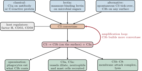
Complement is a system of some thirty plasma proteins, of the serum globulins, that marks and destroys microbes by a cascade of proteolytic activations. Three pathways converge on the cleavage of the central protein C3: the classical pathway, started by C1q binding antibodies on a surface (or C-reactive protein, or apoptotic cells); the lectin pathway, started by mannose-binding lectin recognising microbial sugars; and the alternative pathway, in which C3 hydrolyses spontaneously at a low rate and the resulting C3b attaches to any surface it meets. Each pathway builds a C3 convertase, an enzyme that cleaves more C3; the C3b it deposits covalently on the surface builds more convertase in turn — an amplification loop — so that a bacterium is coated with millions of C3b within minutes. C3b is an opsonin: phagocytes carry receptors for it and eat what it coats. The small fragments C3a and C5a are anaphylatoxins that dilate vessels, attract neutrophils and activate mast cells. And C5b assembles C6–C9 into the membrane attack complex, a pore of that lyses Gram-negative bacteria and enveloped viruses. Host cells are spared because they carry regulators — factor H, CD55, CD59 — that dismantle the convertase and block the pore on their own surfaces; microbes, lacking them, are consumed by the loop.
Theorem 15.4 (The complement loop as a kinetic discriminator)
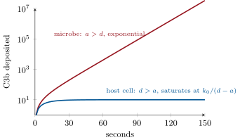
Let be the number of C3b molecules deposited on a surface. C3b is deposited at a small constant rate from the tick-over of C3, and each deposited C3b builds convertase that deposits more at rate per C3b per second, while regulators (host) and decay remove convertase activity at rate per C3b per second:
On a surface where — a microbe, without regulators — grows exponentially with time constant ; on a host surface where , approaches the small steady value . The same molecules, at the same concentrations, deposit a million C3b on a bacterium in a few minutes and a few dozen on the cell beside it: the distinction between self and non-self is made not by recognition but by the sign of .
Proof. The equation is linear with constant coefficients. For the fixed point is ; writing , , so and with , . For this grows without bound (until C3 or surface runs out); for the exponential decays and . With per second, on a bacterium, ; with on a host cell, . ∎
15.3 Inflammation
Definition 15.5 (Acute inflammation)
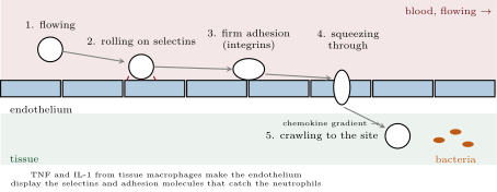
Inflammation is the response of vascularised tissue to injury or infection, orchestrated by cytokines — the small signalling proteins of immune cells, here chiefly TNF, interleukin-1 and interleukin-6 from activated macrophages — and by chemokines, the cytokines that direct migration. Its steps explain Celsus’s signs. Histamine from mast cells, nitric oxide and prostaglandins dilate the arterioles: redness and heat. The endothelium of the venules contracts and leaks plasma, whose proteins — complement, antibodies, clotting factors — flood the tissue: swelling. Bradykinin and prostaglandin E sensitise nerve endings: pain. And the leukocytes arrive by extravasation: TNF and interleukin-1 make the endothelium display selectins, on which passing neutrophils catch and roll; chemokines (interleukin-8) on the endothelial surface activate the neutrophils’ integrins, which bind tightly to endothelial adhesion molecules and stop the cell; and the neutrophil squeezes between the endothelial cells and follows the chemokine gradient into the tissue — the whole sequence in a few minutes, at up to a million cells an hour into an infected site.
Proposition 15.6 (Fever, the acute phase and resolution)
Interleukin-1, TNF and interleukin-6 also act at a distance. In the hypothalamus they induce prostaglandin E, which raises the set point of body temperature: fever, which slows many bacteria, speeds the immune cells’ own reactions, and costs about more metabolism per degree. In the liver they induce the acute-phase proteins — C-reactive protein, which binds bacterial phosphocholine and activates complement and rises a thousandfold within two days, the clinician’s measure of inflammation; fibrinogen; mannose-binding lectin; and hepcidin, which locks iron away from the microbes that need it. In the marrow they release neutrophils and speed their production. Inflammation is meant to end: as the stimulus is cleared, the lipid mediators switch from prostaglandins to lipoxins and resolvins, neutrophils die by apoptosis and are eaten by macrophages, which then switch to a repair programme, secreting growth factors and interleukin-10 — resolution. When the stimulus persists (a tubercle bacillus that cannot be killed, a crystal, a self-antigen) the response becomes chronic: macrophages wall the site in a granuloma, fibroblasts lay down scar, and the tissue is destroyed by its own defence.
Example 15.7 (Sepsis)
Inflammation confined to a splinter is a cure; the same reactions throughout the body are a catastrophe. When bacteria or their lipopolysaccharide reach the blood in quantity, macrophages everywhere release TNF and interleukin-1 at once: every vessel dilates and leaks, blood pressure collapses, clotting is activated in ten thousand capillaries and consumes the clotting factors, and the organs, starved of perfusion, fail — septic shock, which kills a quarter to a half of those it strikes, some eleven million people a year. A few micrograms of lipopolysaccharide injected into a volunteer produce fever, rigors and a fall in blood pressure within two hours; a mouse lacking TLR4 shrugs off a dose a hundred times lethal. The disease is the host’s response, and the antibiotics that kill the bacteria can make it worse for a few hours by releasing more endotoxin.
15.4 Killing
Definition 15.8 (Phagocytosis and the respiratory burst)
A phagocyte binds its prey through receptors for opsonins — C3b, and the constant regions of antibodies — or directly through pattern receptors, wraps it in membrane, and closes the phagosome, which fuses with granules and lysosomes. Killing is by several means at once. The respiratory burst: the enzyme NADPH oxidase, assembled on the phagosome membrane, pumps electrons onto oxygen to make superoxide, which becomes hydrogen peroxide, which myeloperoxidase turns, with chloride, into hypochlorite — bleach, at millimolar concentration inside a compartment a micrometre across. Nitric oxide from inducible NO synthase, which with superoxide gives peroxynitrite. Acid, to pH 5. Lysozyme, proteases, lactoferrin that starves the prey of iron, and defensins that puncture it. A neutrophil that cannot eat what it finds — a fungal hypha, a clump — can expel its own chromatin as a sticky net of DNA and granule proteins, a neutrophil extracellular trap, and die doing it. Children who inherit a defective NADPH oxidase (chronic granulomatous disease) suffer recurrent abscesses and granulomas from bacteria and fungi that ordinary phagocytes kill without difficulty: the burst is not optional.
Definition 15.9 (Natural killer cells and interferon)
Viruses hide inside cells, where complement and phagocytes cannot reach them. Natural killer cells patrol for cells that have stopped displaying MHC class I — the “missing self” of a cell whose viral or tumour occupant has switched off antigen presentation to escape T cells (Chapter 16) — and for stress proteins that infected and transformed cells put on their surface; inhibitory receptors for MHC I and activating receptors for the stress ligands are summed, and a cell that tips the balance is killed within minutes by perforin and granzymes injected into it, which trigger its apoptosis. Type I interferons ( and ) are made by any cell whose RIG-I or cGAS has detected viral nucleic acid; secreted, they bind receptors on neighbouring cells and, through the JAK–STAT pathway, induce hundreds of genes that put those cells into an antiviral state: a kinase that shuts down translation when it meets double-stranded RNA, a nuclease that degrades RNA, proteins that block viral entry and assembly, and more MHC I to show T cells what is inside. The cell that made the interferon may die; its neighbours are forewarned. Interferon was discovered by Isaacs and Lindenmann (1957) as the factor in the medium of virus-infected cells that made fresh cells resistant, and it is the reason a cell infected by one virus resists a second.
Method 15.10 (Measuring inflammation and phagocyte function)
At the bedside: (1) the C-reactive protein in serum, by immunoassay — below at rest, tens in a viral infection, hundreds in bacterial sepsis, falling within days of effective treatment; (2) the neutrophil count, raised in bacterial infection, with immature forms released early from the marrow; (3) the erythrocyte sedimentation rate, a slow proxy for fibrinogen. For the phagocytes: (4) the nitroblue tetrazolium or dihydrorhodamine test, in which neutrophils stimulated in vitro reduce a dye if their NADPH oxidase works — the diagnosis of chronic granulomatous disease in a morning; (5) a chemotaxis assay, counting cells that migrate through a filter toward a chemokine.
15.5 From innate to adaptive, and across life
Proposition 15.11 (The dendritic cell’s decision)
Adaptive immunity does not start itself. A dendritic cell in a tissue eats what it finds; if its pattern receptors are triggered — by microbe or by damage — it matures: it stops eating, migrates through the lymphatics to the draining lymph node, displays the peptides of what it ate on MHC molecules, and puts up costimulatory molecules (B7) and cytokines that tell a T cell recognising the peptide to respond, and how (Chapter 16). A T cell that sees its peptide on a dendritic cell without costimulation is switched off. Innate recognition thus decides whether an antigen is worth a response, and the cytokines the dendritic cell makes — decided by which pattern receptors fired — decide whether the response is aimed at viruses, bacteria or worms. An adjuvant is a pattern-receptor ligand added to a vaccine to supply this signal; the alum and the lipid nanoparticles of modern vaccines work by triggering it.
Remark 15.12 (Innate immunity everywhere, and its memory)
Every multicellular organism has innate immunity, and plants, fungi and invertebrates have nothing else. Plants recognise flagellin and chitin with receptor kinases and mount a two-tier response (Chapter 22); insects have Toll and antimicrobial peptides; even bacteria have restriction enzymes and CRISPR (Chapter 6). The dogma that innate immunity has no memory has softened: a monocyte that has met -glucan or the BCG vaccine is epigenetically reprogrammed (Chapter 1) to respond more strongly to unrelated microbes for months — trained immunity, which explains why BCG lowers infant mortality from infections other than tuberculosis. And the system’s excess is a disease of our time: sterile inflammation driven by uric acid crystals (gout), cholesterol crystals in the arterial wall (atherosclerosis), amyloid in the brain, and the low-grade inflammation of obesity and old age.
15.6 Exercises
Exercise 15.1 ★
List four barriers and five cell types of innate immunity, with one function each.
Solution
Solution of Exercise 15.1.
Barriers: skin keratin (mechanical), airway mucus and cilia (trapping and clearance), stomach acid (kills swallowed microbes), lysozyme and defensins in secretions (lyse and puncture), the commensal microbiome (occupies niches). Cells: neutrophils (kill bacteria by phagocytosis), macrophages (eat, clean up, raise the alarm), dendritic cells (carry antigen to lymph nodes and start adaptive responses), mast cells (release histamine, inflame), natural killer cells (kill infected and transformed cells).
Exercise 15.2 ★
What makes a molecule a good pathogen-associated molecular pattern? Give three examples and the receptor for each.
Solution
Solution of Exercise 15.2.
Shared by a whole class of microbes, essential to them (so it cannot be lost to escape), and absent from the host. Lipopolysaccharide — TLR4; flagellin — TLR5; double-stranded RNA — TLR3 (and RIG-I in the cytosol); unmethylated CpG DNA — TLR9; peptidoglycan fragments — NOD proteins; -glucan — dectin-1.
Exercise 15.3 ★
Explain each of Celsus’s four signs by a mediator and a vascular or neural change.
Solution
Solution of Exercise 15.3.
Redness: arteriolar dilation by histamine, nitric oxide and prostaglandins. Heat: the same increased blood flow. Swelling: venular leak of plasma after endothelial contraction (histamine, C3a, C5a). Pain: bradykinin and prostaglandin E sensitising nociceptors, plus pressure from the swelling.
Exercise 15.4 ★
Name the three complement pathways, what starts each, and the three outcomes of C3 cleavage.
Solution
Solution of Exercise 15.4.
Classical: C1q binding antibody (or C-reactive protein) on a surface. Lectin: mannose-binding lectin on microbial sugars. Alternative: spontaneous C3 tick-over and C3b deposition on any unprotected surface. Outcomes: C3b opsonises for phagocytosis; C3a and C5a inflame and recruit; C5b starts the membrane attack complex that lyses.
Exercise 15.5 ★★
In the complement model with per second, and on a microbe, how many C3b are deposited after ? After ? On a host cell with and , what is the steady state?
Solution
Solution of Exercise 15.5.
with : at , ; at , . Host cell: .
Exercise 15.6 ★★
Order the events of extravasation, name the molecule class at each step, and predict the phenotype of a child whose neutrophils lack integrins (leukocyte adhesion deficiency).
Solution
Solution of Exercise 15.6.
Rolling on selectins (lectins binding carbohydrate); chemokine signalling that activates integrins; firm adhesion of integrins to endothelial adhesion molecules (ICAM); transmigration between endothelial cells; chemotaxis along the gradient. Without integrins the neutrophils roll but never stop or cross: they pile up in the blood (very high counts), infections recur without pus forming, wounds heal badly, and the umbilical cord separates late.
Exercise 15.7 ★★
A patient’s neutrophils fail the dihydrorhodamine test. What is the diagnosis, which reaction is missing, and which infections do you expect? Why are granulomas formed?
Solution
Solution of Exercise 15.7.
Chronic granulomatous disease: the NADPH oxidase is defective, so no superoxide, hydrogen peroxide or hypochlorite is made in the phagosome. Infections by organisms that destroy their own hydrogen peroxide (catalase-positive): Staphylococcus aureus, Aspergillus, Burkholderia, Serratia, Nocardia. Granulomas form because macrophages engulf microbes they cannot kill and wall them off, with continual recruitment, in a chronic lesion.
Exercise 15.8 ★★
Explain missing-self recognition and predict what happens to a tumour cell that (a) loses MHC I to escape T cells, (b) keeps MHC I but displays stress ligands.
Solution
Solution of Exercise 15.8.
Inhibitory receptors count MHC I; activating receptors count stress ligands; the cell dies if activation outweighs inhibition. (a) Losing MHC I removes the inhibition: the natural killer cell kills it — the cost of escaping T cells. (b) With MHC I kept but stress ligands displayed, activation can still win and the cell is killed; tumours therefore often shed or suppress the stress ligands.
Exercise 15.9 ★★
Why does a vaccine of pure protein induce little immunity, and what does an adjuvant add? Relate to Janeway’s argument.
Solution
Solution of Exercise 15.9.
A pure protein carries no pathogen pattern, so the dendritic cells that take it up do not mature, present it without costimulation, and the T cells that recognise it are switched off or ignore it. An adjuvant — alum, a lipid, a Toll-like receptor ligand — triggers pattern receptors, matures the dendritic cell and supplies the second signal. This is Janeway’s point: the adaptive system responds to antigen only when the innate system says the antigen came with a microbe.
Exercise 15.10 ★★★
Fever costs of resting metabolism per degree and slows a bacterium’s doubling from at to at . Over a day of infection, compute the bacterial population from with and without fever (ignoring killing), and the extra energy for a person. Is fever worth it? What does the answer depend on?
Solution
Solution of Exercise 15.10.
Without fever, doublings: (an absurdity that shows killing, not growth, decides the outcome). With fever, doublings: — a factor fewer bacteria for the neutrophils to face. Cost: a day, a large meal. Worth it when the pathogen is slowed and the host has reserves; not when the pathogen grows as well at , or when the host is starving, or when the fever itself approaches dangerous temperatures.
Exercise 15.11 ★★★
Some bacteria bind factor H to their surface; others cleave C3b or shed their capsule. Using Theorem 15.4, explain each as a change of or , and say which is the most economical defence for the microbe.
Solution
Solution of Exercise 15.11.
Binding factor H recruits the host’s own regulator: rises above and the loop decays on the microbe as on a host cell. Cleaving C3b removes deposited convertase: again a rise in . A capsule presents a surface on which convertase forms poorly, lowering , and hides the C3b that is deposited from phagocyte receptors. Cheapest: one surface protein that binds factor H, which the host supplies free and which does the work.
Exercise 15.12 ★★★
Argue, with the mechanisms of this chapter, why the same response is adaptive at a splinter and lethal in the bloodstream, and why drugs that block TNF help rheumatoid arthritis but raise the risk of tuberculosis.
Solution
Solution of Exercise 15.12.
At a splinter the dilation, leak and recruitment involve a few millilitres of vessels and deliver defence where it is needed; the same mediators released into the whole circulation dilate every vessel and leak every capillary, so the pressure collapses and clotting is triggered everywhere — the physiology is identical, the scale is lethal. TNF drives the synovial inflammation of rheumatoid arthritis, so blocking it relieves the disease; but TNF is also what keeps macrophages organised in the granulomas that contain dormant tubercle bacilli, and blocking it lets latent tuberculosis escape.
15.7 Problem: A Splinter
Problem 15.1
Weekend problem — ten thousand bacteria under the skin raced against the neutrophils sent to meet them, coated with complement by the arithmetic of the loop, fought with fever at its metabolic price, and, in the bloodstream, turned into the physiology of shock, ending on the bacteria left after six hours, the C3b on each one and the cost of a day’s fever
Data: bacteria inoculated, doubling every ; neutrophils arrive from at per hour, each killing bacteria then dying. Complement: per second per bacterium, on the bacteria, on host cells; a bacterium’s surface holds at most C3b. Fever: costs of an metabolism and lengthens the bacterial doubling time to . Sepsis: bacteria in of blood, lipopolysaccharide molecules per bacterium; TLR4 signalling saturates at mol/L of lipopolysaccharide.
Part I — The race.
- How many bacteria at , when the first neutrophils arrive, if none have been killed?
- Between and , how many neutrophils arrive and how many bacteria can they kill? Compare with the bacterial growth in that hour from the count of question 1.
- Continue hour by hour (growth, then killing at the end of each hour) to . When does the population peak, and how many remain at ?
- Repeat with a neutrophil arrival delayed to . What happens by ? Comment on the value of speed.
- How many dead neutrophils accumulate by the time the bacteria are gone in question 3? What is pus, and why does it need to be cleared?
- A person with a tenth of the normal neutrophil count (neutropenia after chemotherapy): recompute question 3 and explain why such patients are given antibiotics at the first fever.
Part II — The coat.
- From the theorem, how many C3b are deposited on one bacterium after , and ?
- When does the surface saturate?
- On a host cell beside it, what is the steady-state number of C3b?
- The bacterium acquires a capsule that halves , so that . Recompute the C3b at . What does the capsule buy?
- How many C3b does the whole inoculum of bacteria carry at saturation, and what fraction is that of the C3 molecules in a litre of plasma?
- A patient lacks C3. Which pathways are affected, and what infections follow? A patient lacks C9: why is the phenotype milder, and confined to one genus?
Part III — Fever.
- How much extra energy does two days of fever at cost, in megajoules and in grams of fat ()?
- Redo question 1 at fever temperature: how many bacteria at ? By what factor has fever reduced the population the neutrophils meet?
- In the model of question 3, with the longer doubling time, when are the bacteria gone?
- C-reactive protein rises from to in two days. With a plasma volume of and a molecular mass of , how many molecules has the liver made, and how many per second?
- Antipyretic drugs block prostaglandin synthesis. What do they do to the set point, the comfort of the patient and, by the numbers above, to the infection?
- Why is a fever of dangerous when is not, and what stops the set point rising indefinitely?
Part IV — Shock.
- Lipopolysaccharide molecules in the blood of the septic patient, and their concentration in mol/L.
- Compare with the saturating concentration for TLR4. Are the body’s macrophages all activated?
- Each of the body’s macrophages releases TNF molecules a second for an hour. How many moles of TNF, and what concentration in of extracellular fluid? (TNF acts at .)
- Explain the fall in blood pressure and the failure of the kidneys from the local mechanisms of inflammation.
- The antibiotic given at admission kills the bacteria within an hour. Why may the patient worsen before improving, and what limits the usefulness of anti-TNF antibodies in sepsis?
- A mouse strain lacks TLR4. Predict its response to injected lipopolysaccharide and to a live Gram-negative infection, and explain the apparent paradox.
- Summarise: the bacteria remaining at (question 3), the C3b on one bacterium at (question 7), and the cost of two days’ fever (question 13).
Solution
Solution of Problem 15.1.
1. One hour is doublings: . 2. neutrophils kill up to ; the bacteria grow only from to : killing outstrips growth fivefold. 3. Hour 2: before the kill, none after. The population peaks at about near and is zero from then on; none remain at . 4. Arrival at : then. Hour 4: ; hour 5: ; hour 6: and rising — the neutrophils no longer keep up, and an abscess forms. Two hours’ delay turns a cure into a chronic infection. 5. neutrophils die killing, plus the that arrived and die anyway within a day. Pus is dead neutrophils, dead bacteria and liquefied tissue; macrophages clear it by eating the corpses, or it must drain. 6. Arrivals an hour, kill : hour 2, ; hour 3, ; hour 4, — growing without limit. The patient cannot contain a trivial inoculum, so antibiotics must do the killing from the first sign. 7. : , ; , ; , . 8. : . 9. . 10. , times fewer. The capsule buys minutes — time to multiply, and protection until antibodies arrive to start the classical pathway on the capsule itself. 11. C3b: one millionth of the plasma’s C3. 12. Without C3 all three pathways stop at their common step: no opsonisation, no anaphylatoxins, no lysis — severe recurrent infections with encapsulated bacteria. Without C9 only the terminal pore is lost; opsonisation and inflammation work, and the phenotype is recurrent Neisseria infections, the one genus that phagocytes control poorly and lysis controls well. 13. , about of fat. 14. One doubling in the hour: instead of , a factor . 15. Hour 2: before the kill, none after — gone at , as before; fever’s contribution matters when the neutrophil supply is marginal, as in questions 4 and 6. 16. ; mol molecules; over , a second. 17. They lower the set point back toward , the patient feels better, and the bacteria double a little faster; in this model the neutrophils still win, and the evidence that antipyretics worsen ordinary infections is weak. 18. Above about proteins begin to denature, enzymes fail and neurons misfire (seizures); is lethal. The set point is held below that by cryogens — interleukin-10, vasopressin, glucocorticoids — and by the ceiling of prostaglandin action in the hypothalamus. 19. molecules mol in : . 20. Equal to the saturating concentration: every macrophage in the body is maximally activated at once. 21. molecules mol; in , — four thousand times the active concentration. 22. Every arteriole dilates and every venule leaks: the blood volume pools in the widened vessels and escapes into the tissues, so the pressure falls; the kidneys, underperfused, stop filtering; and clotting triggered in thousands of capillaries blocks them and consumes the clotting factors. 23. Killed Gram-negatives release their whole lipopolysaccharide at once, a bolus for TLR4, so the cytokine surge can worsen for hours. Anti-TNF fails because by the time of shock the cascade has passed TNF — interleukin-1, interleukin-6 and coagulation are already running — and because TNF is needed to control the bacteria: timing, not the target, is the problem. 24. Injected lipopolysaccharide does nothing to the TLR4-null mouse, which survives doses lethal to normal mice; a live Gram-negative infection kills it, because it cannot detect the bacteria early and mount the inflammation that contains them. The paradox dissolves: the detector that causes the shock is the detector that provides the defence. 25. No bacteria remain at (all killed by ); about C3b per bacterium at ; two days’ fever costs , some of fat.